

Turkish Journal of Hematology Volume: 31 - Issue: 4

You also want an ePaper? Increase the reach of your titles
YUMPU automatically turns print PDFs into web optimized ePapers that Google loves.
Turk J Hematol 2014;<strong>31</strong>:342-356<br />
Arıkan Akdağlı S, et al: Diagnosis <strong>of</strong> IFI in Hematological Malignancies<br />
thermally dimorphic fungi causing endemic mycosis, and<br />
the other yeasts with clinical importance [Blastoschizomyces<br />
(Saprochaete), Rhodotorula, Trichosporon, etc.] and for the<br />
diagnosis <strong>of</strong> infections caused by these fungi. If the fact that<br />
Turkey is not in an endemic region for thermally dimorphic<br />
fungi is considered, excluding possible travel-related<br />
infections, isolation <strong>of</strong> fungi causing endemic mycosis from<br />
blood does not carry primary importance. Fungi other<br />
than those causing endemic mycoses and expected to be<br />
isolated in blood culture can be isolated in blood culture<br />
at varying rates in our country, with Candida spp. being the<br />
leading isolate. However, the sensitivity <strong>of</strong> blood culture in<br />
candidemia diagnosis is generally below the desired level.<br />
Autopsy studies show that candidemia can be diagnosed by<br />
blood culture in 50-70% <strong>of</strong> cases [10,11].<br />
Different methods or parameters are being tested<br />
to enhance the sensitivity <strong>of</strong> blood culture in fungemia<br />
diagnosis, and the leading methods are lysis centrifugation<br />
method and use <strong>of</strong> fungal media. The lysis centrifugation<br />
method is especially recommended for the identification<br />
<strong>of</strong> dimorphic fungi in blood culture [12]. However,<br />
studies published in previous years, particularly those<br />
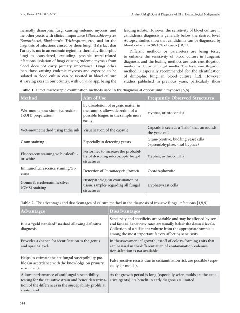
Table 1. Direct microscopic examination methods used in the diagnosis <strong>of</strong> opportunistic mycoses [5,6].<br />
Method Aim <strong>of</strong> Use Frequently Observed Structures<br />
Wet-mount potassium hydroxide<br />
(KOH) preparation<br />
Wet-mount method using India ink<br />
Gram staining<br />
Fluorescent staining with calc<strong>of</strong>luor-white<br />
Immun<strong>of</strong>luorescence staining/Giemsa<br />
Gomori’s methenamine silver<br />
(GMS) staining<br />
By dissolution <strong>of</strong> organic matter in<br />
the sample, allows detection <strong>of</strong> a<br />
possible fungus in the sample more<br />
easily<br />
Visualization <strong>of</strong> the capsule<br />
Especially in detecting yeasts<br />
Performed to increase the probability<br />
<strong>of</strong> detecting microscopic fungal<br />
structures<br />
Detection <strong>of</strong> Pneumocystis jirovecii<br />
Histopathological examination <strong>of</strong><br />
tissue samples regarding all fungal<br />
structures<br />
Hyphae, arthroconidia<br />
Capsule is seen as a “halo” that surrounds<br />
the yeast cell<br />
Gram-positive, budding yeast cells<br />
(+pseudohyphae, -real hyphae)<br />
Hyphae, arthroconidia<br />
Cyst/trophozoite<br />
Hyphae/yeast cells<br />
Table 2. The advantages and disadvantages <strong>of</strong> culture method in the diagnosis <strong>of</strong> invasive fungal infections [4,8,9].<br />
Advantages<br />
It is a “gold standard” method allowing definitive<br />
diagnosis.<br />
Provides a chance for identification to the genus<br />
and species level.<br />
Helps to estimate the antifungal susceptibility pr<strong>of</strong>ile<br />
(in accordance with the knowledge on primary<br />
resistance).<br />
Allows performance <strong>of</strong> antifungal susceptibility<br />
testing for the causative strain and hence determination<br />
<strong>of</strong> the differences in the susceptibility pr<strong>of</strong>ile at<br />
strain level.<br />
Disadvantages<br />
Sensitivity and specificity are variable and may be affected by several<br />
factors. Sensitivity rates are usually below the desired levels.<br />
Collection <strong>of</strong> a sufficient volume from the appropriate sample is<br />
among the most important factors affecting sensitivity.<br />
In the assessment <strong>of</strong> growth, cut<strong>of</strong>f <strong>of</strong> colony-forming units that<br />
can be used in the differentiation <strong>of</strong> contamination-colonization-infection<br />
is not available.<br />
False positive results due to contamination risk are possible (especially<br />
for molds).<br />
As the growth period is long (especially when molds are the causative<br />
agents), its benefit in early diagnosis is limited.<br />
344


