

Turkish Journal of Hematology Volume: 31 - Issue: 4

You also want an ePaper? Increase the reach of your titles
YUMPU automatically turns print PDFs into web optimized ePapers that Google loves.
Turk J Hematol 2014;<strong>31</strong>:408-410<br />
Anoun S, et al: Primary Splenic Angiosarcoma<br />
angiosarcomas arising in the spleen have a unique propensity<br />
for bone marrow metastasis, but this is unfortunately not well<br />
documented in the literature [8]. We have found 3 cases in<br />
the literature in which primary splenic angiosarcoma was<br />
extended to the bone marrow.<br />
The KI-67 proliferation index determines prognosis.<br />
Because splenic angiosarcoma is a rare tumor, no specific<br />
regimen <strong>of</strong> chemotherapy has been employed in enough cases<br />
to enable the drawing <strong>of</strong> a conclusion as to effects on survival<br />
[6].<br />
Despite best efforts, the prognosis for this diagnosis is poor,<br />
with mean survival ranging from 10.3 to 14.4 months [9].<br />
Conclusion<br />
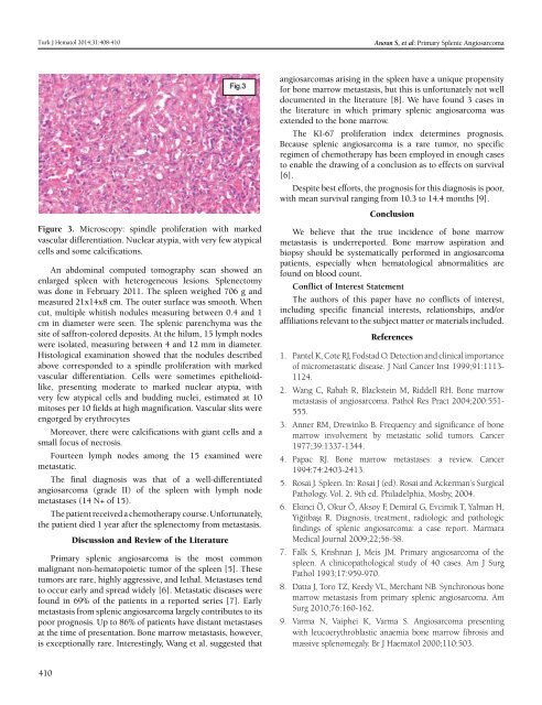
Figure 3. Microscopy: spindle proliferation with marked<br />
vascular differentiation. Nuclear atypia, with very few atypical<br />
cells and some calcifications.<br />
An abdominal computed tomography scan showed an<br />
enlarged spleen with heterogeneous lesions. Splenectomy<br />
was done in February 2011. The spleen weighed 706 g and<br />
measured 21x14x8 cm. The outer surface was smooth. When<br />
cut, multiple whitish nodules measuring between 0.4 and 1<br />
cm in diameter were seen. The splenic parenchyma was the<br />
site <strong>of</strong> saffron-colored deposits. At the hilum, 15 lymph nodes<br />
were isolated, measuring between 4 and 12 mm in diameter.<br />
Histological examination showed that the nodules described<br />
above corresponded to a spindle proliferation with marked<br />
vascular differentiation. Cells were sometimes epithelioidlike,<br />
presenting moderate to marked nuclear atypia, with<br />
very few atypical cells and budding nuclei, estimated at 10<br />
mitoses per 10 fields at high magnification. Vascular slits were<br />
engorged by erythrocytes<br />
Moreover, there were calcifications with giant cells and a<br />
small focus <strong>of</strong> necrosis.<br />
Fourteen lymph nodes among the 15 examined were<br />
metastatic.<br />
The final diagnosis was that <strong>of</strong> a well-differentiated<br />
angiosarcoma (grade II) <strong>of</strong> the spleen with lymph node<br />
metastases (14 N+ <strong>of</strong> 15).<br />
The patient received a chemotherapy course. Unfortunately,<br />
the patient died 1 year after the splenectomy from metastasis.<br />
Discussion and Review <strong>of</strong> the Literature<br />
Primary splenic angiosarcoma is the most common<br />
malignant non-hematopoietic tumor <strong>of</strong> the spleen [5]. These<br />
tumors are rare, highly aggressive, and lethal. Metastases tend<br />
to occur early and spread widely [6]. Metastatic diseases were<br />
found in 69% <strong>of</strong> the patients in a reported series [7]. Early<br />
metastasis from splenic angiosarcoma largely contributes to its<br />
poor prognosis. Up to 86% <strong>of</strong> patients have distant metastases<br />
at the time <strong>of</strong> presentation. Bone marrow metastasis, however,<br />
is exceptionally rare. Interestingly, Wang et al. suggested that<br />
We believe that the true incidence <strong>of</strong> bone marrow<br />
metastasis is underreported. Bone marrow aspiration and<br />
biopsy should be systematically performed in angiosarcoma<br />
patients, especially when hematological abnormalities are<br />
found on blood count.<br />
Conflict <strong>of</strong> Interest Statement<br />
The authors <strong>of</strong> this paper have no conflicts <strong>of</strong> interest,<br />
including specific financial interests, relationships, and/or<br />
affiliations relevant to the subject matter or materials included.<br />
References<br />
1. Pantel K, Cote RJ, Fodstad O. Detection and clinical importance<br />
<strong>of</strong> micrometastatic disease. J Natl Cancer Inst 1999;91:1113-<br />
1124.<br />
2. Wang C, Rabah R, Blackstein M, Riddell RH. Bone marrow<br />
metastasis <strong>of</strong> angiosarcoma. Pathol Res Pract 2004;200:551-<br />
555.<br />
3. Anner RM, Drewinko B. Frequency and significance <strong>of</strong> bone<br />
marrow involvement by metastatic solid tumors. Cancer<br />
1977;39:1337-1344.<br />
4. Papac RJ. Bone marrow metastases: a review. Cancer<br />
1994:74:2403-2413.<br />
5. Rosai J. Spleen. In: Rosai J (ed). Rosai and Ackerman’s Surgical<br />
Pathology. Vol. 2. 9th ed. Philadelphia, Mosby, 2004.<br />
6. Ekinci Ö, Okur Ö, Aksoy F, Demiral G, Evcimik T, Yalman H,<br />
Yiğitbaşı R. Diagnosis, treatment, radiologic and pathologic<br />
findings <strong>of</strong> splenic angiosarcoma: a case report. Marmara<br />
Medical <strong>Journal</strong> 2009;22;56-58.<br />
7. Falk S, Krishnan J, Meis JM. Primary angiosarcoma <strong>of</strong> the<br />
spleen. A clinicopathological study <strong>of</strong> 40 cases. Am J Surg<br />
Pathol 1993;17:959-970.<br />
8. Datta J, Toro TZ, Keedy VL, Merchant NB. Synchronous bone<br />
marrow metastasis from primary splenic angiosarcoma. Am<br />
Surg 2010;76:160-162.<br />
9. Varma N, Vaiphei K, Varma S. Angiosarcoma presenting<br />
with leucoerythroblastic anaemia bone marrow fibrosis and<br />
massive splenomegaly. Br J Haematol 2000;110:503.<br />
410


