

Turkish Journal of Hematology Volume: 31 - Issue: 4

You also want an ePaper? Increase the reach of your titles
YUMPU automatically turns print PDFs into web optimized ePapers that Google loves.
Özsan N, et al: Intravascular Large B Cell Lymphoma<br />
Turk J Hematol 2014;<strong>31</strong>:403-407<br />
The patient was hospitalized in the hematology department<br />
with a diagnosis <strong>of</strong> lymphoma. No enlarged superficial lymph<br />
nodes were found on physical examination. Positron emission<br />
tomography-computed tomography (PET-CT) showed an<br />
increase in fluorodeoxyglucose signals in both kidneys, which<br />
was interpreted as consistent with an inflammatory origin.<br />
Upper abdominal ultrasonography revealed mild splenomegaly<br />
(135 mm). The patient was given prednisolone at 1 g/day for<br />
10 days, when bone marrow biopsy was performed. Bone<br />
marrow biopsy revealed no infiltration <strong>of</strong> lymphoma from<br />
either morphology or immunohistochemistry. R-CHOP<br />
chemotherapy was planned, but the patient refused to receive<br />
therapy and was discharged at his request. He received no<br />
therapy for IVLBCL and died from the disease 8 months after<br />
the diagnosis.<br />
Discussion and Review <strong>of</strong> the Literature<br />
In the current World Health Organization classification <strong>of</strong><br />
hematopoietic neoplasms, IVLBCL is defined as a rare type<br />
<strong>of</strong> extranodal large B-cell lymphoma characterized by the<br />
selective infiltration <strong>of</strong> neoplastic cells in the lumina <strong>of</strong> vessels<br />
and capillaries, with the exception <strong>of</strong> large arteries and veins<br />
[3]. The disease is widely disseminated in extranodal sites: the<br />
bone marrow, central nervous system, skin, lungs, liver, and<br />
spleen are the most common sites <strong>of</strong> involvement [4]. Lymph<br />
node infiltration and lymphadenopathy are usually absent<br />
[5]. The clinical signs and symptoms are variable, related<br />
to the site <strong>of</strong> involvement. Neoplastic cells are rarely seen in<br />
bone marrow and peripheral blood smears and so IVLBCL<br />
is very difficult to diagnose; most <strong>of</strong> the cases reported<br />
have been confirmed by autopsy or cutaneous biopsies [6].<br />
IVLBCL diagnosed in prostate specimens is extremely rare<br />
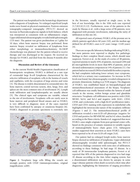
Figure 2. Neoplastic large cells filling the vessels are<br />
strongly positive with CD20 (immunoperoxidase, original<br />
magnification: x200).<br />
in the literature, usually reported as single cases; to the<br />
best <strong>of</strong> our knowledge, this is the 10th such case reported<br />
[1,7,8,9,10,11,12]. Furthermore, none <strong>of</strong> the patients in the<br />
study by Murase et al., with a large series <strong>of</strong> 96 patients, were<br />
diagnosed with prostatic involvement, indicating the rarity <strong>of</strong><br />
infiltration in this site [6].<br />
All reported cases <strong>of</strong> primary IVLBCL <strong>of</strong> the prostate are in<br />
elderly patients aged above 60 years [10], and the median age<br />
defined for all IVLBCL cases is 67 years (range: 13-85 years)<br />
[4].<br />
There are no specific laboratory findings indicating IVLBCL,<br />
but most patients were reported to display few pathologic<br />
findings in their complete blood counts, which should raise<br />
suspicion. Ferreri et al., in the study <strong>of</strong> a series <strong>of</strong> 38 patients,<br />
reported anemia in nearly 65% <strong>of</strong> patients, increased LDH and<br />
β2 microglobulin levels in more than 80% <strong>of</strong> patients, and an<br />
elevated sedimentation component in 14% <strong>of</strong> patients [1]. Our<br />
patient had an increased LDH level and mild thrombocytopenia.<br />
He had complaints indicating lower urinary tract symptoms,<br />
which led to a urinary tract examination. No increase in PSA<br />
levels was found, but ultrasonography revealed enlargement in<br />
prostatic dimensions, leading to p-TUR surgery. The diagnosis<br />
<strong>of</strong> IVLBCL was established based on histopathological and<br />
immunohistochemical assessment <strong>of</strong> the biopsy. Neoplastic<br />
cell infiltration was totally limited within the lumens <strong>of</strong> small<br />
vessels in the stroma, within benign acinar and glandular<br />
structures. Neoplastic cell infiltration was positive with CD20,<br />
MUM1, bcl-6, and bcl-2 and negative with ALK1, CD10,<br />
CD30, and cytokeratin, with a high Ki-67 proliferation index.<br />
CD34 and CD<strong>31</strong> staining with expression in endothelial cells<br />
highlighted the intravascular nature <strong>of</strong> the neoplastic infiltrate<br />
(Figures 1 and 2). The immunohistochemical pr<strong>of</strong>ile <strong>of</strong><br />
infiltration revealed a non-GCB cell phenotype (negative for<br />
CD10 and positive for IRF4/MUM1 and bcl-6) when classified<br />
according to the Hans criteria. Kanda et al. suggested that most<br />
IVLBCL cases might originate from post-germinal center cells,<br />
based on the presence <strong>of</strong> somatic mutation in variable regions<br />
<strong>of</strong> immunoglobulin heavy chain genes [13]. Subsequent<br />
studies supported their assertion as most IVLBCL cases have<br />
been reported to be <strong>of</strong> non-GCB cell origin [5,6].<br />
Our patient had no enlarged lymph nodes; bone marrow<br />
biopsy showed no infiltration <strong>of</strong> lymphoma. Upper abdominal<br />
ultrasonography revealed mild splenomegaly, but PET-CT<br />
showed no findings consistent with extra sites <strong>of</strong> infiltration;<br />
according to these findings, he had stage 1E disease. In a<br />
study <strong>of</strong> 38 intravascular lymphoma patients, Ferreri et al.<br />
reported that 40% <strong>of</strong> the patients diagnosed in vivo had stage<br />
1E disease according to the Ann Arbor staging system, but<br />
a disseminated infiltration was shown by autopsy in some<br />
stage I disease patients when death occurred a short time after<br />
diagnosis, highlighting the limitations <strong>of</strong> staging procedures<br />
405


