

Turkish Journal of Hematology Volume: 33 - Issue: 4

Create successful ePaper yourself
Turn your PDF publications into a flip-book with our unique Google optimized e-Paper software.
Patıroğlu T and Akar HH; HLA (A, B, and DRB1) Alleles and Acute Lymphoblastic Leukemia<br />
Turk J Hematol 2016;<strong>33</strong>:<strong>33</strong>9-345<br />
non-self and self-proteins. The HLA class I molecules (A, B, and<br />
C) present intracellular antigens to CD8 + T cells, while class II<br />
molecules (DR, DQ, and DP) present extracellular antigens to<br />
CD4 + T cells, which activate macrophages and B cells. HLA has a<br />
major role in regulating host responses to infections. It has been<br />
hypothesized that the HLA alleles may have an important role in<br />
predisposal to ALL [6]. The HLA genes are the most polymorphic<br />
genes in the human genome [7]. An association between ALL and<br />
HLA alleles has been shown in the literature; however, the data<br />
are not conclusive so far [8,9,10,11,12,13]. There are no identified<br />
consistent leukemia-associated HLA class I genes to date, but<br />
investigations <strong>of</strong> HLA class II genes such as DRB3, DRB4, and<br />
DRB5 have demonstrated consistent associations in patients<br />
with leukemia [14]. Genome-wide association studies have<br />
also identified some other risk alleles in different genes such as<br />
CDKN2A, PIP4K2A, GATA3, ARID5B, and CEBPE in patients with<br />
ALL [15]. In this study, we aimed to evaluate the association <strong>of</strong><br />
HLA alleles, haplotypes, and homozygosity in patients with ALL.<br />
Materials and Methods<br />
Study Population<br />
This retrospective study was performed at the HLA Tissue Typing<br />
Laboratory <strong>of</strong> Erciyes University in Kayseri, Turkey. Ninety pediatric<br />
ALL patients (58 male patients and 32 female patients, 76 patients<br />
with B-cell and 14 patients with T-cell ALL, aged 7 months to 16<br />
years) and 126 age- and sex-matched unrelated healthy controls<br />
(72 males and 54 females, aged 1-18 years) were enrolled in this<br />
study, all <strong>of</strong> <strong>Turkish</strong> ethnic origin. Participants in the control<br />
group were selected from among unrelated healthy donors who<br />
were studied for HLA alleles for transplantation (for solid organ<br />
or hematological malignancies). In the 90 patients with ALL, risk<br />
groups were as follows: 29 patients in the SRG, 37 in the MRG,<br />
and 24 in the HRG.<br />
Human Leukocyte Antigen Typing<br />
Whole venous blood specimens were collected in 2KE tubes with<br />
EDTA for all participants. Genomic DNAs were obtained using<br />
the BioRobot EZ1 (QIAGEN, Hilden, Germany). Genotyping <strong>of</strong><br />
HLA alleles was done as low-resolution typing by the polymerase<br />
chain reaction with sequence-specific oligonucleotide probe<br />
(PCR-SSOP) method (Gen-Probe Lifecodes, Stanford, CA, USA).<br />
MATCH IT DNA s<strong>of</strong>tware version 1.2.0 was used for HLA allele<br />
interpretation.<br />
Statistical Analysis<br />
Statistical analyses were performed using SPSS 22. The association<br />
<strong>of</strong> alleles, haplotypes, and homozygosity was compared with the<br />
chi-square test (χ 2 ). Two groups were in accordance with Hardy-<br />
Weinberg equilibrium (p>0.005). The Bonferroni correction test<br />
was performed for multiple comparisons in risk groups. A value<br />
<strong>of</strong> p≤0.05 was accepted to be statistically significant.<br />
Results<br />
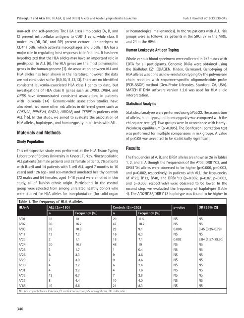
The frequencies <strong>of</strong> A, B, and DRB1 alleles are shown as 2n in Tables<br />
1, 2, and 3. Although the frequencies <strong>of</strong> the A*03, DRB1*03, and<br />
DRB1*04 alleles were observed to be higher (p=0.006, p=0.003,<br />
and p=0.002, respectively) in patients with ALL, the frequencies<br />
<strong>of</strong> A*23, B*13, B*40, and DRB1*13 (p=0.002, p=0.07, p=0.002,<br />
and p=0.003, respectively) were observed to be lower. In the<br />
second step, we evaluated the frequency <strong>of</strong> haplotypes (Table<br />
4). The A*02/B*35/DRB1*13 haplotype was found to be higher in<br />
Table 1. The frequency <strong>of</strong> HLA-A alleles.<br />
HLA-A ALL (2n=180) Controls (2n=252) p-value OR (95% CI)<br />
A*01<br />
A*02<br />
A*03<br />
A*11<br />
A*23<br />
A*24<br />
A*25<br />
A*26<br />
A*29<br />
A*30<br />
A*31<br />
A*32<br />
A*<strong>33</strong><br />
A*68<br />
n Frequency (%) n Frequency (%)<br />
18<br />
30<br />
<strong>33</strong><br />
13<br />
2<br />
30<br />
3<br />
6<br />
7<br />
4<br />
4<br />
12<br />
8<br />
10<br />
10<br />
16.7<br />
18.8<br />
7.2<br />
1.1<br />
16.7<br />
1.7<br />
3.3<br />
3.9<br />
2.2<br />
2.2<br />
6.7<br />
4.4<br />
5.6<br />
ALL: Acute lymphoblastic leukemia, CI: confidence interval, NS: nonsignificant, OR: odds ratio.<br />
29<br />
47<br />
23<br />
16<br />
18<br />
48<br />
1<br />
9<br />
9<br />
6<br />
4<br />
7<br />
10<br />
21<br />
11.5<br />
18.7<br />
9.1<br />
6.3<br />
7.1<br />
19<br />
0.4<br />
3.6<br />
3.6<br />
2.4<br />
1.6<br />
2.8<br />
4.0<br />
8.3<br />
NS<br />
NS<br />
0.006<br />
NS<br />
0.002<br />
NS<br />
NS<br />
NS<br />
NS<br />
NS<br />
NS<br />
NS<br />
NS<br />
NS<br />
NS<br />
NS<br />
0.45 (0.25-0.79)<br />
NS<br />
6.84 (1.57-29.90)<br />
NS<br />
NS<br />
NS<br />
NS<br />
NS<br />
NS<br />
NS<br />
NS<br />
NS<br />
340


