Indian Academy of Forensic Medicine (IAFM) - Official website of IAFM
Indian Academy of Forensic Medicine (IAFM) - Official website of IAFM
Indian Academy of Forensic Medicine (IAFM) - Official website of IAFM

Create successful ePaper yourself
Turn your PDF publications into a flip-book with our unique Google optimized e-Paper software.
J <strong>Indian</strong> Acad <strong>Forensic</strong> Med. Jan- March 2012, Vol. 34, No. 1 ISSN 0971-0973<br />
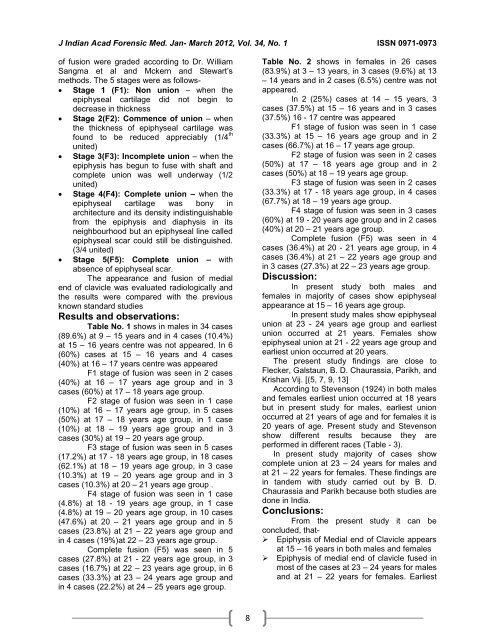
<strong>of</strong> fusion were graded according to Dr. William<br />
Sangma et al and Mckern and Stewart’s<br />
methods. The 5 stages were as follows-<br />
Stage 1 (F1): Non union – when the<br />
epiphyseal cartilage did not begin to<br />
decrease in thickness<br />
Stage 2(F2): Commence <strong>of</strong> union – when<br />
the thickness <strong>of</strong> epiphyseal cartilage was<br />
found to be reduced appreciably (1/4 th<br />
united)<br />
Stage 3(F3): Incomplete union – when the<br />
epiphysis has begun to fuse with shaft and<br />
complete union was well underway (1/2<br />
united)<br />
Stage 4(F4): Complete union – when the<br />
epiphyseal cartilage was bony in<br />
architecture and its density indistinguishable<br />
from the epiphysis and diaphysis in its<br />
neighbourhood but an epiphyseal line called<br />
epiphyseal scar could still be distinguished.<br />
(3/4 united)<br />
Stage 5(F5): Complete union – with<br />
absence <strong>of</strong> epiphyseal scar.<br />
The appearance and fusion <strong>of</strong> medial<br />
end <strong>of</strong> clavicle was evaluated radiologically and<br />
the results were compared with the previous<br />
known standard studies<br />
Results and observations:<br />
Table No. 1 shows in males in 34 cases<br />
(89.6%) at 9 – 15 years and in 4 cases (10.4%)<br />
at 15 – 16 years centre was not appeared. In 6<br />
(60%) cases at 15 – 16 years and 4 cases<br />
(40%) at 16 – 17 years centre was appeared<br />
F1 stage <strong>of</strong> fusion was seen in 2 cases<br />
(40%) at 16 – 17 years age group and in 3<br />
cases (60%) at 17 – 18 years age group.<br />
F2 stage <strong>of</strong> fusion was seen in 1 case<br />
(10%) at 16 – 17 years age group, in 5 cases<br />
(50%) at 17 – 18 years age group, in 1 case<br />
(10%) at 18 – 19 years age group and in 3<br />
cases (30%) at 19 – 20 years age group.<br />
F3 stage <strong>of</strong> fusion was seen in 5 cases<br />
(17.2%) at 17 - 18 years age group, in 18 cases<br />
(62.1%) at 18 – 19 years age group, in 3 case<br />
(10.3%) at 19 – 20 years age group and in 3<br />
cases (10.3%) at 20 – 21 years age group .<br />
F4 stage <strong>of</strong> fusion was seen in 1 case<br />
(4.8%) at 18 - 19 years age group, in 1 case<br />
(4.8%) at 19 – 20 years age group, in 10 cases<br />
(47.6%) at 20 – 21 years age group and in 5<br />
cases (23.8%) at 21 – 22 years age group and<br />
in 4 cases (19%)at 22 – 23 years age group.<br />
Complete fusion (F5) was seen in 5<br />
cases (27.8%) at 21 - 22 years age group, in 3<br />
cases (16.7%) at 22 – 23 years age group, in 6<br />
cases (33.3%) at 23 – 24 years age group and<br />
in 4 cases (22.2%) at 24 – 25 years age group.<br />
8<br />
Table No. 2 shows in females in 26 cases<br />
(83.9%) at 3 – 13 years, in 3 cases (9.6%) at 13<br />
– 14 years and in 2 cases (6.5%) centre was not<br />
appeared.<br />
In 2 (25%) cases at 14 – 15 years, 3<br />
cases (37.5%) at 15 – 16 years and in 3 cases<br />
(37.5%) 16 - 17 centre was appeared<br />
F1 stage <strong>of</strong> fusion was seen in 1 case<br />
(33.3%) at 15 – 16 years age group and in 2<br />
cases (66.7%) at 16 – 17 years age group.<br />
F2 stage <strong>of</strong> fusion was seen in 2 cases<br />
(50%) at 17 – 18 years age group and in 2<br />
cases (50%) at 18 – 19 years age group.<br />
F3 stage <strong>of</strong> fusion was seen in 2 cases<br />
(33.3%) at 17 - 18 years age group, in 4 cases<br />
(67.7%) at 18 – 19 years age group.<br />
F4 stage <strong>of</strong> fusion was seen in 3 cases<br />
(60%) at 19 - 20 years age group and in 2 cases<br />
(40%) at 20 – 21 years age group.<br />
Complete fusion (F5) was seen in 4<br />
cases (36.4%) at 20 - 21 years age group, in 4<br />
cases (36.4%) at 21 – 22 years age group and<br />
in 3 cases (27.3%) at 22 – 23 years age group.<br />
Discussion:<br />
In present study both males and<br />
females in majority <strong>of</strong> cases show epiphyseal<br />
appearance at 15 – 16 years age group.<br />
In present study males show epiphyseal<br />
union at 23 - 24 years age group and earliest<br />
union occurred at 21 years. Females show<br />
epiphyseal union at 21 - 22 years age group and<br />
earliest union occurred at 20 years.<br />
The present study findings are close to<br />
Flecker, Galstaun, B. D. Chaurassia, Parikh, and<br />
Krishan Vij. [(5, 7, 9, 13]<br />
According to Stevenson (1924) in both males<br />
and females earliest union occurred at 18 years<br />
but in present study for males, earliest union<br />
occurred at 21 years <strong>of</strong> age and for females it is<br />
20 years <strong>of</strong> age. Present study and Stevenson<br />
show different results because they are<br />
performed in different races (Table - 3).<br />
In present study majority <strong>of</strong> cases show<br />
complete union at 23 – 24 years for males and<br />
at 21 – 22 years for females. These findings are<br />
in tandem with study carried out by B. D.<br />
Chaurassia and Parikh because both studies are<br />
done in India.<br />
Conclusions:<br />
From the present study it can be<br />
concluded, that-<br />
Epiphysis <strong>of</strong> Medial end <strong>of</strong> Clavicle appears<br />
at 15 – 16 years in both males and females<br />
Epiphysis <strong>of</strong> medial end <strong>of</strong> clavicle fused in<br />
most <strong>of</strong> the cases at 23 – 24 years for males<br />
and at 21 – 22 years for females. Earliest