
Sie wollen auch ein ePaper? Erhöhen Sie die Reichweite Ihrer Titel.
YUMPU macht aus Druck-PDFs automatisch weboptimierte ePaper, die Google liebt.
RESEARCH ARTICLE<br />
<strong>Biology</strong> Open (2015) 00, 1-8 doi:10.1242/bio.012195<br />
In this paper, a comparative study is performed for the stings of<br />
honey bees and paper wasps. Their chemical constitutes, structures,<br />
and properties were experimentally investigated, and their refined<br />
insertion skills were also compared. It is found that the stings of<br />
honey bees and paper wasps, though with similar constituents and<br />
biological functions, have distinctly different structures and<br />
insertion skills.<br />
RESULTS<br />
The bodies of honey bees and paper wasps have a similar waist-like<br />
appearance but they differ in shapes and sizes, as shown in Fig. 1.<br />
For instance, a bee has hairy abdomens and flat legs (Fig. 1A), while<br />
a wasp has sleek abdomens and round legs (Fig. 1C). The abdomen<br />
ventral of a honey bee always keeps flat during penetration, while a<br />
paper wasp can swiftly bend the abdomen into a highly curved<br />
morphology when it attacks. As a thin junction between their thorax<br />
and abdomen, the waist allows flexible movements of the abdomen<br />
with respect to the thorax. The bodies of the honey bees are<br />
measured to be 90.5±29.5 mg in weight and 11.7±1.3 mm in length,<br />
while the paper wasps are 105.4±20.6 mg in weight and 14.7±<br />
1.7 mm in length.<br />
The stings of honey bees and paper wasps are commonly held<br />
inside a chamber at the rear end of their abdomens. A honey bee can<br />
only defense and stab the intruders at its ventral side (Fig. 1B,<br />
supplementary material Movie S1), while a paper wasp can attack<br />
the enemies at both the ventral (Fig. 1D, supplementary material<br />
Movie S2) and dorsal (Fig. 1E, supplementary material Movie S3)<br />
sides by flexibly spinning and bending its abdomen. Therefore, it is<br />
dangerous to hold the wings of a paper wasp by fingers. The<br />
different shapes and flexibilities of the abdomens of the two species<br />
might affect their striking scopes, and the morphologies of the stings<br />
are also adaptive to their different attacking features.<br />
Fig. 1. Honey bee and paper wasp morphology. (A) A honey bee, (C) a<br />
paper wasp, and (B,D,E) the stings of the two species at the maximum thrust,<br />
which are indicated by the yellow arrows. Scale bars=5 mm.<br />
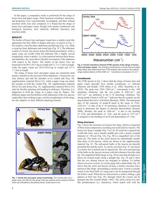
Fig. 2. Fourier transform infrared (FTIR) spectra of the stings of honey<br />
bees and paper wasps. The chemical compositions of honey bee and paper<br />
wasp stings were determined from FTIR analysis with spectra in the optical<br />
range (wavenumbers) of 650–4000 cm −1 recorded at a resolution of 4 cm −1 .<br />
Constituents<br />
The FTIR spectra in Fig. 2 shows that the stings of honey bees and<br />
paper wasps basically have similar constituents, i.e. chitosan<br />
(Darmon and Rudall, 1950; Khan et al., 2002; Kumirska et al.,<br />
2010). The peak near 3284–3286 cm −1 corresponds to the –OH<br />
stretching vibrations, and the two peaks at 2962 cm −1 and<br />
2927 cm −1 are attributed to the C–H stretching vibrations. The<br />
strong amide-I band at 1628 cm −1 indicates that the samples are the<br />
deacetylated derivative of β-chitin (Rinaudo, 2006). The absorption<br />
ratio of the intensity of amide-II band in the range of 1520–<br />
1529 cm −1 to that of the C–H stretching vibrations is commonly<br />
used to determine the degree of chitosan deacetylation (Kasaai,<br />
2008). Besides, the peak at 1448 cm −1 is due to the bending<br />
of =CH 2 and deformation of –CH 3 , and the peak at 1377 cm −1<br />
is assigned to the bending of ≡CH and deformation of –CH 3 .<br />
Sting structures<br />
Fig. 3 shows the structures of a honey bee sting, which is comprised<br />
of three main components, including one stylet and two lancets. The<br />
honey bee sting is straight (Fig. 3A,C,E). Its stylet has a tapered tip,<br />
a bulb-like base, and a slender middle part with a nearly constant<br />
diameter of ∼86 μm (Fig. 3A). Fig. 3B is a magnified view of the<br />
stylet tip. The barbs on the stylet dorsal are substantially smaller<br />
than those of the lancets, which are shown in the supplementary<br />
material Fig. S1. The sub-apical barbs of the lancets are laterally<br />
protruded beyond the stylet. As can be seen from Fig. 3C, the lancet<br />
of a honey bee is barbed near its tapered tip. The sizes and spacings<br />
of the barbs on the lancets increase with their distance from the sting<br />
tip. The magnified SEM image in Fig. 3D reveals that all barbs on a<br />
lancet are located neatly along a straight line. Fig. 3E and F show<br />
that the two abreast lancets are arranged in tandem. To further<br />
observe its cross section, we cut the sting with a sharp blade. Fig. 3G<br />
shows that the stylet and lancets all have a hollow structure and a<br />
meniscus-like shape, surrounding a circular canal. Venom is usually<br />
stored in the basal bulb of a sting and it can be transported through<br />
the hollow canal. When the two lancets have a relative sliding, a gap<br />
appears at their tips (Fig. 3F), through which the venom can be<br />
pumped into the wounds (Dade, 1962). The stylet carries two raillike<br />
protuberances (i.e. rhachises), which match well with the<br />
<strong>Biology</strong> Open<br />
2