
Erfolgreiche ePaper selbst erstellen
Machen Sie aus Ihren PDF Publikationen ein blätterbares Flipbook mit unserer einzigartigen Google optimierten e-Paper Software.
RESEARCH ARTICLE<br />
<strong>Biology</strong> Open (2015) 00, 1-8 doi:10.1242/bio.012195<br />
smaller than those on the honey bee stings. To avoid their barbs<br />
being anchored by tissue fibers, the lancets assume a spiral shape.<br />
When submerged in the victim, the originally curved sting will be<br />
straightened. The barbs, hidden in the broad stylet, have little<br />
interaction with the substrate. In comparison with the honey bee<br />
stings, the paper wasp stings are easier to be extracted from the<br />
wound. Therefore, both the intrinsic curved shape of the sting and<br />
the spiral morphologies of its two lancets are crucial for the paper<br />
wasp stings to rapidly penetrate into and readily extract from the<br />
attacked body.<br />
Following the above results and discussions, some similarities<br />
and differences of the stings of honey bees and paper wasps are<br />
summarized in Table 1.<br />
CONCLUSION<br />
We have experimentally investigated and compared the stings of<br />
honey bees and paper wasps from the viewpoints of chemical<br />
compositions, geometric morphologies, and biological functions.<br />
Both kinds of stings are mainly comprised of chitosan, and each<br />
sting has a stylet and two barbed lancets, which are connected by a<br />
sliding interlocking mechanism. The honey bee stings are relatively<br />
straight and have laterally stretched barbs, while the paper wasp<br />
stings have an intrinsic curved shape and have smaller barbs hidden<br />
in the stylet. The paper wasp stings can be easily retracted from the<br />
victim substrate, while the removal of the honey bee stings is more<br />
difficult due to their externally protruded barbs. Both honey bees<br />
and paper wasps have refined insertion skills adaptive to their stings<br />
with different sizes and morphologies. This study might be helpful<br />
to gain insights into the relations among the chemical compositions,<br />
geometric structures, mechanical properties, and biological<br />
functions of the insect stings. The results may also provide<br />
inspirations for the design of bioinspired stings and microneedles.<br />
MATERIALS AND METHODS<br />
The care and use of the experimental animals complied with the institutional<br />
and national animal welfare laws, guidelines, and policies. Honey bees (Apis<br />
cerana Fabricius) and paper wasps (Polistes sp.) were collected at Beijing.<br />
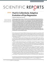
Fig. 1 shows a honey bee, a paper wasp, and the stings of the two species at<br />
the maximum thrust, which are indicated by the yellow arrows. The masses<br />
and body lengths of 10 honey bees and 10 paper wasps randomly selected<br />
were measured. The mass was determined by an electronic balance, and the<br />
body length was measured from the forehead to the tip of abdomen by using<br />
a digital caliper.<br />
The stings of honey bees and paper wasps were taken out from the fresh<br />
cadavers by extruding their abdomens with a pair of forceps. The stings were<br />
submerged in distilled water, cleaned by the ultrasonic method, and then<br />
naturally dried in airtight glassware. To determine their chemical<br />
compositions, the Fourier transform infrared (FTIR) spectra of the stings<br />
were obtained by using a Nicolet 6700 FTIR spectrophotometer (Nicolet<br />
Instrument Company, USA). The spectra in the optical range (wavenumbers)<br />
of 650–4000 cm −1 were recorded at a resolution of 4 cm −1 . The sting<br />
microstructures were observed using scanning electron microscope (SEM,<br />
Quanta FEG 450, FEI, USA). The stings were cut by sharp blades for crosssection<br />
observations. The different components in each sting were separated<br />
from each other with a forceps in order to observe their more detailed<br />
structures. All samples were gold sputtered before observation.<br />
The quasi-static penetration–extraction tests of the stings were carried out<br />
on a silica gel (non-fibrous substrate) and a piece of porcine skin (fibrous<br />
tissue) in the dorsal region. The silica gel samples, with Young’s modulus<br />
1–2.8 MPa and tensile strength 3.5–15 MPa (Shergold and Fleck, 2005),<br />
were made into a cuboid of 2 mm in depth. The fresh skin samples, obtained<br />
by removing the subcutaneous tissues, were roughly comprised of epidermis<br />
and dermis. The Young’s modulus and tensile strength of the skin samples<br />
are 0.3–1.0 MPa and 10–20 MPa, respectively (Shergold and Fleck, 2005).<br />
A microforce test system (JQN04C, Powereach, China) was used to examine<br />
the force–displacement relation during the penetration–extraction process<br />
of a sting. The sting was immobilized at the upper end, while the silica<br />
gel/porcine skin was fixed on the mobile platform. Insertion and pull-out of<br />
the sting were controlled by moving the platform upwards and downwards,<br />
respectively. The real-time motion of the sting was synchronously recorded<br />
by using a CCD camera assembled with micro-lens. Before the tests, the<br />
sting foreparts were adjusted to be perpendicular to the substrate surface<br />
through the micro-lens. The loading rate was set as 1 μm s −1 .<br />
The slow motion analysis of the penetration behaviors of honey bees and<br />
paper wasps was performed by using a high-speed video camera (Fastcam<br />
Mini UX100, Photron, Japan). The honey bee and paper wasp, handled by a<br />
forceps, were guided to pierce a polydimethylsiloxane (PDMS) bulk by, e.g.<br />
alarm pheromone and physical stimulation. The PDMS substrate was<br />
prepared by mixing the curing agent (Sylgard 184, Dow Corning, USA) and<br />
base with a weight ratio of 1:30. The mixture was degassed for 1–2 hto<br />
remove excess bubbles, and cured at 60°C for 3 h. The Young’s modulus of<br />
the PDMS (1:30) bulk is 0.3±0.01 MPa (Lim and Chaudhri, 2004). The<br />
PDMS (1:30) bulk was semi-transparent such that the sting motion could be<br />
directly observed and recorded. The insertion skills of honey bees and paper<br />
wasps were examined by comparing their features of penetration. The angle<br />
of the sting deviated from the normal of the substrate surface, referred to as<br />
the penetration angle θ, was measured.<br />
Competing interests<br />
The authors declare no competing or financial interests.<br />
Author contributions<br />
X.-Q.F., H.-P.Z., G.-J.M., and C.-W.W. designed and directed this study. Z.-L.Z. and<br />
K.Y. carried out the experiments. X.-Q.F., Z.-L.Z., and H.-P.Z. contributed to the<br />
analysis of experimental results. X.-Q.F. and Z.-L.Z. contributed to writing and<br />
revising the manuscript.<br />
Funding<br />
This work is sponsored by the National Natural Science Foundation of China (Grant<br />
Nos. 11432008 and 11372162), the 973 Program of MOST (2012CB934001), and<br />
Tsinghua University (20121087991).<br />
Supplementary material<br />
Supplementary material available online at<br />
http://bio.biologists.org/lookup/suppl/doi:10.1242/bio.012195/-/DC1<br />
References<br />
Akre, R. D., Greene, A., MacDonald, J. F., Landolt, P. J. and Davis, H. G. (1981).<br />
The Yellowjackets of America North of Mexico. Washington, DC, USA: United<br />
States Department of Agriculture.<br />
Aoyagi, S., Izumi, H. and Fukuda, M. (2008). Biodegradable polymer needle with<br />
various tip angles and consideration on insertion mechanism of mosquito’s<br />
proboscis. Sens. Actuator. A Phys. 143, 20-28.<br />
Azar, T. and Hayward, V. (2008). Estimation of fracture toughness of soft tissue<br />
from needle insertion. Biomed. Simul., Proc., eds Bello, F., Edward, E., Lect.<br />
Notes Comput. Sci. 5104, 166-175.<br />
Barham, O. M. (2007). A mathematical model for the mechanics of a mosquito bite<br />
with applications to microneedle design. MS Thesis, Department of Mechanical<br />
and Aerospace Engineering, North Carolina State University, USA.<br />
Brett, P. N., Parker, T. J., Harrison, A. J., Thomas, T. A. and Carr, A. (1997).<br />
Simulation of resistance forces acting on surgical needles. Proc. Inst. Mech. Eng.<br />
211, 335-347.<br />
Cardinal, S. and Packer, L. (2007). Phylogenetic analysis of the corbiculate Apinae<br />
based on morphology of the sting apparatus (Hymenoptera: Apidae). Cladistics<br />
23, 99-118.<br />
Cho, W. K., Ankrum, J. A., Guo, D., Chester, S. A., Yang, S. Y., Kashyap, A.,<br />
Campbell, G. A., Wood, R. J., Rijal, R. K., Karnik, R. et al. (2012).<br />
Microstructured barbs on the North American porcupine quill enable easy tissue<br />
penetration and difficult removal. Proc. Natl. Acad. Sci. USA 109, 21289-21294.<br />
Dade, H. A. (1962). Anatomy and Dissection of the Honeybee. Cardiff, UK:<br />
International Bee Research Association.<br />
Darmon, S. E. and Rudall, K. M. (1950). Infra-red and X-ray studies of chitin. Disc.<br />
Faraday Soc. 9, 251-260.<br />
Das, S. and Ghatak, A. (2011). Puncturing of soft gels with multi-tip needles.<br />
J. Mater. Sci. 46, 2895-2904.<br />
Davis, S. P., Landis, B. J., Adams, Z. H., Allen, M. G. and Prausnitz, M. R. (2004).<br />
Insertion of microneedles into skin: measurement and prediction of insertion force<br />
and needle fracture force. J. Biomech. 37, 1155-1163.<br />
<strong>Biology</strong> Open<br />
7