A guide to the deep-water sponges of - NMFS Scientific Publications ...
A guide to the deep-water sponges of - NMFS Scientific Publications ...
A guide to the deep-water sponges of - NMFS Scientific Publications ...
- No tags were found...

You also want an ePaper? Increase the reach of your titles
YUMPU automatically turns print PDFs into web optimized ePapers that Google loves.
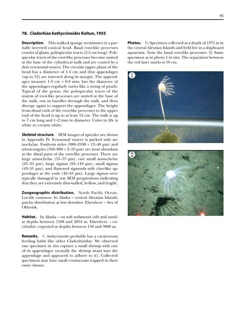
78. Cladorhiza bathycrinoides Koltun, 1955<br />
Description. This stalked sponge terminates in a partially<br />
inverted conical head. Basal root-like processes<br />
consist <strong>of</strong> glassy, polyspicular tracts (2.5 cm long). Polyspicular<br />
tracts <strong>of</strong> <strong>the</strong> root-like processes become united<br />
at <strong>the</strong> base <strong>of</strong> <strong>the</strong> cylindrical stalk and are coated by a<br />
thin ec<strong>to</strong>somal veneer. The circular upper plane <strong>of</strong> <strong>the</strong><br />
head has a diameter <strong>of</strong> 1.4 cm and thin appendages<br />
(up <strong>to</strong> 12) are inserted along its margin. The appendages<br />
measure 1.9 cm × 0.8 mm, but <strong>the</strong> diameter <strong>of</strong><br />
<strong>the</strong> appendages regularly varies like a string <strong>of</strong> pearls.<br />
Typical <strong>of</strong> <strong>the</strong> genus, <strong>the</strong> polyspicular tracts <strong>of</strong> <strong>the</strong><br />
system <strong>of</strong> root-like processes are united at <strong>the</strong> base <strong>of</strong><br />
<strong>the</strong> stalk, run in bundles through <strong>the</strong> stalk, and <strong>the</strong>n<br />
diverge again <strong>to</strong> support <strong>the</strong> appendages. The height<br />
from distal ends <strong>of</strong> <strong>the</strong> root-like processes <strong>to</strong> <strong>the</strong> upper<br />
end <strong>of</strong> <strong>the</strong> head is up <strong>to</strong> at least 12 cm. The stalk is up<br />
<strong>to</strong> 7 cm long and 1–2 mm in diameter. Color in life is<br />
white or creamy white.<br />
Skeletal structure. SEM images <strong>of</strong> spicules are shown<br />
in Appendix IV. Ec<strong>to</strong>somal veneer is packed with anisochelae.<br />
Fusiform styles (980–2100 × 12–40 µm) and<br />
tylostrongyles (350–900 × 2–10 µm) are most abundant<br />
at <strong>the</strong> distal parts <strong>of</strong> <strong>the</strong> root-like processes. There are<br />
large anisochelae (55–75 µm), rare small anisochelae<br />
(25–35 µm), large sigmas (95–110 µm), small sigmas<br />
(43–55 µm), and flattened sigmoids with claw-like appendages<br />
at <strong>the</strong> ends (42–45 µm). Large sigmas were<br />
typically damaged in our SEM preparations indicating<br />
that <strong>the</strong>y are extremely thin-walled, hollow, and fragile.<br />
Zoogeographic distribution. North Pacific Ocean.<br />
Locally common. In Alaska – central Aleutian Islands;<br />
patchy distribution at low densities. Elsewhere – Sea <strong>of</strong><br />
Okhotsk.<br />
Habitat. In Alaska – on s<strong>of</strong>t sediments (silt and sand)<br />
at depths between 1108 and 2854 m. Elsewhere – eurybathic;<br />
reported at depths between 150 and 3800 m.<br />
Remarks. C. bathycrinoides probably has a carnivorous<br />
feeding habit like o<strong>the</strong>r Cladorhizidae. We observed<br />
one specimen in situ capture a small shrimp with one<br />
<strong>of</strong> its appendages (actually <strong>the</strong> shrimp swam in<strong>to</strong> <strong>the</strong><br />
appendage and appeared <strong>to</strong> adhere <strong>to</strong> it). Collected<br />
specimens may have small crustaceans trapped in <strong>the</strong>ir<br />
outer tissues.<br />
95<br />
Pho<strong>to</strong>s. 1) Specimen collected at a depth <strong>of</strong> 1371 m in<br />
<strong>the</strong> central Aleutian Islands and held live in a shipboard<br />
aquarium. Note <strong>the</strong> basal root-like processes. 2) Same<br />
specimen as in pho<strong>to</strong> 1 in situ. The separation between<br />
<strong>the</strong> red laser marks is 10 cm.