Data sheet DBCO-Cy5 - Jena Bioscience
Data sheet DBCO-Cy5 - Jena Bioscience
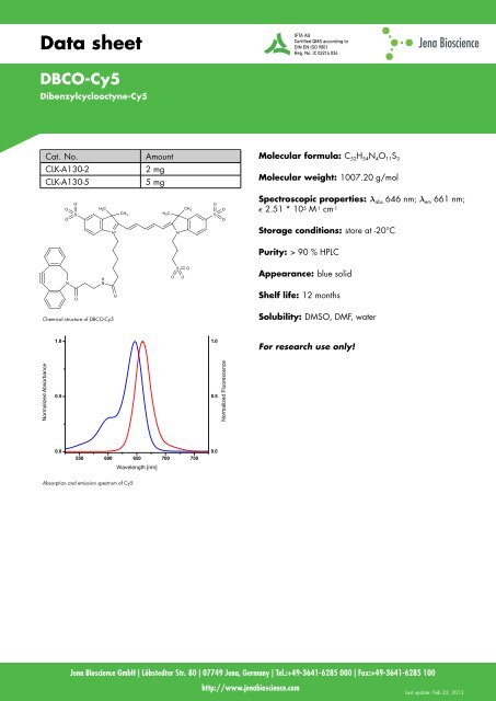
Data sheet DBCO-Cy5 - Jena Bioscience

Create successful ePaper yourself
Turn your PDF publications into a flip-book with our unique Google optimized e-Paper software.
<strong>Data</strong> <strong>sheet</strong><br />
<strong>DBCO</strong>-<strong>Cy5</strong><br />
Dibenzylcyclooctyne-<strong>Cy5</strong><br />
Normalized Absorbance<br />
Cat. No. Amount<br />
CLK-A130-2 2 mg<br />
CLK-A130-5 1.0<br />
5 mg<br />
O<br />
O<br />
N<br />
O<br />
S –<br />
H 3 C<br />
H<br />
N<br />
N +<br />
CH 3<br />
CH 3<br />
H 3 C – S<br />
O<br />
450 O<br />
500 550 600 650<br />
Chemical structure of <strong>DBCO</strong>-<strong>Cy5</strong><br />
Normalized Absorbance<br />
0.5<br />
0.0<br />
1.0<br />
0.5<br />
0.0<br />
Wavelength [nm]<br />
550 600 650 700 750<br />
Absorption and emission spectrum of <strong>Cy5</strong><br />
Wavelength [nm]<br />
O<br />
N<br />
S –<br />
O<br />
O<br />
1.0<br />
O<br />
0.5<br />
0.0<br />
1.0<br />
0.5<br />
0.0<br />
Normalized Fluorescence<br />
O<br />
O<br />
Normalized Fluorescence<br />
Molecular formula: C 52H 54N 4O 11S 3<br />
Molecular weight: 1007.20 g/mol<br />
Spectroscopic properties: λ abs 646 nm; λ em 661 nm;<br />
ɛ 2.51 * 10 5 M -1 cm -1<br />
Storage conditions: store at -20°C<br />
Purity: > 90 % HPLC<br />
Appearance: blue solid<br />
Shelf life: 12 months<br />
Solubility: DMSO, DMF, water<br />
For research use only!<br />
<strong>Jena</strong> <strong>Bioscience</strong> GmbH | Löbstedter Str. 80 | 07749 <strong>Jena</strong>, Germany | Tel.:+49-3641-6285 000 | Fax:+49-3641-6285 100<br />
http://www.jenabioscience.com<br />
Last update: Feb 22, 2013
<strong>Data</strong> <strong>sheet</strong><br />
<strong>DBCO</strong>-<strong>Cy5</strong><br />
Dibenzylcyclooctyne-<strong>Cy5</strong><br />
Product Features and Benefits:<br />
<strong>DBCO</strong>-<strong>Cy5</strong> is an azide-reactive fluorescent dye that is<br />
well suited for detection and labeling of chemically,<br />
enzymatically or metabolically azide-modified<br />
biopolymers or peptides. The <strong>DBCO</strong> group reacts with<br />
an azide to produce a stable triazole, which is also<br />
referred to the Cu(I)-free or strain-promoted click reaction.<br />
Recommended Assay Procedure:<br />
Live Cell Labeling<br />
• Grow mammalian cells in an appropriate medium<br />
with an azide-derivatized metabolite (e.g.<br />
ManNAz) at 37°C in 5 % CO 2.<br />
• Wash the cells two times with D-PBS containing 1 %<br />
FBS.<br />
• Prepare a 5 mM stock solution of <strong>DBCO</strong>-<strong>Cy5</strong> in a<br />
water-miscible solvent such as DMSO or DMF by<br />
adding 0.394 ml of solvent to 2 mg vial or 0.985<br />
ml to a 5 mg vial and vortex to dissolve all solid.<br />
• Label the azide-modified cells at room temperature<br />
in the dark for 30 - 60 min with 5 to 30 µM of<br />
<strong>DBCO</strong>-<strong>Cy5</strong> in D-PBS containing 1 % FBS.<br />
• Wash the cells four times with D-PBS containing 1 %<br />
FBS.<br />
• Fix the cells with 4 % formaldehyde in D-PBS for 20<br />
min at room temperature.<br />
• Wash the cells with D-PBS.<br />
• Optional step: Counterstain the cells for 15 min at<br />
room temperature with Hoechst 33342 in D-PBS.<br />
• Wash the cells two times with D-PBS.<br />
• Image the cells.<br />
Lysing Cells<br />
Note: Do not use DTT, TCEP or β-mercaptoethanol,<br />
because they will reduce the azide.<br />
• Prepare lysis buffer (100 mM Tris buffer pH 8.0<br />
containing 1 % (w/v) SDS).<br />
• Optional step: Add protease and phosphatase<br />
inhibitors to the lysis buffer.<br />
• Suspend cells in the lysis buffer (50 µl lysis buffer<br />
per 10 6 cells) and heat to 75°C. If using a 6-well<br />
plate you need 500 µl lysis buffer per 100 mm dish<br />
and 200 µl lysis buffer per well.<br />
• Sonicate the lysate briefly to shear DNA and reduce<br />
the viscosity of the solution.<br />
• Vortex the lysate for 5 min.<br />
• Centrifuge the cell lysate at 16,000 g at 4°C for 10<br />
min.<br />
• Transfer the supernatant to a clean tube and<br />
determine the protein concentration if required.<br />
Ideally, the protein concentration should be 1-2<br />
mg/ml.<br />
• Prepare 1 M solution of iodoacetamide by adding 3<br />
ml of DMSO to IAA labeled vial (provided with a<br />
lysis labeling kit).<br />
• Block cysteine thiols in lysate by addition of<br />
iodoacetamide stock solution to a final<br />
concentration of 15 mM, agitate mildly for 30 min.<br />
• Prepare a 5 mM stock solution of <strong>DBCO</strong>-<strong>Cy5</strong> by<br />
adding 0.394 ml of <strong>DBCO</strong> to 2 mg vial or 0.985<br />
ml to 5 mg vial.<br />
• Label the cell lysate by addition of <strong>DBCO</strong>-<strong>Cy5</strong> to a<br />
final concentration of 20 µM. Protect from light and<br />
agitate mildly for 30 min at room temperature.<br />
• Prepare a 50 mM stock solution of stop buffer by<br />
adding 3 ml of water to Stop Reagent labeled vial<br />
(provided with a lysis labeling kit).<br />
• Stop reaction by addition of stop buffer to a final<br />
concentration of 100 µM, agitate briefly for 20 min.<br />
• Load 10 µg of protein on 12 % Tris-Tricine<br />
SDS-PAGE gel.<br />
<strong>Jena</strong> <strong>Bioscience</strong> GmbH | Löbstedter Str. 80 | 07749 <strong>Jena</strong>, Germany | Tel.:+49-3641-6285 000 | Fax:+49-3641-6285 100<br />
http://www.jenabioscience.com<br />
Last update: Feb 22, 2013
<strong>Data</strong> <strong>sheet</strong><br />
<strong>DBCO</strong>-<strong>Cy5</strong><br />
Dibenzylcyclooctyne-<strong>Cy5</strong><br />
• Image the gel by fluorescence scanning with<br />
detection for <strong>Cy5</strong>, AF 647 or DyLight 647.<br />
• Stain the gel with Coomassie-stain according to the<br />
manufacture’s protocol and image cells.<br />
Selected References:<br />
Ngo et al. (2012) State-selective metabolic labeling of cellular proteins.<br />
ACS Chemical Biology. 7(8):1326.<br />
Rubino et al. (2012) Chemoselective Modification of Viral Surfaces via<br />
Bioorthogonal Click Chemistry. J. Vis. Exp. 66:4246.<br />
Yao et al. (2012) Fluorophore Targeting to Cellular Proteins via<br />
Enzyme-Mediated Azide Ligation and Strain-Promoted Cycloaddition. J.<br />
Am. Chem. Soc. 134:4246.<br />
Best et al. (2009) Click chemistry and bioorthogonal reactions:<br />
unprecedented selectivity in the labeling of biological molecules.<br />
Biochemistry. 48(28):6571.<br />
Ning et al. (2008) Visualizing metabolically labeled glycoconjugates of<br />
living cells by copper-free and fast Huisgen cycloaddition. Anweg.<br />
Chem. Int. Ed. 47(12):2253.<br />
Baskin et al. (2007) Copper-free click chemistry for dynamic in vivo<br />
imaging. PNAS. 104(43):16793.<br />
Dieterich et al. (2007) Labeling, detection and identification of newly<br />
synthesized proteomes with bioorthogonal non-canonical amino acid<br />
tagging. Nature Protocols. 2(3):532.<br />
<strong>Jena</strong> <strong>Bioscience</strong> GmbH | Löbstedter Str. 80 | 07749 <strong>Jena</strong>, Germany | Tel.:+49-3641-6285 000 | Fax:+49-3641-6285 100<br />
http://www.jenabioscience.com<br />
Last update: Feb 22, 2013