No 3 - Polish Journal of Microbiology
No 3 - Polish Journal of Microbiology
No 3 - Polish Journal of Microbiology

You also want an ePaper? Increase the reach of your titles
YUMPU automatically turns print PDFs into web optimized ePapers that Google loves.
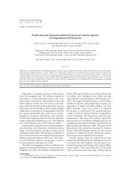
3 Function <strong>of</strong> H. pylori ggt domains<br />
205<br />
and Xho I to be inserted into the baculovirus transfer<br />
vector digested with the same enzymes. After the plasmids<br />
were transferred into the Bm-dH10 bacteria, the<br />
bacmid dNAs were extracted, respectively. To confirm<br />
the fragment inserts, PCR was used to amplify the fragments.<br />
The results indicated that the HpGT fragments<br />
were inserted into the viral genome.<br />
To construct the fusing fragments with eGFP, the<br />
eGFP fragments were amplified and were digested with<br />
Xho I and Hind III, and then ligated to the recombinant<br />
vectors containing the HpGT fragment.<br />
A<br />
B<br />
Fig. 2a and 2b. detection <strong>of</strong> expression products <strong>of</strong> the fusing fragment<br />
<strong>of</strong> ggt and gfp in baculovirus by Western-blot.<br />
The lysates <strong>of</strong> BmN cells and <strong>of</strong> cells infected with wild baculovirus<br />
(BmNPV) were the negative controls. In Figure 2a, the products <strong>of</strong> the<br />
full length ggt fusing with eGFP gene were detected, and in Figure 2b, the<br />
products <strong>of</strong> the fragments <strong>of</strong> ggt fusing with eGFP gene were detected.<br />
Fig. 1. Observation <strong>of</strong> HpGT products fusing<br />
with eGFP under fluorescent microscope<br />
(200-fold).<br />
The GGT-d380a-eGFP products had different<br />
location in BmN cells. Bm-eGFP recombinant<br />
baculovirus, with egfp gene instead <strong>of</strong> the polyhedron<br />
gene, was a control.<br />
The recombinant bacmid dNAs were transfected<br />
into BmN cells to generate budded viruses. After 72 h<br />
infection, the symptoms <strong>of</strong> viral infection were observed<br />
using an inverted phase microscope and the medium<br />
was collected as viral stock. Green fluorescence was<br />
observed in the infected cells when the HpGT fragments<br />
fusing eGFP were expressed (Fig. 1).<br />
Confirmation <strong>of</strong> fusing expression. At <strong>of</strong> yet, the<br />
HpGT antibody has not been raised. Expression <strong>of</strong><br />
the HpGT fragments was tested by tagging with eGFP<br />
gene. Using eGFP antibody, expression was confirmed<br />
by Western blot (Fig. 2a and 2b). After that, the location<br />
<strong>of</strong> the HpGT fragments in the cells was observed under<br />
an inverted fluorescent microscope. Interestingly, the<br />
fragments that lost N-terminal 380aa were concentrated<br />
in the cytoplasm (Fig. 1).<br />
GGT activity assay. The test <strong>of</strong> GGT activity assay<br />
showed that all the fragments used in this paper had<br />
GGT activity, even after deleting the N-terminal 380aa<br />
Fig. 3. GGT activity detection <strong>of</strong> the expression products<br />
<strong>of</strong> the fusing fragment <strong>of</strong> the ggt in baculovirus.<br />
The Bm-GFP virus was a negative control.