Plant basal resistance - Universiteit Utrecht
Plant basal resistance - Universiteit Utrecht
Plant basal resistance - Universiteit Utrecht

You also want an ePaper? Increase the reach of your titles
YUMPU automatically turns print PDFs into web optimized ePapers that Google loves.
<strong>Plant</strong> <strong>basal</strong> <strong>resistance</strong>: genetics, biochemistry<br />
and impacts on plant-biotic interactions<br />
Shakoor Ahmad
<strong>Plant</strong> <strong>basal</strong> <strong>resistance</strong>: genetics, biochemistry and<br />
impacts on plant-biotic interactions<br />
Shakoor Ahmad
Most of the research described in this thesis was conducted at the Department of Biological<br />
Chemistry and Crop Protection, Rothamsted Research, Harpenden, Herts, AL5 2JQ, United<br />
Kingdom and was funded by the UK Biotechnology and Biological Sciences Research<br />
Council (BBSRC) Institute Career Path Fellowship (grant no. BB/E023959/1) to J.T.<br />
Printed by: Proefschriftmaken.nl || Printyourthesis.com<br />
Layout: Shakoor Ahmad<br />
ISBN: 978-90-8891-423-2
<strong>Plant</strong> <strong>basal</strong> <strong>resistance</strong>: genetics, biochemistry and impacts on<br />
plant-biotic interactions<br />
Basale ziekteresistentie: genetica, biochemie en de effecten op<br />
plant-biotische interacties<br />
(met een samenvatting in het Nederlands)<br />
Proefschrift<br />
ter verkrijging van de graad van doctor aan de <strong>Universiteit</strong> <strong>Utrecht</strong> op gezag van de rector<br />
magnificus, prof. dr. G. J. van der Zwaan, ingevolge het besluit van het college voor promoties<br />
in het openbaar te verdedigen op maandag 18 juni 2012 des middags te 12.45 uur<br />
door<br />
Shakoor Ahmad<br />
geboren op 14 april 1976<br />
te Bannu, Pakistan
Promotor: Prof. dr. Ir. C.M.J. Pieterse<br />
Co-promotoren: Dr. J.Ton<br />
Dr. R.Gordon-Weeks
Contents<br />
Chapter 1 General introduction and thesis outline 9<br />
Chapter 2 Genetic dissection of <strong>basal</strong> defence responsiveness<br />
in acces sions of Arabidopsis thaliana 31<br />
Chapter 3 Benzoxaziniod metabolites regulates innate immunity<br />
against aphids and fungi in maize 59<br />
Chapter 4 Benzoxaziniods in root exudates of maize attract<br />
Pseudomonas putida to the rhizosphere 85<br />
Chapter 5 General discussion 107<br />
References 123<br />
Summary 139<br />
Samenvatting 141<br />
Acknowledgments 145<br />
Curriculum vitae 147<br />
List of publications 149
10<br />
CHAPTER 1<br />
General Introduction<br />
Adapted from:<br />
Ahmad S, Gordon-Weeks R, Pickett J, Ton J.<br />
Natural variation in priming of <strong>basal</strong> <strong>resistance</strong>: from evolutionary origin to agricultural<br />
exploitation. Molecular <strong>Plant</strong> Pathology 11(6), 817-827 (2010).
THE PLANT IMMUNE SYSTEM<br />
11<br />
General introduction<br />
<strong>Plant</strong>s are constantly interacting with potentially hostile organisms, such as viruses,<br />
bacteria, fungi, oomycetes, nematodes, and insects. Over the course of millions of years of<br />
co-evolution, these plant-microbe interactions have shaped the plant immune system. This<br />
regulatory system is complex and involves multiple inducible defence mechanisms, which<br />
are active at different stages of colonization by the plant attacker (Figure 1; Ton et al., 2009).<br />
To establish a successful parasitic interaction, pathogens and herbivores need to penetrate<br />
the host tissue. <strong>Plant</strong> viruses mostly depend on vectors to penetrate plant tissues, but many<br />
fungi, oomycetes and aphids can penetrate cell walls directly, whereas pathogenic bacteria<br />
often depend on natural openings, such as stomata or wound sites. At this stage of infection,<br />
a rapid closure of the stomata can form a first pre-invasive defence barrier (Melotto et<br />
al., 2008). After successful entry of the plant tissue, plant attackers often face an early-<br />
acting, post-invasive defence barrier that is marked by localized defence responses, such<br />
as the accumulation of reactive oxygen species, defence gene induction and deposition of<br />
callose-rich papillae (Eulgem et al., 1999; Flors et al., 2005; Torres et al., 2006). Upon further<br />
colonization, plants undergo a large-scale transcriptional and metabolic reprogramming<br />
that coincides with the biosynthesis of defence regulatory hormones, such as salicylic acid<br />
(SA) or jasmonic acid (JA), and complementary long-distance signals to regulate a broad<br />
spectrum of local and systemic defence mechanisms (Heil and Ton, 2008).<br />
The default mode of the plant immune system: non-host <strong>resistance</strong><br />
The most common type of disease <strong>resistance</strong> in plants is non-host <strong>resistance</strong>, which is<br />
effective against a very wide range of attackers and provides the most effective form of<br />
plant defence (Mysore and Ryu, 2004). Non-host <strong>resistance</strong> can result from the fact that<br />
the host plant simply does not provide the right environment for the attacking pathogen.<br />
However, in addition to this passive form of non-host <strong>resistance</strong>, research over the past<br />
decade has uncovered that non-host <strong>resistance</strong> also relies on active inducible defence<br />
mechanisms (Lipka et al., 2005). This non-host defence response is typically activated by<br />
conserved microbial features, such as flagellin, chitin, glycoproteins or lipopolysaccharides,<br />
which are referred to as “pathogen-associated molecular patterns” (PAMPs, synonymously<br />
called MAMPs for “microbe-associated molecular patterns”). Defence responses to<br />
herbivores can be triggered by herbivore-associated molecular patterns (HAMPs; Mithoefer<br />
and Boland, 2008), but are more commonly triggered by the perception of endogenous<br />
plant elicitors that are released upon tissue damage, which are called damage-associated<br />
molecular patterns (DAMPs; Heil, 2009; Heil et al., 2012). Much about the perception<br />
machinery of HAMPs remains unknown (Mithoefer and Boland, 2008). PAMPS and DAMPS<br />
are thought to be recognised by plasma membrane-localised pattern-recognition receptors
Chapter 1<br />
(PRRs; Gomez-Gomez and Boller, 2000; Scheer and Ryan, 2002; Huffaker et al., 2006; Miya<br />
et al., 2007). Although immune response triggered by these defence elicitors are commonly<br />
referred to as PAMP-triggered immunity (PTI), the term ‘pattern-triggered immunity’ would<br />
be more appropriate as it more collectively reflects responses to PAMPs, MAMPs, DAMPs<br />
and HAMPs. The PTI response is associated with a wide range of quickly activated defence<br />
mechanisms, such as localized callose deposition, reactive oxygen species accumulation and<br />
single cell death responses (Schwessinger and Zipfel, 2008).<br />
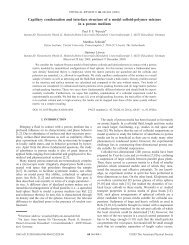
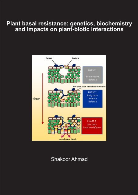
Figure 1: Induced plant defence is a multi-layered phenomenon involving a multitude of defence<br />
mechanisms that are activated at different stages of the plant-microbe interaction. Upon first<br />
contact with a microbial pathogen, plants can express pre-invasive defence mechanisms. A wellknown<br />
example is the rapid closure of stomata upon recognition of pathogen-associated molecular<br />
patterns (PAMPs). When the invading pathogen is capable of penetrating into the host tissue, it faces<br />
a second layer of inducible plant defences. This relatively early post-invasive defence is marked by<br />
accumulation of reactive oxygen species (ROS), often directly followed by deposition of callose-rich<br />
papillae. If the attacking pathogen is able to suppress and/or evade this early post-invasive defence<br />
barrier, it will encounter a third layer of inducible defences. This relatively late post-invasive defence is<br />
associated with the activation a wide range of defence mechanisms that are under control by de novo<br />
produced signalling hormones, such as salicylic acid. Late post-invasive defence is also associated with<br />
the generation of vascular long-distance signals that can prime systemic plant parts against upcoming<br />
pathogen attack. Red cells indicate defence-expressing cells and orange cells indicate those that are<br />
being successfully parasitized. The figure is adopted from Ton et al. (2009).<br />
12
13<br />
General introduction<br />
Co-evolution between virulent pathogens and plant immunity “zigzags” between <strong>basal</strong><br />
<strong>resistance</strong> and effector-triggered immunity<br />
As mentioned above, PTI is a nonspecific defence response and is able to stop the majority<br />
of hostile microbes. Virulent pathogens, however, have evolved the ability to suppress PTI<br />
through the use of pathogen effectors (Jones and Dangl, 2006). This effector-triggered<br />
susceptibility (ETS) reduces the efficiency of the plant immune response to <strong>basal</strong> <strong>resistance</strong>,<br />
which is insufficient to provide effective protection against disease. To counteract ETS, selected<br />
plant varieties have evolved <strong>resistance</strong> (R) proteins, which can detect pathogen effectors<br />
directly, or can guard the targets of pathogen effectors, thereby indirectly recognizing the<br />
activity of effectors (McDowell and Woffenden, 2003). Activation of R proteins often gives<br />
rise to a hypersensitive response (HR) that can block virulent pathogens at relatively early<br />
stages of infection. This so called effector-triggered immunity (ETI) is extremely effective<br />
against biotrophic pathogens and has, therefore, been studied extensively over the past<br />
decades. However, a major limitation of ETI is that it only protects against specific races<br />
of biotrophic pathogens (Lukasik and Takken, 2009), whereas it can be ineffective or even<br />
disease-promoting in response to necrotrophic pathogens (Kliebenstein and Rowe, 2008).<br />
Moreover, avirulent biotrophs are under constant selective pressure to break ETI, which<br />
limits the durability of this defence strategy. Pathogens can break ETI by evolving alternative<br />
effectors that suppress ETI, or that are no longer recognized by R proteins (Abramovitch<br />
et al., 2006; Fu et al., 2007; Cui et al., 2009; Houterman et al., 2009). Consequently, ETI is<br />
reverted to <strong>basal</strong> <strong>resistance</strong>, thereby imposing further selection pressure on the host plant<br />
to evolve improved R proteins that are capable of recognising the newly evolved effectors.<br />
The resulting arms race between plants and their (a)virulent pathogens manifests as an<br />
on-going oscillation in the effectiveness of plant defence and is referred to as the “zigzag”<br />
model (Jones and Dangl, 2006).<br />
PRIMING OF DEFENCE: AN ALTERNATIVE DEFENCE STRATEGY TO COPE WITH BIOTIC<br />
STRESS<br />
Although ETI can be extremely effective against biotrophic pathogens, plants can also<br />
counteract pathogens through a sensitization of their <strong>basal</strong> immune system. This priming of<br />
defence causes a faster and stronger induction of defensive mechanisms upon subsequent<br />
attack (Conrath et al., 2006; Frost et al., 2008). Priming of defence, also known as<br />
sensitization of defence, is a physiological state that enables plants to respond to a low<br />
level of environmental stress in a more efficient manner (Conrath, 2011). Similar to PTI,<br />
priming of defence is effective against a broad spectrum of plant attackers, suggesting that<br />
primed <strong>resistance</strong> is at least partially based on an augmented expression of PTI mechanisms.<br />
However, some forms of defence priming have also been shown to reduce lesion formation
Chapter 1<br />
by avirulent pathogens (Ross, 1961; Hoffland et al., 1996), suggesting that priming can<br />
boost both PTI and ETI mechanisms. Since <strong>basal</strong> <strong>resistance</strong> has been defined as the sum of<br />
<strong>resistance</strong> by PTI and ETI, minus the susceptibility by ETS (Jones and Dangl, 2006), priming<br />
of defence can best be defined as an augmented capacity to express <strong>basal</strong> <strong>resistance</strong><br />
mechanisms (Figure 2A). If the augmented <strong>basal</strong> defence response precedes the delivery of<br />
pathogen effectors, priming can provide full immunity against otherwise virulent pathogens<br />
(Figures 2B). Indeed, this has been reported for some forms of chemically-induced priming<br />
(Zimmerli et al., 2000; Conrath et al., 2006). In most cases, however, primed defence<br />
expression slows down the colonisation by virulent pathogens to a larger extent than<br />
<strong>basal</strong> <strong>resistance</strong> (Conrath et al., 2006). Most priming-inducing stimuli can trigger defence<br />
mechanisms directly if applied in higher doses. For instance, relatively high soil-drench<br />
concentrations of beta-aminobutyric acid (BABA) trigger PR-1 gene induction directly in<br />
Arabidopsis, whereas lower concentrations of BABA merely prime the induction of PR-1<br />
(Van Hulten et al., 2006). Furthermore, transient induction of direct defence can give rise<br />
to longer-lasting priming of defence (Bruce et al., 2007; Heil and Ton, 2008). Hence, many<br />
induced <strong>resistance</strong> phenomena are based on a combination of direct defence and priming<br />
and their relative contribution depends on the dose of the <strong>resistance</strong> inducing stimulus and<br />
the time point after induction.<br />
Biologically induced priming of defence<br />
Priming of defence can be induced by various biological agents and is often expressed in plant<br />
parts distal from the initial site of stimulation. For example, localised attack by pathogenic<br />
microbes can elicit a broad-spectrum systemic acquired <strong>resistance</strong> (SAR) response that is<br />
associated with priming of defence responses (Kohler et al., 2002; Conrath et al., 2006; Jung<br />
et al., 2009; Conrath, 2011). SAR is triggered by localised pathogen attack and develops in<br />
uninfected distal parts of the plant as against a broad spectrum of pathogens (Durrant and<br />
Dong, 2004). During this process, leaves/tissue under pathogen attack produce a systemic<br />
signal which is transported to uninfected distal plant parts, where it primes the tissues<br />
for SA-dependent defences (Jung et al., 2009). The 1 st systemic study of SAR in Nicotiana<br />
benthamiana demonstrated that the phenomenon lasts for up to 20 days after primary<br />
infection (Ross, 1961). Studies in the following decades have mostly focused on the onset of<br />
SAR, which requires accumulation of plant stress hormone, SA and an intact NPR1 protein<br />
(Durrant and Dong, 2004). More recent studies have revealed that SAR establishment<br />
requires additional signals, which precede systemic accumulation of SA, such as jasmonates<br />
(Truman et al., 2007) and indole-derived metabolites (Truman et al., 2010). The exact nature<br />
of the mobile SAR signal, however, remains debatable, even within the same Arabidopsis-<br />
based pathosystem (Attaran et al., 2009). Apart from MeSA (Vlot et al., 2008), glycerolipids<br />
(Chaturvedi et al., 2008), azelaic acid (Jung et al., 2009), and glycerol-3-phosphate (Chanda<br />
14
15<br />
General introduction<br />
et al., 2011) have been reported to act as mobile signals. As a possible explanation for this<br />
controversy, Liu et al. (2011) recently suggested that SAR is mediated by an interaction<br />
between two mobile signals: MeSA and a complex formed between the lipid transfer protein<br />
DIR1 and glycerolipid and/or lipid derivatives. Hence, the signalling pathways controlling<br />
systemic defence priming during SAR are mediated by complex signalling networks.<br />
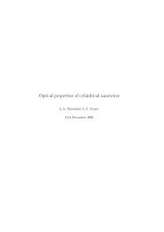
Figure 2: Priming of <strong>basal</strong> <strong>resistance</strong> provides protection against virulent pathogens. (A) Basal<br />
<strong>resistance</strong> against virulent pathogens results from a residual level of host defence after defence<br />
suppression by disease-promoting pathogen effectors (blue arrows). Priming of <strong>basal</strong> <strong>resistance</strong><br />
leads to a faster and stronger induction of <strong>basal</strong> defence mechanisms, providing enhanced <strong>resistance</strong><br />
against the invading pathogen. In most cases, priming of <strong>basal</strong> <strong>resistance</strong> cannot prevent the delivery<br />
of pathogen effectors entirely, and thereby only slows down the introgression of the pathogen<br />
(‘moderately primed defence response’). However, if the primed defence response precedes the<br />
delivery of pathogen effectors, this defence strategy can prevent pathogen infection and provide<br />
full protection against otherwise virulent pathogens (‘strongly primed defence response’). Red plant<br />
cells indicate the expression of <strong>basal</strong> defence mechanisms. (B) The ‘zigzag’ model describes <strong>basal</strong><br />
<strong>resistance</strong> as the sum of pathogen-associated molecular pattern (PAMP)-triggered immunity (PTI)<br />
and weak effector-triggered immunity (ETI) minus effector-triggered susceptibility (ETS) (Jones and<br />
Dangl, 2006). Apart from newly evolved R proteins that recognize effectors or their activities, ETS can<br />
be counteracted by the priming of defence, causing faster and stronger induction of <strong>basal</strong> defence<br />
mechanisms after pathogen attack. A moderately primed defence response merely augments the PTI/<br />
ETI response, but would still allow ETS to take place (shown in orange), whereas a strongly primed<br />
defence reaction can prevent ETS entirely (shown in red).
Chapter 1<br />
Selective non-pathogenic root-colonizing microbes can induce systemic defence<br />
priming as well (Van Wees et al., 2008). The resulting disease <strong>resistance</strong> is commonly<br />
referred to as induced systemic <strong>resistance</strong> (ISR). For instance, non-pathogenic rhizobacteria<br />
can prime Arabidopsis against a wide range of plant pathogens, like bacteria, oomycetes,<br />
fungi, viruses and even herbivores (Pieterse et al., 1996; Ton et al., 2002; Van Wees et al.,<br />
2008). A more recent example by (Verhagen et al., 2010) showed that beneficial bacteria<br />
such as Pseudomonas fluorescens CHA0 and Pseudomonas aeruginosa 7NSK2 mediate<br />
ISR in grapevine through potentiating oxidative burst and phytoalexin production (i.e.<br />
resveratrol and viniferin) after attack by Botrytis cinerea. Arbuscular micorhizal fungi (AMF)<br />
can also induce defence priming in plants (Pozo and Azcón-Aguilar, 2007; Pozo et al., 2009).<br />
The interaction between Arabidopsis and Pseudomonas fluorescens WCS417r has served<br />
as a biological model system to study the signalling transduction pathways controlling ISR.<br />
Research on this model system revealed P. fluorescens WCS417r–mediated ISR in Arabidopsis<br />
requires responsiveness to jasmonate and ethylene (ET) and is dependent on NPR1 (Pieterse<br />
et al., 1998). A transcriptome analysis for P. fluorescens WCS417r-inducible genes led to the<br />
identification of the ISR responsive transcription factor gene MYB72 (Verhagen et al., 2004).<br />
Subsequent analysis of mutant lines in MYB72 revealed that this transcription factor gene<br />
plays a critical role for the early signalling events leading to elicitation of the systemic signal<br />
(Van der Ent et al., 2008). The same authors reported that P. fluorescens WCS417r–mediated<br />
priming in Arabidopsis thaliana against Hyaloperonospora arabidopsidis, an oomycete<br />
pathogen that is unaffected by JA- and ET-dependent defences (Thomma et al., 1998; Ton et<br />
al., 2002), is based on priming of callose deposition, which requires intact abscisic acid (ABA)<br />
signalling (Van der Ent et al., 2009).<br />
Apart from defence priming against pathogens, systemic defence priming can also<br />
be effective against herbivore attack. When plants are subjected to damage by herbivorous<br />
insects, they emit a complex blend of airborne chemical signals, known as volatile organic<br />
compounds (VOCs). VOCs serve primarily to attract natural enemies of the herbivore<br />
(Turlings and Ton, 2006), but they have also been shown to induce defence priming in<br />
systemic tissues and even neighbouring plants (Engelberth et al., 2004; Heil and Silva<br />
Bueno, 2007; Ton et al., 2007; Heil and Ton, 2008). Three green leaf volatiles, (Z)-3-hexenal,<br />
(Z)-3-hexen-1-ol, and (Z)-3-hexenyl acetate, in particular have been linked to elicitation of<br />
defence priming by herbivore-induced VOCs (Engelberth et al., 2004). VOC-induced priming<br />
augments JA-inducible defences (Frost et al., 2008). Interestingly, however, only a sub-set<br />
of JA-dependent genes is responsive to priming by VOCs in maize (Ton et al., 2007). The<br />
latter observation suggests that priming-inducing VOCs target specific components in the JA<br />
response pathway.<br />
16
Chemically induced defence priming<br />
17<br />
General introduction<br />
Induced <strong>resistance</strong> by pathogens, rhizobacteria and herbivores can be mimicked by<br />
selective chemical agents (Oostendorp et al., 2001). Table I lists an overview of publications<br />
reporting defence priming in response to exogenous application of chemical compounds.<br />
In addition to the pathogen-, herbivore-, or damage associated patterns triggering the<br />
above-mentioned biological priming responses, defence priming can also be mimicked by<br />
endogenous plant signalling metabolites, such as JA, SA, and functional analogues thereof<br />
(Kauss et al., 1994; Mur et al., 1996; Kohler et al., 2002). Treatment with thiamine (Vitamin<br />
B1) has been reported to prime Arabidopsis, causing augmented accumulation of hydrogen<br />
peroxide (H 2 O 2 ), callose-rich papillae, and PR-1 transcript following pathogen infection<br />
(Ahn et al., 2007). Recently, cytokinins have emerged as another plant-endogenous priming<br />
signal. These plant hormones control specification of cells, maintenance of meristematic<br />
cells, shoot formation and development of plant vasculature, but recent evidence suggests<br />
that these compounds also regulate defence in plants (Choi et al., 2011). Interestingly, the<br />
defensive targets of cytokinins seem to depend on the plant species under investigation.<br />
Whereas cytokinins prime for JA-controlled defences in poplar (Dervinis et al., 2010), they<br />
prime for SA-dependent gene expression in Arabidopsis (Choi et al., 2011), and they were<br />
recently reported to control pathogen-induced biosynthesis of JA- and SA-independent<br />
phytoalexins in tobacco (Großkinsky et al., 2011). Finally, azelaic acid has been shown to<br />
act as a long-distance priming signal during the onset of SAR. Upon exogenous application,<br />
this dicarboxylic acid primes Arabidopsis for SA-dependent defences and confers systemic<br />
<strong>resistance</strong> against P. syringae (Jung et al., 2009).<br />
There are also xenobiotic chemicals that can trigger defence priming in plants. For<br />
instance, the chemical Probenazole (PBZ; 3-allyloxy-1,2-benzisothiazole-1,1-dioxide), which<br />
is the active ingredient in Oryzemate, has been used widely in Asia to induce <strong>resistance</strong> in<br />
rice against Magnaporthe grisea. Its mode of action relies on enhanced biosynthesis of SA<br />
(Yoshioka et al., 2001; Iwai et al., 2007), which by itself serves as an endogenous priming signal.<br />
PBZ is metabolised by plants into saccharin (1,2-benzisothiazole-1,1-dioxide), a compound<br />
that is best known for its application as an artificial sweetener. However, saccharin can also<br />
induce <strong>resistance</strong> in plants against various diseases (Oostendorp et al., 2001; Boyle and<br />
Walters, 2006; Srivastava et al., 2011). In barley, saccharin induces <strong>resistance</strong> to powdery<br />
mildew fungus, which is associated with priming of cinnamyl alcohol dehydrogenase activity<br />
(Walters et al., 2008). Another xenobiotic chemical capable of inducing <strong>resistance</strong> through<br />
defence priming is BABA. Application of this non-protein amino acid protects against an<br />
exceptionally broad spectrum of plant diseases (Jakab et al., 2001), including crop diseases<br />
that are difficult to control by conventional strategies of disease management, such as late<br />
blight disease (Liljeroth et al., 2010). BABA is active at relatively low concentrations and acts<br />
in an enantiomer-specific manner (Cohen, 2002). These findings suggest that BABA mimics
Chapter 1<br />
an endogenous plant signalling metabolite, or that it activates a plant regulatory protein<br />
controlling multiple immune responses simultaneously. Indeed, research on BABA-induced<br />
defence priming in Arabidopsis revealed that BABA not only mimics SAR-related priming<br />
of SA-dependent defences, but it also primes for pathogen-induced deposition of callose-<br />
containing papillae (Zimmerli et al., 2000; Ton et al., 2005). This priming of cell wall defence<br />
functions independently of SA and JA, but requires intact biosynthesis and perception of the<br />
plant hormone ABA (Ton and Mauch-Mani, 2004; Van der Ent et al., 2009)<br />
Table I. Chemicals that trigger priming of defence in plants after exogenous application.<br />
Chemical Stimulus Primed defence response <strong>Plant</strong> Species Reference<br />
Benzothiadiazole<br />
(BTH)<br />
PAL gene induction Arabidopsis (Kohler et al., 2002)<br />
Probenazole SA-inducible genes Rice (Iwai et al., 2007)<br />
Saccharin Cinnamyl alcohol dehydrogenase<br />
activity<br />
Beta amino butyric<br />
acid (BABA)<br />
Thiamine<br />
(Vitamin B1)<br />
SA-inducible genes and<br />
callose deposition<br />
ROS accumulation, callose<br />
deposition, and SAinduced<br />
expression<br />
18<br />
Barley (Boyle and Walters, 2006)<br />
Arabidopsis (Zimmerli et al., 2000; Ton<br />
and Mauch-Mani, 2004)<br />
Arabidopsis (Ahn et al., 2007)<br />
Cytokinins SA-inducible genes Arabidopsis (Choi et al., 2011)<br />
JA-inducible genes Poplar (Dervinis et al., 2010)<br />
Scopoletin and Capsidiol Tobacco (Großkinsky et al., 2011)<br />
Azelaic acid SA-inducible genes Arabidopsis (Jung et al., 2009)<br />
Quercetin ROS accumulation, callose<br />
deposition, and PR1 gene<br />
induction<br />
Molecular mechanisms of priming<br />
Arabidopsis (Jia et al., 2010)<br />
In contrast to research on innate plant defences that are directly responsive to pathogens<br />
and herbivores, the majority of research on priming of defence has remained limited to a<br />
description of the phenomenon after treatment with <strong>resistance</strong>-inducing agents, along with<br />
an assessment of its effectiveness in terms of disease <strong>resistance</strong> (Conrath et al., 2006; Frost<br />
et al., 2008). Only a few research groups have begun to address the mechanistic basis of<br />
defence priming (Conrath, 2011). Consequently, there are still many open questions about<br />
defence priming in plants, particularly with respect to the signalling mechanisms controlling<br />
the onset and long-term maintenance of the phenomenon.
19<br />
General introduction<br />
enhanced accumulation of signalling proteins. One common hypothesis postulates<br />
that priming is based on an increased accumulation of inactive defence signalling proteins,<br />
thereby providing enhanced defence signalling capacity (Conrath et al., 2006). Subsequent<br />
exposure to environmental stress would then lead to a faster and stronger defence<br />
signalling cascade, ultimately resulting in the augmented defence response. Indeed, SAR-<br />
related priming is associated with an increased accumulation of two inactive MAP protein<br />
kinases, MPK3 and MPK6, which show enhanced kinase activity upon secondary stress<br />
application (Beckers et al., 2009). Simultaneously, a genome-wide profiling of transcription<br />
factor (TF) genes was performed, which demonstrated that induction of ISR-related priming<br />
is associated with augmented expression of JA-regulatory TF genes (Van der Ent et al.,<br />
2009). The same study showed that BABA-induced priming was associated with enhanced<br />
expression of WRKY transcription factor genes, which encode transcription factors that<br />
regulate SA-induced defence gene transcription (Van Verk et al., 2011). Although enhanced<br />
accumulation of defence-related signalling TFs can contribute to a faster and stronger<br />
transcriptional activation of defence genes after pathogen attack, these signalling proteins<br />
typically have a limited half-life. Hence, their enhanced accumulation after application of<br />
a single priming stimulus does not provide a satisfactory explanation for the long-lasting<br />
nature of priming phenomena.<br />
epigenetic mechanisms. A recent study showed that SAR-related priming in<br />
Arabidopsis is associated with post-translational changes of histone H3 and H4 tails at gene<br />
promoters of defence-regulatory transcription factor genes (Jaskiewicz et al., 2011). Although<br />
these chromatin modifications were monitored relatively shortly after SAR induction, such<br />
epigenetic regulatory mechanism provides an attractive explanation for the long-lasting<br />
nature of the priming phenomenon. Indeed, recent evidence demonstrated that priming<br />
is an epigenetic phenomenon. Three independent research groups demonstrated that the<br />
primed defence state in Arabidopsis can be transmitted to following generations from iso-<br />
genic plant lines (Luna et al., 2012; Rasmann et al., 2012; Slaughter et al., 2012). Moreover,<br />
expression of trans-generational priming of SA-dependent defence genes was associated<br />
with chromatin remodelling at the corresponding gene promoters (Luna et al., 2012),<br />
whereas priming of JA-dependent defence requires intact biogenesis of small interfering<br />
RNAs (Rasmann et al., 2012).<br />
glycolylation of secondary metabolites. It is also conceivable that secondary<br />
metabolites contribute to long-lasting priming of defence. The chemical defence capacity<br />
of plants can be enhanced by an increased accumulation of inactive defence metabolite<br />
conjugates, such as glucosinolates and plant hormone-glucosides. Consequently, pathogen-<br />
or wounding-induced activity of hydrolytic glucosidase enzymes would lead to a faster and<br />
greater release of active aglycone metabolites.<br />
signalling cross-talk. Priming can also result from a shift in the cross-talk balance
Chapter 1<br />
between defence signalling pathways. For instance, suppression of the SA-dependent<br />
pathway by mycorrhizal fungi results in a potentiation of JA-dependent defences (Pozo and<br />
Azcón-Aguilar, 2007), while trans-generational priming of the SA response in Arabidopsis<br />
coincides with a repression of JA response (Luna et al., 2012) Interestingly augmented levels<br />
of JA have been associated with primed callose deposition in grapevine against Plasmopara<br />
(Hamiduzzaman et al., 2005). Similarly priming of papillae formation was observed in the<br />
roots of mycorrhiza-infected tomatoes with Phytophthora (Cordier et al., 1998). These<br />
observations point to a mechanism by which suppression of the SA-response results in a<br />
beneficial side effect: systemic priming of JA-dependent defences and callose deposition.<br />
reduced scavenging capacity of reactive oxygen species. Several recent studies<br />
have pointed to an important role of reactive oxygen species (ROS) in priming of defence.<br />
Thiamine (vitamin B 1 ) induces <strong>resistance</strong> against Pseudomonas syringae pv. tomato<br />
DC3000 (PstDC3000), which is associated with hydrogen peroxide (H 2 O 2 )-dependent<br />
priming of defence genes and callose deposition (Ahn et al., 2007). Vitamin B 2 (riboflavin)<br />
induces a phenotypically similar <strong>resistance</strong> response that is associated with priming of<br />
ROS production, callose deposition and SA-inducible genes (Zhang et al., 2009). The plant<br />
secondary metabolite quercetin has also been demonstrated to induce SA- and NPR1-<br />
dependent <strong>resistance</strong> against PstDC3000, which is associated with augmented deposition<br />
of ROS, callose, PR1 and PAL gene transcripts (Jia et al., 2010). A recent study by Mukherjee<br />
et al. (2010) provided a plausible mechanism for ROS-dependent regulation of priming. The<br />
authors performed a phenotypic analysis of different alleles of the ascorbic acid deficient<br />
mutant vtc1 and demonstrated that the enhanced disease <strong>resistance</strong> of this mutant is<br />
based on priming of pathogen-induced accumulation of ROS, SA and NPR1 gene transcripts<br />
(Mukherjee et al., 2010). The authors suggested that the reduced ROS scavenging capacity<br />
of vtc1 causes constitutive priming of pathogen-induced H 2 O 2 , thereby causing augmented<br />
SA accumulation and enhanced defence induction.<br />
Costs & benefits of priming<br />
The full development of an inducible defence response requires energy and, therefore,<br />
involves costs on growth and reproduction. Apart from allocation costs, costs can also arise<br />
from toxicity of the defence to the plant’s own metabolism, or when the defence response<br />
affects the plant’s interaction with beneficial organisms (Heil, 2002). It is commonly accepted<br />
that plants only express inducible defences if the benefits (i.e. protection against the<br />
attackers) outweigh the associated costs (Heil, 2002; Walters and Boyle, 2005). Van Hulten<br />
et al. (2006) conducted a laboratory study to compare the costs and benefits of defence<br />
priming versus direct induction of defence in Arabidopsis. By using low doses of BABA to<br />
induce priming and high doses of either BABA or BTH to induce defence expression directly,<br />
it was found that priming is associated with relatively minor costs on plant growth and seed<br />
20
21<br />
General introduction<br />
set. Moreover, the protective benefits of priming outweighed its costs under conditions of<br />
high disease pressure. It was thus concluded that priming is a cost-efficient defence strategy<br />
in disease-imposing environments. Interestingly, the outcome of this laboratory study<br />
was subsequently tested under agronomical field conditions by Walters et al. (2008), who<br />
subjected saccharin-primed barley to varying degrees of disease by the hemi-biotrophic<br />
fungus Rhynchosporium secalis and monitored fitness levels by plant growth and grain<br />
yield. As predicted, primed plants displayed significantly higher fitness than un-primed<br />
plants, thereby extending our laboratory demonstration that priming is a beneficial defence<br />
strategy in hostile environment.<br />
PLANT DEFENCE STRATEGIES AND THEIR ADAPTIVE VALUES IN HOSTILE<br />
ENVIRONMENTS<br />
Naturally occurring plant species can often be sub-divided into genetically distinct geographic<br />
varieties. Although these so-called ecotypes are similar enough to be considered as one<br />
species, they differ genetically in some traits due to variant selection pressures from their<br />
environments of origin. In this context, plant defence strategies can have adaptive values<br />
that vary according to the environmental conditions (Figure 3).<br />
The concept that priming of defence provides benefits in hostile environments<br />
suggests that plants in these environments are under pressure to evolve a constitutively<br />
enhanced responsiveness of <strong>basal</strong> defence mechanisms. Since priming protects against a<br />
wide variety of diseases and pests (Conrath et al., 2006), this selection pressure would be most<br />
pronounced under pressure by a wide range of different pathogens and herbivores (Figure<br />
3; strategy A). There are, however, alternative defence strategies that could provide similar<br />
or even greater benefits, depending on the nature of the environment. For instance, PAMPs<br />
from plant-beneficial microbes have been demonstrated to trigger defensive responses (Van<br />
Loon et al., 2008), suggesting that plants with primed defence responsiveness to PAMPs<br />
may risk compromising their interaction with plant-beneficial micro-organisms. Indeed,<br />
various studies have reported negative impacts of SA-dependent <strong>resistance</strong> on rhizobial<br />
and mycorrhizal symbioses with legumes (Stacey et al., 2006; Jin et al., 2009; Faessel et<br />
al., 2010). Hence, there could be a counteracting selection against constitutive priming to<br />
maintain associations with mycorrhiza or N-fixing bacteria. Therefore, an increased ability<br />
to attract and interact with micro-organisms that are capable of suppressing pathogens<br />
directly through nutrient competition or antibiosis (Handelsman and Stabb, 1996; Weller<br />
et al., 2002), could be an alternative defence strategy in hostile environments (Figure 3;<br />
strategy B). In support of this, Rudrappa et al. (2008) demonstrated that Arabidopsis can<br />
attract disease-suppressing rhizobacteria through exudation of L-malic acid, which is<br />
further boosted by aboveground infection by P. syringae pv. tomato. Secondly, priming
Chapter 1<br />
rarely provides complete protection against one pathogen or pathogen race, whereas ETI<br />
typically does. Hence, ETI would be more efficient in environments with disease pressure<br />
from one predominant pathogen species (Figure 3; strategy C). Thirdly, although priming<br />
is less costly than direct induction of defence, it is still associated with minor costs under<br />
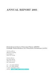
Figure 3: Model of plant defence strategies and their adaptive values under different biotic stress<br />
conditions. <strong>Plant</strong>s in environments with relatively high disease pressure from a wide array of different<br />
attackers benefit from constitutive priming of <strong>basal</strong> <strong>resistance</strong> mechanisms, which provide broadspectrum<br />
protection against pests and diseases (strategy A). However, this defence strategy may<br />
affect the plant’s ability to associate with plant-beneficial microbes, such as mycorrhizae, N-fixing<br />
bacteria or plant-growth promoting rhizobacteria. In this situation, plants would benefit more from<br />
an increased ability to attract and associate with plant-beneficial microbes with disease-suppressing<br />
traits (strategy B). <strong>Plant</strong>s in environments with a constant pressure from one dominant biotrophic<br />
pathogen benefit from effector-triggered immunity (ETI; strategy C). ETI can be broken and give rise<br />
to an ongoing “zigzag” evolution, as described by Jones and Dangl (2006). Inducible defence priming<br />
upon perception of stress-indicating signals provides a cost-efficient adaptation to environments<br />
with variable degrees of disease pressure (strategy D). Because priming of defence and induction of<br />
defence are both associated with costs on plant growth and reproduction, a relatively un-responsive<br />
immune system would be beneficial in environments with relatively low disease pressure (strategy E).<br />
22
23<br />
General introduction<br />
conditions of low disease pressure (Van Hulten et al., 2006). Consequently, plants exposed<br />
to variable levels of disease pressure would benefit from an inducible priming response<br />
(Figure 3; strategy D). As variable degrees of disease pressure are the reality in many natural<br />
plant environments, priming mainly manifests as an inducible <strong>resistance</strong> response. Finally,<br />
the selection for any of the above defence strategies is likely to be influenced by the plant’s<br />
abiotic environment. For example, the plant hormone ABA not only controls tolerance to<br />
abiotic stress, but also plays a multifaceted role in the fine-tuning of <strong>resistance</strong> to diseases<br />
and pests (Ton et al., 2009).<br />
Most plants are capable of expressing combinations of different defence strategies.<br />
The importance of each of these strategies depends on the environment. For instance, many<br />
plant-beneficial micro-organisms have the ability to protect plants through a combination<br />
of direct disease suppression and induction of defence priming in the host plant (ISR;<br />
Van Wees et al., 2008; Zamioudis and Pieterse, 2011). Colonisation by these microbes<br />
causes a constitutive level of systemic priming that is phenotypically similar to genetically<br />
acquired priming, thus combining the advantages of two defence strategies: direct disease<br />
suppression by plant-beneficial microbes and constitutive defence priming. Furthermore,<br />
the expression of one defence strategy can give rise to induction of another. For example,<br />
localised expression of PTI results in the development of SAR (Mishina and Zeier, 2007),<br />
which is largely based on priming of defence (Jung et al., 2009; Kohler et al., 2002).<br />
Natural selection for constitutively primed <strong>basal</strong> <strong>resistance</strong>?<br />
Although the above examples justify the general conclusion that natural variation in<br />
responsiveness of <strong>basal</strong> <strong>resistance</strong> mechanisms is prevalent, this does not necessarily prove<br />
that hostile environments select for constitutively primed immune systems. In order to<br />
demonstrate that constitutive defence priming has evolved from inducible priming under<br />
constant levels of disease pressure, more evidence is required from both the molecular<br />
level and the plant community level (Shindo et al., 2007). For instance, patterns of single<br />
nucleotide polymorphisms in alleles contributing to natural variation in <strong>basal</strong> <strong>resistance</strong><br />
could provide indications of past selective pressures. If the degree of nucleotide diversity<br />
deviates from the estimated diversity under neutral selection, this could be interpreted<br />
as evidence for environmental selection pressures. Typically, reduced levels of nucleotide<br />
polymorphisms indicate selective sweeps, during which newly evolved gene variants<br />
outcompete others (Nielsen, 2005). On the other hand, enhanced levels of nucleotide<br />
polymorphisms suggest balancing selection, which maintains ancient genetic variation<br />
(Mitchell-Olds and Schmitt, 2006). Although these methods provide useful indications for<br />
past selective forces on genes, fitness assays under different disease pressures would still<br />
be necessary to establish what gene variants provide which adaptive phenotypes. As was<br />
outlined by Holub (2007), a major challenge for the future is to apply currently available
Chapter 1<br />
genetic resources for Arabidopsis (i.e. fully genotyped recombinant mapping populations<br />
or association mapping populations) to field experimentation. For instance, Arabidopsis<br />
mapping populations in which defence traits segregate could be grown under different<br />
field conditions with varying degrees of disease pressure by one or multiple pathogens<br />
and/or herbivores. Subsequent fitness evaluation may reveal defence-regulating QTLs that<br />
provide selective benefits under specified environmental conditions. With more and more<br />
Arabidopsis accessions being genome-sequenced, another promising approach arises from<br />
genome-wide association mapping approaches, which are based on associations between<br />
phenotypes and DNA sequence variants within individuals or isogenic populations (Nordborg<br />
and Weigel, 2008; Atwell et al., 2010). Particularly if defence phenotypes can be related to<br />
ecological stress parameters from the accessions’ geographical origins, this technique has<br />
the potential to assign measurable ecological significance to defence regulatory alleles.<br />
SECONDARY DEFENCE METABOLITES: BIOSYNTHETIC ORIGINS AND CHEMICAL<br />
CLASSIFICATION<br />
Irrespective of the type of <strong>resistance</strong> response expressed in plants, secondary metabolites<br />
are ubiquitous “tools” in plant defence. Figure 4 provides a generic overview of the<br />
biosynthesis pathways involved in the production of defence secondary compounds in plants.<br />
Unlike primary metabolites, which play a role in the process of photosynthesis, respiration,<br />
solute transport, nutrient assimilation and differentiation, secondary metabolites have no<br />
recognised role in plant processes that are essential for growth. The distribution of secondary<br />
metabolites across the plant kingdom is diverse and varies between plant species and taxa.<br />
Based on chemical structure, plant secondary metabolites can be divided into four major<br />
groups: terpenes, phenolics, nitrogen- and sulphur-containing compounds and oxyilipins.<br />
Figure 4 shows a generic overview of the different biochemical pathways controlling these<br />
plant compounds.<br />
Terpenes<br />
This class of metabolites is immensely diverse and includes more than 30,000 lipophilic<br />
compounds (Kennedy and Wightman, 2011). Their structure includes one or more 5-carbon<br />
isoprene (C 5 H 8 ) units, which are synthesized in plants by both the mevalonate and dexy-d-<br />
xylulose pathways (Rohmer, 1999). Classifications of terpenoids are based on the number<br />
of isoprene units they contain. Hemiterpenes incorporate 1 isoprene unit, monoterpenes<br />
incorporate 2 units, sesquiterpenes incorporate 3 units, diterpenes incorporate 4 units,<br />
sesterpenes comprise 5 units, triterpenes include 6 units, and tetraterpenes incorporate 8<br />
units. Terpenes exhibit a broad range of ecological roles in the plant kingdom. Their roles<br />
include antimicrobial properties, attraction of pollinator, parasitoic or predator insects, and<br />
24
activities as allelopathic chemicals (De Almeida et al., 2010; Martino et al., 2010).<br />
Phenolics<br />
25<br />
General introduction<br />
Figure 4: A simplified scheme of the major biosynthetic pathways controlling plant secondary<br />
metabolites with representative examples and structures from each class. Secondary metabolites<br />
are derived from different pathways: the shikimate pathway, the malonic acid pathway, mavalonic<br />
acid pathway, the MEP/DOXP (2-C-methyl-D-erythritol 4-phosphate/1-deoxy-D-xylulose 5-phosphate)<br />
pathway, and the oxylipin pathway. Based on structural characteristics, the resulting secondary<br />
metabolites can be divided into four main classes: nitrogen (N) - and sulphur (S) containing compounds,<br />
phenolic compounds, terpenes, and oxylipins.<br />
Approximately 10,000 plant secondary metabolites are known to contain phenolic ring<br />
structures (Kennedy and Wightman, 2011). These are derivatives of the pentose phosphate<br />
pathway, the shikimate pathway, and the phenylpropanoid pathway. Structurally, natural<br />
phenolic compounds from plants have at least 1 aromatic hydrocarbon ring with one or more<br />
hydroxyl groups attached. Phenolics range from simple low molecular weight compounds<br />
like phenylpropanoids, coumarins, and benzoic acid derivatives, to more complex structures<br />
like flavanoids, stilbenes, and tannins. Phenolics play diverse roles in plant defence, such<br />
as responding to bacteria/fungal attack, providing scent/colour/flavour to attract beneficial<br />
insects/deter herbivores, and acting as semiochemicals during interactions with plant-<br />
beneficial microbes (Treutter, 2006).<br />
Sulphur and Nitrogen containing secondary metabolites<br />
This is a large group of secondary metabolites containing more than 15,000 molecules. They<br />
include alkaloids, cyanogenic glucosides, and non-protein amino acids. S- and N-containing
Chapter 1<br />
metabolites are biosynthesised from common amino acids. For example, N-containing<br />
alkaloids are usually synthesized from aspartic acid, lysine, tyrosine and tryptophan<br />
(Facchini, 2001) These compounds play important roles in plant-pest interactions in different<br />
plant families , such as Brassicaceae, Alliaceae and Asteraceae (Burow et al., 2008). Two<br />
well-known examples of S- and N-containing defence metabolites are glucosinolates in<br />
Brassicaceae and the Alliins in Alliaceae (Burow et al., 2008). S- and N-containing secondary<br />
metabolites offer an array of defence compounds that activate direct and/or indirect<br />
defences against a broad range of harmful microbes/insects.<br />
Oxylipins<br />
Oxylipins encompass a large family of oxygenated metabolites that are derived from fatty<br />
acids. Oxylipins are best known for their role in plant defence signalling pathways (Blée,<br />
2002), and are produced by oxidation of fatty acids, mainly linolenic acid and linoleic acid,<br />
followed by secondary modification (Vicente et al., 2011). The oxylipin biosynthesis pathway<br />
converts linoleic acid or linolenic acid into hydroperoxide substrates, such as 9-HPOD<br />
(hydroperoxy-octadecadienoic acids), 9-HPOT (hydroperoxy-octadecatrienoic acids), 13-<br />
HPOT and 13-HPOD. These compounds are subsequently utilized by different pathway<br />
branches that are under control by HPL-hydroperoxide lyase, AOS-allene oxide synthase,<br />
DES-divinyl synthase, and P0X-peroxygenase, respectively (Figure 4). The AOS pathway<br />
generates the plant defence hormone JA, which is essential for activation of direct and<br />
indirect defences against necrotrophic pathogens and insects (Pozo et al., 2005). In addition<br />
to jasmonates, the oxylpin pathway produces antimicrobial leaf aldehydes or divinyl ethers<br />
and herbivore-induced volatiles, such as green leaf volatiles (Liavonchanka and Feussner,<br />
2006).<br />
SECONDARY DEFENCE METABOLITES: FUNCTIONAL CLASSIFICATION<br />
The function of secondary metabolites in plant defence ranges from direct to indirect.<br />
Metabolites can act directly as anti-proliferative agents for pathogenic microorganisms<br />
(González-Lamothe et al., 2009). Secondary metabolites can also act as feeding deterrents<br />
against herbivores, during which the metabolites offer a bitter taste, or are directly toxic to<br />
the herbivore (Michael, 2003). On the other hand, secondary metabolites can contribute to<br />
plant defence indirectly, by stimulating the interaction with disease-suppressing organisms.<br />
This form of defence includes certain tritrophic interactions, where herbivore-infested plants<br />
emit volatile metabolites that attract natural enemies of the attacking herbivore (Turlings<br />
and Ton, 2006). Volatile metabolites also function to attract pollinating insects, which by<br />
themselves can have an herbivory-suppressing effect (Tautz and Rostas, 2008).<br />
On the basis of their activity, secondary defence metabolites can roughly be<br />
26
27<br />
General introduction<br />
divided into three general classes: phytoanticipins and phytoalexins, which contribute to<br />
direct defence, and semiochemicals, which function in indirect defence. Phytoanticipins<br />
are constitutively produced and are commonly stored in the inactive glycosulated form,<br />
whereas phytoalexins are inducible defence metabolites that are synthesised de novo upon<br />
pathogen and/or insect attack (VanEtten et al., 1994). As is demonstrated in chapters III and<br />
IV of this thesis, some secondary metabolites fulfil multiple tasks in plant defence.<br />
Phytoalexins<br />
Phytoalexins are low molecular weight secondary metabolites with antimicrobial properties<br />
and are synthesized in plants in response to environmental stress. Phytoalexins are widely<br />
distributed among crop species, have broad-spectrum antimicrobial effects, and are<br />
commonly used as biochemical markers for expression of plant defence (Ahuja et al., 2012).<br />
Camalexin (3-thiazol-2-yl-indole) is the major phytoalexins in Arabidopsis and is derived from<br />
tryptophan. Depending on attacking pathogen, different signalling pathways are involved in<br />
the activation of camalexin biosynthesis (Heck et al., 2003; Denby et al., 2005; Rowe et<br />
al., 2010). Camalexin biosynthesis itself is regulated via a MAPK signalling cascade (Ren et<br />
al., 2008; Xu et al., 2008). Mao et al., (2011) demonstrated that induction of camalexin<br />
is controlled by a MPK3- and MPK6-dependent signalling cascade via phosphorylation of<br />
the WRKY33 transcription factor, which in turn binds to the promoter of the camalexin<br />
biosynthesis gene PAD3. Camalexin is effective against broad range of biotrophic and<br />
necrotrophic fungi and oomycetes (Glazebrook et al., 1997; Van Baarlen et al., 2007; Sanchez-<br />
Vallet et al., 2010; Schlaeppi et al., 2010), but is not effective against hemi-biotrophic P.<br />
syringae bacteria and generalist insects, such as Myzus persicae and Spodoptera littoralis<br />
(Ahuja et al., 2012). While only two phytoalexins are known to be induced by pathogens in<br />
Arabidopsis (camalexin and rapalexin A; Pedras and Adio, 2008)), the range of phytoalexins<br />
found in crops is typically more diverse. Phytoalexins have been studied in Brassicaceae,<br />
Fabaceae, Solanaceae, Vitaceae and Poaceae. The most recent are kauralexins and zealexins<br />
from Zea mays (Huffaker et al., 2011; Schmelz et al., 2011).<br />
Phytoanticipins<br />
In contrast to phytoalexins, which are induced in response to environmental stress,<br />
phytoanticipins are present in pre-existing quantities. These low-molecular weight<br />
compounds are present either in their active aglycone form, or they are converted into<br />
an inactive form by glucosyltransferase activity. Phytoanticipons encompass a diverse<br />
group of secondary metabolises and can be annotated to several structural groups, such as<br />
terpenoids (e.g. sclareol, episclareol), N- and S-containing metabolites (e.g. benzoxzenone,<br />
glucosinolate) and aromatics (e.g. sakuranetin). Some compounds can be classified as both<br />
phytoalexins and phytoanticipins, such as the flavanone sakuranetin. This compound is
Chapter 1<br />
constitutively produced in blackcurrant leaves, but is pathogen-inducible in rice leaves after<br />
infection (Kodama et al., 1988).<br />
Glucosinolates are N- and S- containing indolic phytoanticipins that are<br />
exclusively found in Brassicaceae. Upon tissue damage, glucosinolates are hydrolysed by<br />
endogenous β-thioglucoside glucohydrolases, also known as myrosinases, which results in<br />
the accumulation of toxic metabolites, such as isothiocyanates, thiocyanates and nitriles<br />
(Halkier and Gershenzon, 2006). Glucosinolates can be directly toxic, but their activity is<br />
mostly based on deterrence of plant attackers, including mammals, birds, insects, mollusks,<br />
nematodes, bacteria and fungi (Halkier and Gershenzon, 2006).<br />
The most abundant class of phytoanticipins in Poaceae are benzoxazinones<br />
(BXs). These phenolic compounds have broad-spectrum defence activity against insects,<br />
nematodes, bacteria and insects (Niemeyer, 1988, 2009). Their biosynthesis originates from<br />
indole and is mostly under developmental control, which leads to accumulation of less<br />
inactive BX-glucosides in the vacuole (Frey et al., 2009). BX-glucosides are hydrolysed by<br />
β-glucosidases upon tissue disruption, which leads to the release of biocidal aglycone BXs<br />
(Nikus and Jonsson, 1999).<br />
Semiochemicals<br />
Semiochemicals are naturally produced low molecular weight compounds used as signals<br />
in communication between organisms. Based on their effects, they can be divided into:<br />
pheromones (chemical cues used for intra-species communication), kairomones (chemcials<br />
used for host identification and location), allomones (defence secretions which only<br />
serve the producing organism itself) and allelochemicals (signalling chemicals used for<br />
communication between individuals of different species). Semiochemicals can be volatile<br />
or non-volatile. Volatile semiochemicals can act over long distances, while non-volatile<br />
semiochemicals more likely act over shorter ranges (Romeis and Zebitz, 1997). <strong>Plant</strong>-<br />
derived semiochemicals can originate from wide range of biosynthetic pathways, but<br />
predominantly come from the lipoxygenase and isoprenoid pathways. Well know examples<br />
of above-ground semiochemicals are monoterpenes, such as (E)-ocimene, sesquiterpenes,<br />
such as germacrene D, (E)-β-farnesene, and the aromatic compounds methyl salicylate. The<br />
emission of volatile semio-chemicals signals is not restricted to above-ground plant parts,<br />
the sesquiterpene (E)-β-caryophyllene was found to be released from maize root upon<br />
feeding by the Western Corn Rootworm, which can attract entomapathogenic nematodes<br />
(Rasmann et al., 2005).<br />
28
OUTLINE OF THIS THESIS<br />
29<br />
General introduction<br />
Chapters 2 and 3 address different aspects of plant <strong>basal</strong> <strong>resistance</strong>, whereas Chapter 4<br />
describes the role of a defence-related metabolite during plant-rhizobacteria interactions.<br />
In Chapter 2, six different Arabidopsis ecotypes were tested for their responsiveness of<br />
relatively early post-invasive defence (marked by PAMP-induced callose deposition) and<br />
relatively late post-invasive defence (marked by SA-induced PR-1 induction). This analysis<br />
revealed considerable natural variation in the responsiveness of these post-invasive<br />
defence layers. Surprisingly, there was an inverse relationship between early and late-acting<br />
defence responses amongst these accessions: those that were primed to activate PAMP-<br />
induced callose were relatively un-responsive in their activation of the SA-inducible PR-1<br />
gene, and vice versa. To explore the genetic basis of this natural variation, we analysed 164<br />
recombinant inbred lines from a cross between accession Bur-0 and Col-0 and identified<br />
QTLs influencing both early and late defences. One QTL controlling SA responsiveness was<br />
found to contribute to <strong>basal</strong> <strong>resistance</strong> against P. syringae pv. tomato.<br />
Chapter 3 describes the role of maize BXs during expression of <strong>basal</strong> <strong>resistance</strong><br />
against aphids and fungi, using mutants in the first biosynthetic step of BX biosynthesis.<br />
Mutants in the BX1 gene were more susceptible to cereal aphids and northern blight<br />
fungus. Treatment with the fungal/insect-derived PAMP chitosan stimulated the conversion<br />
of 2,4-dihydroxy-7-methoxy-2H-1,4-benzoxazin-3(4H)-one-glucoside (DIMBOA-glc) into<br />
N-O-methylated 2-hydroxy-4,7-dimethoxy-1,4-benzoxazin-3-one-glucoside (HDMBOA-glc)<br />
and DIMBOA, which was particularly pronounced in apoplastic fractions. Furthermore, bx1<br />
mutants were strongly reduced in chitosan-induced callose, and infiltration with DIMBOA,<br />
but not HDMBOA-glc, elicited callose deposition.<br />
In chapter 4, the role of BXs in maize-rhizobacteria interactions is described.<br />
Chromatographic analysis revealed that DIMBOA is the dominant BX compound in root<br />
exudates of maize. Growth analysis of the rhizobacterial strain Pseudomonas putida<br />
KT2440, a competitive colonizer of the maize rhizosphere with plant-beneficial traits,<br />
revealed that P. putida KT2440 is relatively tolerant to DIMBOA and accelerates DIMBOA<br />
breakdown. Transcriptome analysis of P. putida KT2440 after exposure to DIMBOA revealed<br />
increased transcription of genes controlling benzoate catabolism and chemotaxis. Bacterial<br />
chemotaxis assays confirmed motility of P. putida KT2440 cells towards DIMBOA. Moreover,<br />
BX-deficient bx1 mutants of maize allowed less bacterial colonization than roots of wild type<br />
plants when cultivated in soil that had been supplemented with P. putida KT2440 bacteria.<br />
This difference was also apparent in a competitive (non-sterilised) soil environment,<br />
demonstrating that DIMBOA acts as a belowground plant semio-chemical, which recruits<br />
plant-beneficial rhizobacteria from the soil.<br />
Finally, in Chapter 5, the results of my PhD work are discussed in a context of the
Chapter 1<br />
latest insights. This Chapter also presents some additional results, which have not been<br />
presented in the experimental Chapters 2 to 4.<br />
30
32<br />
CHAPTER 2<br />
Genetic dissection of <strong>basal</strong> defence responsiveness in<br />
accessions of Arabidopsis thaliana<br />
Shakoor Ahmad 1, 2, Marieke Van Hulten 2 , Janet Martin 1 , Corné M.J. Pieterse 2, 3 ,<br />
Saskia C.M. Van Wees 2 and Jurriaan Ton 1<br />
1 Rothamsted Research, Centre of Sustainable Pest and Disease Management, West<br />
Common, AL5 2JQ, Harpenden, Herts, United Kingdom.<br />
2 <strong>Plant</strong>-Microbe Interactions, Institute of Environmental Biology, Faculty of Science,<br />
<strong>Utrecht</strong> University, P.O. Box 800.56, 3508 TB <strong>Utrecht</strong>, The Netherlands.<br />
3 Centre for BioSystems Genomics, P.O. Box 98, 6700 AB Wageningen, The Netherlands.<br />
These authors contributed equally<br />
<strong>Plant</strong>, Cell & Environment (2011), 34 (7): 1191-1206.
ABSTRACT<br />
33<br />
Genetic dissection of <strong>basal</strong> defence responsiveness<br />
Basal <strong>resistance</strong> involves a multitude of pathogen- and herbivore-inducible defence<br />
mechanisms, ranging from localized callose deposition to systemic defence gene induction<br />
by salicylic acid (SA) and jasmonic acid (JA). In this study, we have explored and dissected<br />
genetic variation in the responsiveness of <strong>basal</strong> defence mechanisms within a selection<br />
of Arabidopsis accessions. Responsiveness of JA-induced PDF1.2 gene expression was<br />
associated with enhanced <strong>basal</strong> <strong>resistance</strong> against the necrotrophic fungus Plectosphaerella<br />
cucumerina and the herbivore Spodoptera littoralis. Conversely, accessions showing<br />
augmented PR-1 induction upon SA treatment were more resistant to the hemi-<br />
biotrophic pathogen Pseudomonas syringae, and constitutively expressed defence-related<br />
transcription factor (TF) genes. Unexpectedly, accessions with primed responsiveness to SA<br />
deposited comparatively little callose after treatment with microbe-associated molecular<br />
patterns. A quantitative trait locus (QTL) analysis identified two loci regulating flagellin-<br />
induced callose and one locus regulating SA-induced PR-1 expression. The latter QTL was<br />
found to contribute to <strong>basal</strong> <strong>resistance</strong> against P. syringae. None of the defence regulatory<br />
QTLs influenced plant growth, suggesting that the constitutive defence priming conferred<br />
by these loci is not associated with major costs on plant growth. Our study demonstrates<br />
that natural variation in <strong>basal</strong> <strong>resistance</strong> can be exploited to identify genetic loci that prime<br />
the plant’s <strong>basal</strong> defence arsenal.
Chapter 2<br />
INTRODUCTION<br />
The plant immune system governs a wide range of defence mechanisms that are activated<br />
after recognition of pathogen-associated molecular patterns (PAMPs). This PAMP-triggered<br />
immunity (PTI) protects the plant against the majority of potentially harmful micro-<br />
organisms (Jones and Dangl, 2006). However, a small minority of virulent pathogens have<br />
evolved ways to suppress PTI by using effectors that interfere with PTI signalling components<br />
(Nomura et al., 2005), rendering the host plant susceptible. To counteract this effector-<br />
triggered susceptibility (ETS), plants have co-evolved the ability to recognize and respond<br />
to these pathogen effectors (Jones and Dangl, 2006). This immune response is dependent<br />
on specific <strong>resistance</strong> (R) proteins that can recognize the presence or activity of effectors,<br />
resulting in effector-triggered immunity (ETI). Pathogens that are resisted by ETI can break<br />
this immune response by evolving alternative effectors that suppress ETI, or that are no<br />
longer recognized by the host’s R proteins (Abramovitch et al., 2006; Fu et al., 2007; Cui et<br />
al., 2009; Houterman et al., 2009). In this situation, ETI is reverted to <strong>basal</strong> <strong>resistance</strong>, which<br />
is too weak to protect against disease, thereby putting the susceptible host plant under<br />
selective pressure to evolve alternative R proteins. The resulting arms race between plants<br />
and their (a)virulent pathogens manifests as an ongoing oscillation in the effectiveness of<br />
plant defence and is referred to as the zigzag model (Jones and Dangl, 2006).<br />
PTI, ETI and <strong>basal</strong> <strong>resistance</strong> involve multiple defensive mechanisms that are<br />
activated at different stages of infection. Induced defence can already be active before the<br />
host tissue is colonized. Rapid closure of stomata can form a first pre-invasive defence barrier<br />
against bacterial pathogens (Melotto et al., 2006; Melotto et al., 2008). After successful<br />
entry of the host tissue, plant attackers often encounter early-acting post-invasive defence<br />
barriers, such as accumulation of reactive oxygen species, followed by depositions of<br />
callose-rich papillae (Eulgem et al., 1999; Ton et al., 2009; Luna et al., 2011). Upon further<br />
colonization, plants undergo a large-scale transcriptional reprogramming that coincides with<br />
the generation of long-distance defence signals and de novo biosynthesis of the regulatory<br />
plant hormones salicylic acid (SA) and jasmonic acid (Heil and Ton, 2008). This relatively<br />
late-acting post-invasive defence involves expression of wide range of local and systemic<br />
defence mechanisms. Hence, induced defence is a multilayered phenomenon that includes<br />
a wide range of <strong>resistance</strong> mechanisms, which are regulated by a complex cellular signalling<br />
network (Pieterse et al., 2009).<br />
Arabidopsis thaliana displays substantial natural variation in <strong>basal</strong> <strong>resistance</strong><br />
against a variety of pathogens, such as Pseudomonas syringae pv. tomato DC3000 (Kover and<br />
Schaal, 2002; Perchepied et al., 2006; Van Poecke et al., 2007), Erysiphe pathogens (Adam et<br />
al., 1999), Fusarium graminearum (Chen et al., 2006), Plectosphaerella cucumerina (Llorente<br />
et al., 2005), Botrytis cinerea (Denby et al., 2004) and Alternaria brassicicola (Kagan and<br />
34
35<br />
Genetic dissection of <strong>basal</strong> defence responsiveness<br />
Hammerschmidt, 2002). Quantitative trait locus (QTL) mapping of this natural variation have<br />
identified novel regulatory loci. Llorente et al. (2005) revealed that genetic variation in <strong>basal</strong><br />
<strong>resistance</strong> to P. cucumerina is largely determined by the ERECTA gene, which encodes for a<br />
LRR receptor like kinase protein. QTL analysis of natural variation in <strong>basal</strong> <strong>resistance</strong> against<br />
Pseudomonas syringae pv. tomato DC3000 (Pst DC3000) has identified various QTLs that<br />
mapped to genomic regions containing putative R and/or PRR genes (Kover et al., 2005;<br />
Perchepied et al., 2006). This suggests that natural variation in <strong>basal</strong> <strong>resistance</strong> against Pst<br />
DC3000 is based on differences in the perception of the pathogen. However, downstream<br />
signal transduction components can contribute to natural variation in <strong>basal</strong> <strong>resistance</strong><br />
as well. For instance, variation in <strong>basal</strong> <strong>resistance</strong> against necrotrophic fungi has been<br />
reported to originate from accumulation levels of the phytoalexin camalexin (Kagan and<br />
Hammerschmidt, 2002; Denby et al., 2004), which are due to variations in signalling, rather<br />
than synthesis per se (Denby et al., 2004). Furthermore, Koornneef et al. (2008) reported<br />
natural variation between Arabidopsis accessions in the level of cross-talk between SA and<br />
JA signalling, suggesting that differences in signalling downstream of plant hormones can<br />
contribute to natural variation in <strong>basal</strong> <strong>resistance</strong>.<br />
The relative weakness of <strong>basal</strong> <strong>resistance</strong> imposes selective pressure on plants<br />
to evolve alternative defensive strategies (Ahmad et al., 2010). Apart from ETI, plants<br />
have evolved the ability to enhance their <strong>basal</strong> defence capacity after perception of<br />
selected environmental signals. This so-called priming of defence results in a faster and/<br />
or stronger expression of <strong>basal</strong> <strong>resistance</strong> upon subsequent attack by pathogenic microbes<br />
or herbivorous insects (Conrath et al., 2006). Priming is typically induced by signals that<br />
indicate upcoming stress, such as localised attack by pathogens (Van Wees et al., 1999;<br />
Jung et al., 2009), or wounding-induced volatiles that are released by neighbouring, insect-<br />
infested plants (Engelberth et al., 2004; Ton et al., 2007). However, there are also examples<br />
where interactions with plant beneficial microorganisms trigger defence priming, such as<br />
non-pathogenic rhizobacteria (Van Wees et al., 1999; Verhagen et al., 2004; Pozo et al.,<br />
2008) or mycorrhizal fungi (Pozo et al., 2009). Finally, most biologically induced priming<br />
phenomena can be mimicked by applications of chemicals, such as low doses of SA (Mur<br />
et al., 1996), methyl jasmonate (MeJA; Kauss et al., 1994) and β-aminobutyric acid (BABA;<br />
Jakab et al., 2001). The primed defence state is associated with enhanced expression of<br />
defence regulatory protein kinases that remain inactive until a subsequent stress stimulus<br />
is perceived, (Conrath et al., 2006; Beckers et al., 2009). Furthermore, we recently<br />
demonstrated that induction of rhizobacteria- and BABA-induced priming coincides with<br />
enhanced expression of defence-regulatory transcription factor (TF) genes (Van der Ent<br />
et al., 2009). Accumulation of these signalling proteins can contribute to an augmented<br />
induction of defence-related genes after pathogen attack.<br />
Previously, we demonstrated that priming of defence is associated with minor
Chapter 2<br />
fitness costs when compared to expression of induced defence (Van Hulten et al., 2006). In<br />
addition, we found that the costs of priming are outweighed by the benefits of protection<br />
under conditions of disease pressure (Van Hulten et al., 2006). Together, these findings<br />
suggest that defence priming entails a beneficial defence strategy in hostile environments.<br />
Accordingly, it can be predicted that selected plant accessions have adapted to hostile<br />
environments by acquiring a constitutively primed immune system (Ahmad et al., 2010).<br />
This hypothesis prompted us to investigate whether natural variation in <strong>basal</strong> <strong>resistance</strong> of<br />
Arabidopsis is associated with variation in responsiveness of <strong>basal</strong> defence mechanisms. To<br />
this end, we selected six Arabidopsis accessions that had previously been reported to differ in<br />
<strong>basal</strong> <strong>resistance</strong> against Pst DC3000 (Supplementary information Table S1) and tested them<br />
for <strong>basal</strong> <strong>resistance</strong> against different attackers and responsiveness to exogenously applied<br />
JA, SA, and PAMPs. We show that natural variation in <strong>basal</strong> <strong>resistance</strong> against pathogens and<br />
herbivores is associated with variation in the sensitivity of <strong>basal</strong> defence responses. Further<br />
genetic dissection of this variation identified two QTLs controlling PAMP-induced callose<br />
and one QTL regulating SA-induced defence gene induction and <strong>basal</strong> <strong>resistance</strong> against Pst<br />
DC3000.<br />
MATERIALS AND METHODS<br />
Cultivation of plants, pathogens, and herbivores<br />
Arabidopsis accessions Col-0, Can-0, No-0, Bur-0, Sf-2 and Ws-2 (Supplementary information<br />
Table S1) from the Nottingham Arabidopsis Stock Centre (UK) were grown in sand for 2<br />
weeks and subsequently transferred to 60-mL pots containing a compost soil/sand mixture,<br />
as described previously by Pieterse et al. (1998). <strong>Plant</strong>s were cultivated in a growth<br />
chamber with an 8-h day (24°C) and 16-h (20°C) night cycle at 60-70% relative humidity<br />
(RH). Pseudomonas syringae pv. tomato DC3000 (Pst DC3000; Whalen et al., 1991) and<br />
luxCDABE-tagged P. syringae pv. tomato DC3000 (Pst DC3000-lux; Huckelhoven, 2007) were<br />
cultured as described by Van Wees et al. (1999) and P. cucumerina was cultured as described<br />
by Ton and Mauch-Mani (2004). Spodoptera littoralis eggs were provided by Dr. Ken Wilson<br />
(Lancester University, UK) and reared on artificial diet as described (Shorey and Hale, 1965).<br />
Pseudomonas syringae pv. tomato DC3000 bioassays<br />
Five-week-old plants were inoculated by dipping the leaves in a bacterial suspension<br />
containing 10 8 colony-forming units (CFU).mL -1 in 10 mM MgSO 4 and 0.01% (v/v) Silwet<br />
L-77 (Van Meeuwen Chemicals BV, Weesp, the Netherlands), or by pressure infiltration<br />
of a bacterial suspension containing 5 × 10 5 colony-forming units.mL -1 in 10 mM MgSO 4 .<br />
After inoculation, plants were maintained at 100% RH. At 4 days after dip-inoculation, the<br />
percentage of diseased leaves per plant was determined (n=35). Leaves were scored as<br />
36
37<br />
Genetic dissection of <strong>basal</strong> defence responsiveness<br />
diseased when showing water-soaked lesions surrounded by chlorosis. Bacterial proliferation<br />
over a 3 day time interval was determined as described by Ton et al. (2005). Colonisation<br />
by bioluminescent Pst DC3000-lux was quantified at 3 days after dip inoculation, using a<br />
liquid nitrogen cooled CCD detector (Princeton Instruments, Trenton, NJ, USA) at maximum<br />
sensitivity. Digital photographs of inoculated leaves were taken under bright light (exposure<br />
time 0.1 s) and in darkness (exposure time 300 s), using WinView/32 software at fixed black<br />
and white contrast settings. Bacterial titres in each plant were expressed as the number of<br />
bioluminescent pixels in their leaves, standardised to the total number of leaf pixels from<br />
bright light pictures, using Photoshop CS3 software as described previously (Luna et al.,<br />
2011).<br />
Plectosphaerella cucumerina bioassays<br />
Five-week-old plants were inoculated by applying 6-μl droplets containing 5 x 10 5 spores mL -1<br />
onto 6 - 8 fully expanded leaves and maintained at 100% RH. Seven days after inoculation,<br />
each leaf was examined for disease severity. Disease rating was expressed as intensity of<br />
disease symptoms: I, no symptoms; II, moderate necrosis at inoculation site; III, full necrosis<br />
size of inoculation droplet, and IV, spreading lesion. Leaves were stained with lactophenol<br />
trypan blue and examined microscopically as described previously (Ton and Mauch-Mani,<br />
2004).<br />
Spodoptera littoralis bioassays<br />
Two independent experiments were performed using 3.5- and 5-week-old plants (n = 45),<br />
divided over three 250 mL-pots per accession. Third-instar S. littoralis larvae of equal size<br />
were selected, starved for 3 h, weighted and divided between the 6 different accessions<br />
(4 caterpillars per pot; 12 caterpillars per genotype). After 18 h of infestation, caterpillars<br />
were re-collected, weighted and plant material was collected for photographic assessment<br />
of leaf damage. Caterpillar regurgitant was collected by anesthetising caterpillars with CO 2<br />
and gently centrifuging at 800-1000 rpm for 5 minute in 50-mL tubes containing fitted sieves<br />
to separate the regurgitant from caterpillars.<br />
Statistical analysis of bioassays<br />
Student’s t-tests, χ 2 tests, ANOVA, and multiple regression analysis were performed using<br />
IBM SPSS statistics 19 software (IBM, SPSS, Middlesex, UK).<br />
RNA blot analysis of hormone-induced gene expression<br />
<strong>Plant</strong> hormone treatments were performed by dipping the rosettes of 5 to 6-week-old plants<br />
in a solution containing 0.01 % (v/v) Silwet L-77 and SA (sodium salicylate), JA, or MeJA at<br />
the indicated concentrations. <strong>Plant</strong>s were placed at 100% RH and leaves from 3 – 5 rosettes
Chapter 2<br />
were collected at 6 h (for SA) and 4 h (for JA or MeJA) after treatment. RNA extraction, RNA<br />
blotting, and labelling of specific probes for PR-1 and PDF1.2 were performed as previously<br />
described by Ton et al. (2002). Equal loading was verified by ethidium bromide staining of<br />
the gels.<br />
Gene expression assays by reverse transcription-quantitative PCR (RT-qPCR)<br />
Basal TF gene expression profiles in accessions were based on 3 biologically replicate samples,<br />
each consisting of 3 to 5 rosettes from 5-week-old plants. TF gene expression profiling of<br />
water- and BABA-treated Col-0 plants were based on 3 similar biologically replicate samples,<br />
collected at 2 days after soil-drench treatment of 4-week-old plants with water or 80 μM<br />
BABA. Analysis of PDF1.2 and VSP2 gene induction was based on 3 biologically replicate<br />
samples, each consisting of 6 leaves of similar age from 3 different plants of 5 weeks old,<br />
which were collected at the indicated time-points after spraying 0.01 % (v/v) Silwet L-77<br />
solution with 0, 200, or 500 μM JA, or after mechanical wounding by forceps (1 wounding<br />
site per leaf), with or without 5 μL caterpillar regurgitant pipetted onto the wounded leaf<br />
areas. Gene expression analysis of RILs was performed by cultivating 15 – 18 plants of each<br />
RIL (maximally 20 RILs per screen) along with both parental accessions. Leaves of 4-week-<br />
old plants were sprayed with water, 200 uM JA, or 0.5 mM SA, each supplemented with<br />
0.01% (v/v) Silwet L-77. At 4 h and 7 h after treatment, 3 biologically replicate samples,<br />
each consisting of 6 leaves from 3 different plants, were collected for analysis of PDF1.2<br />
and PR-1 gene expression, respectively. RNA extraction, cDNA synthesis and qPCR reactions<br />
were performed as described by Van der Ent et al. (2009). Primers were similar as described<br />
previously (Czechowski et al., 2004; Czechowski et al., 2005), or designed using Primer3<br />
software (http://frodo.wi.mit.edu/primer3/input.htm) with a T m between 60.5 and 62, and<br />
a product size
39<br />
Genetic dissection of <strong>basal</strong> defence responsiveness<br />
one day later seedlings were treated with 0.01 mM flg22 or 0.01% chitosan. Cotyledons (8 to<br />
15 from different plants) were collected at 24 h after PAMP treatment, stained with alinine<br />
blue, and quantified for callose intensity as described by (Luna et al., 2011).<br />
Cluster analysis of TF gene expression profiles<br />
TF gene profiles were analysed using TIGR Multi-experiment Viewer (tmev) software (Saeed<br />
et al., 2003). Analyses were based on the log-transformed values of the fold inductions of<br />
each gene, relative to the mean expression value of three independent, un-induced Col-0<br />
samples. Differences in TF gene expression between accessions were tested for statistical<br />
significance using a Student’s t-tests, or a non-parametric Wilcoxon Mann-Whitney test<br />
when values did not follow normal distributions.<br />
QTL mapping analysis<br />
QTL mapping was performed with the Bur-0 x Col-0 core population from INRA Versailles<br />
Genomic Resource Centre (Bouchabke et al., 2008). This mapping population was genotyped<br />
with 87 molecular markers at an average genetic marker distance of 4.4 cM (~1.4 Mb) and<br />
has a global allelic equilibrium of 51.3 % of Col-0 and 48.7 % Bur-0 (http://dbsgap.versailles.<br />
inra.fr/vnat/Documentation/20/DOC.html). GenStat software (12 th edition) was used for<br />
analysis of genetic linkage. Gene expression values (2 ΔCt ), callose intensities, and rosette<br />
diameters for each RIL were standardised to corresponding average values from the parental<br />
accessions in each screen and up-loaded as phenotypic data. After calculation of the genetic<br />
predictors, an initial genome scan produced candidate QTL positions by simple interval<br />
mapping, which were used as cofactors in a subsequent genome scan by composite interval<br />
mapping. A logaritham of odds (LOD) score of 2.94 was used as threshold of significance,<br />
corresponding to a genome-wide significance of P = 0.05 for normally distributed data.<br />
Comparison of Col-0 and Bur-0 genomic sequences<br />
Genomic polymorphisms between Col-0 and Bur-0 were based on the fully sequenced<br />
genome of accession Bur-0 (Ossowski et al., 2008) and visualized using polymorph (http://<br />
polymorph.weigelworld.org/).<br />
RESULTS<br />
Natural variation in JA-induced PDF1.2 expression is associated with <strong>basal</strong> <strong>resistance</strong><br />
against the nectrophic fungus Plectosphaerella cucumerina and the generalist herbivore<br />
Spodoptera littoralis<br />
Six Arabidopsis accessions were selected on the basis of previously reported natural<br />
variation in <strong>basal</strong> <strong>resistance</strong> (Supplementary information Table S1). To test the response of
Chapter 2<br />
these accessions to the defence hormone JA, induction of the JA-responsive marker gene<br />
PDF1.2 was determined at 4 h after treatment with increasing concentrations of MeJA. At<br />
this early time point, the reference accession Col-0 did not yet show detectable levels of<br />
PDF1.2 expression upon treatment with 100 μM MeJA, which typically becomes detectable<br />
on Northern blots around 6 to 8 h after treatment (Koornneef et al., 2008). However,<br />
accession Bur-0 already showed significant levels of PDF1.2 induction upon treatment with<br />
100 μM MeJA, whereas all other accessions failed to mount detectable induction of PDF1.2<br />
(Figure 1a). Hence, accession Bur-0 is primed to activate the PDF1.2 gene upon exogenous<br />
application of MeJA.<br />
Figure 1: Natural variation in defence responsiveness to JA between Arabidopsis accessions. (a)<br />
Northern blot analysis of PDF1.2 gene expression in 5-week-old plants at 4 h after treatment of the<br />
leaves with increasing concentrations of JA. Equal loading was verified by ethidium bromide staining<br />
of the gels. (b) Natural variation in <strong>basal</strong> <strong>resistance</strong> to the nectrotrophic fungus P. cucumerina. Disease<br />
severity was scored at 7 days after drop inoculation with 6-μL droplets of 5 x 10 5 P. cucumerina spores.<br />
mL -1 on leaves of 5-week-old plants. I, no symptoms; II, moderate necrosis at inoculation site; III, full<br />
necrosis size of inoculation droplet, and IV, spreading lesion. Asterisks indicate statistically significant<br />
different distributions of the disease classes compared to the reference accession Col-0 (χ 2 test; α<br />
= 0.05; n = 90 leaves). Colonization by the pathogen and cell death was visualized by lactophenoltrypan<br />
blue staining and light microscopy. Photographs show the mildest (left) and most severe (right)<br />
symptoms observed within each accession. (c) Natural variation in <strong>basal</strong> <strong>resistance</strong> to the generalist<br />
herbivore S. littoralis. Larval weight gain was based on an 18-hour time interval of feeding. Asterisks<br />
indicate statistically significant differences in weight gain compared to the reference accession Col-0<br />
(Fisher’s LSD test; α = 0.05; n=10). Photographs show representative levels of feeding damage in leaves<br />
of similar age. The experiment was repeated with 3.5-week-old plants, yielding similar results.<br />
If the observed variation in JA responsiveness influences <strong>basal</strong> <strong>resistance</strong>, it can be<br />
expected that accession Bur-0 is more resistant to pathogens or herbivores that are resisted<br />
by JA-dependent defences. To test this hypothesis, we quantified <strong>basal</strong> <strong>resistance</strong> of the<br />
six accessions to the necrotrophic fungus P. cucumerina, which is resisted by JA-dependent<br />
40
41<br />
Genetic dissection of <strong>basal</strong> defence responsiveness<br />
defence mechanisms (Berrocal-Lobo et al., 2002). At 6 days after inoculation of 5-week-<br />
old plants, the accessions showed variation in symptom severity. Amongst all accessions,<br />
Ws-2 was most severely affected by the pathogen, whereas accession Bur-0 and, to a lesser<br />
extent, accession Sf-2 were more resistant than Col-0 (Figure 1b). Microscopic analysis of<br />
lactophenol trypan-blue stained leaves confirmed that the differences in disease symptoms<br />
are consistent with differences in tissue damage and colonization by the fungus (Figure 1b).<br />
To examine levels of <strong>basal</strong> <strong>resistance</strong> against JA-resisted herbivores, we quantified larval<br />
weight gain and leaf damage on 5-week-old plants upon 18 h of infestation by Spodoptera<br />
littoralis, a generalist herbivore that is resisted by JA-controlled defences (Mewis et al.,<br />
2005; Bodenhausen, 2007; Bodenhausen and Reymond, 2007). As is shown in Figure 1c,<br />
larval growth on accession Bur-0 was the lowest of all combinations and differed statistically<br />
from the larval growth values on Col-0 plants (Figure 1c). Furthermore, levels of leaf damage<br />
appeared relatively severe on accessions Ws-2 and Col-0, whereas Can-0, No-0 and Sf-2<br />
showed intermediate levels of damage. Consistent with the larval weight gain values,<br />
accession Bur-0 showed the lowest degree of damage by the caterpillars (Figure 1c). The<br />
caterpillar experiment yielded identical results when repeated with 3.5-week-old plants<br />
(data not shown), indicating that the variation in herbivore <strong>resistance</strong> is un-related to<br />
plant age. Hence, the enhanced responsiveness of accession Bur-0 to JA is associated with<br />
increased levels of <strong>basal</strong> <strong>resistance</strong> against attackers that are controlled by JA-dependent<br />
defence mechanisms.<br />
Accession Bur-0 is primed to activate ERF1/ORA59-dependent PDF1.2 but is repressed in<br />
MYC2-dependent induction of VSP2<br />
Induction of PDF1.2 gene expression is regulated by the transcription factors (TFs) ORA59<br />
and ERF1, which integrate JA- and ET-dependent defence signals (Lorenzo et al., 2003;<br />
Pre et al., 2008). Conversely, induction of JA-dependent VSP2 is regulated by MYC2, a<br />
TF that integrates JA- and abscisic acid (ABA)-dependent signals (Lorenzo et al., 2004).<br />
Both branches of the JA response pathway act antagonistically on each other (Lorenzo<br />
et al., 2004; Lorenzo and Solano, 2005). To investigate whether the primed PDF1.2 gene<br />
responsiveness of Bur-0 is caused by a shift in negative cross-talk between ERF1/ORA59-<br />
and MYC2-dependent signalling branches, we monitored expression of PDF1.2 and VSP2<br />
in Col-0 and Bur-0 at different time points after application of JA. Again, accession Bur-0<br />
showed strongly augmented levels of JA-induced PDF1.2 expression in comparison to Col-0<br />
(Figure 2a). In contrast, expression of VSP2 remained consistently lower in Bur-0 at all-time<br />
points after JA application. A similar transcription profile was observed after leaf wounding<br />
and combined treatment of wounding + S. littoralis regurgitant (Figure 2b). The antagonistic<br />
induction profiles of PDF1.2 and VSP2 indicate that accession Bur-0 is primed to activate the<br />
ERF1/ORA59-dependent branch of the JA response, but is repressed in the MYC2-dependent
Chapter 2<br />
JA response. Nevertheless, we did not detect consistent differences in expression levels of<br />
ORA59, ERF1, or MYC2 between Bur-0 and Col-0 (Supplementary information Figure S1a),<br />
nor did we find differences in genomic coding sequences of these TF genes (Supplementary<br />
information Figure S1b). Hence, the differentially regulated JA response in accession Bur-0 is<br />
caused by modulating factors acting up- or downstream of ORA59, ERF1, or MYC2.<br />
Figure 2: Arabidopsis accession Bur-0 is primed to activate PDF1.2 gene expression, but is repressed<br />
in VSP2 gene expression. (a) RT-qPCR analysis of PDF1.2 and VSP2 expression in 5-week-old Col-0 and<br />
Bur-0 plants at different time points after application of increasing concentrations of JA to the leaves.<br />
Data presented are average fold-change values (n=3; ± SEM) relative to the mean expression level in<br />
control-treated Col-0 at 2 hpt. (b) RT-qPCR analysis of PDF1.2 and VSP2 gene expression in 5-weekold<br />
Col-0 and Bur-0 plants at different time points after leaf wounding, or combined treatment of<br />
wounding and S. littoralis regurgitant. Data presented are average fold-change values (n=3; ± SEM)<br />
relative to the mean expression level in control-treated Col-0 at 2 hpt.<br />
Natural variation in SA-induced PR-1 expression is associated with <strong>basal</strong> <strong>resistance</strong> against<br />
the hemi-biotrophipc pathogen Pst DC3000<br />
To assess natural variation in responsiveness to the plant defence hormone SA, the six<br />
accessions were examined for levels of PR-1 gene induction at 6 h after treatment of the<br />
leaves with increasing concentrations of SA. In the first experiment, accessions Bur-0, Can-0<br />
and Sf-2 showed enhanced levels of PR-1 gene induction by SA, whereas accessions Col-<br />
0, No-0 and Ws-2 exhibited relatively moderate levels of PR-1 induction (Figure 3a). In an<br />
independent second experiment, similar results were obtained for all accessions except No-<br />
0, which showed constitutive PR-1 gene expression in the control group. The latter finding<br />
is likely caused by the occasional development of spontaneous lesions in No-0 under our<br />
greenhouse conditions (data not shown). Thus, despite the variable behaviour of accession<br />
42
43<br />
Genetic dissection of <strong>basal</strong> defence responsiveness<br />
No-0, these results demonstrate consistent and substantial natural variation in PR-1 gene<br />
responsiveness to exogenously applied SA.<br />
To examine whether the observed natural variation in SA responsiveness has an<br />
effect on <strong>basal</strong> disease <strong>resistance</strong>, we evaluated <strong>basal</strong> <strong>resistance</strong> against the hemi-biotrophic<br />
pathogen Pst DC3000, which is resisted by SA-dependent defence mechanisms (Ton et al.,<br />
2002; Glazebrook, 2005). <strong>Plant</strong>s were inoculated by dipping the rosettes into a bacterial<br />
suspension and examined for disease symptoms and bacterial proliferation. Accessions<br />
Can-0, No-0, Bur-0, and Sf-2 developed significantly fewer disease symptoms (Figure 3b),<br />
and allowed less bacterial growth in comparison to accession Col-0 (Figure 3c). Conversely,<br />
accession Ws-2 allowed higher levels of bacterial growth than Col-0 (Figures 3b and 3c). To<br />
examine whether these differences are caused by pre-invasive early-acting defence barriers,<br />
we quantified bacterial proliferation upon pressure infiltration of the leaves. This experiment<br />
yielded similar differences in bacterial proliferation between the accessions (Supplementary<br />
information Figure S2), suggesting that the genetic variation in <strong>basal</strong> <strong>resistance</strong> to Pst<br />
DC3000 is based on post-invasive defence mechanisms. Hence, primed SA responsiveness<br />
of accessions Can-0, Bur-0, and Sf-2 is associated with increased levels of <strong>basal</strong> <strong>resistance</strong> to<br />
Pst DC3000.<br />
Figure 3: Natural variation in defence responsiveness to SA between Arabidopsis accessions. (a)<br />
Northern blot analysis of PR-1 gene induction in 5-week-old Arabidopsis accessions at 6 h after<br />
treatment with different concentrations of SA. Equal loading was verified by ethidium bromide<br />
staining of the gels. (b) Disease symptoms caused by P. syringae pv. tomato DC3000 (Pst DC3000)<br />
at 4 days after dipping the leaves in a suspension containing 5 × 10 5 CFU.mL -1 . Values represent the<br />
average percentage of leaves showing chlorotic symptoms per plant (± SEM; n = 25–30). Asterisks<br />
indicate statistically significant differences compared to reference accession Col-0 (Student’s t-test; α<br />
= 0.05). (c) Bacterial proliferation of Pst DC3000 over a 3-day time interval after dip-inoculation of the<br />
leaves. Shown are average values (± SE; n = 5–10). Proliferation was calculated relative to the average<br />
bacterial titer at 30 min after inoculation (dpi). Asterisks indicate statistically significant differences<br />
compared to reference accession Col-0 (Student’s t-test; α = 0.05).
Chapter 2<br />
Accessions with primed PR-1 gene responsiveness express enhanced levels of priming-<br />
related TF genes<br />
Previously, we demonstrated that priming of SA-dependent defence upon treatment with the<br />
chemical priming agent BABA is marked by enhanced expression of 28 defence-regulatory<br />
TF genes (Van der Ent et al., 2009). Because Can-0, Bur-0, and Sf-2 are primed to respond to<br />
SA in comparison to Col-0 (Figure 3a), we used RT-qPCR to examine whether this phenotype<br />
is similarly marked by enhanced expression of TF genes. Out of the 28 TF genes tested, 21,<br />
23 and 24 genes were significantly enhanced in Can-0, Sf-2 and Bur-0, respectively (p
Natural variation in responsiveness of callose deposition to PAMPs<br />
45<br />
Genetic dissection of <strong>basal</strong> defence responsiveness<br />
To examine genetic variation in sensitivity of locally expressed defence responses that are<br />
not directly under control by JA or SA, we quantified callose intensities upon treatment with<br />
the fungal PAMP elicitor chitosan and the bacterial PAMP elicitor flg22. Seedlings of the<br />
six accessions were cultivated in a hydroponic growth medium and examined for levels of<br />
callose deposition at 24 h after mock or PAMP treatment, using a standardised quantification<br />
method as described previously (Luna et al., 2011). Although all accessions showed a<br />
statistically significant increase in callose deposition after chitosan treatment, there was<br />
substantial variation in the intensity of this defence response (Figure 5). On average, Col-<br />
0 and Ws-2 deposited 5-fold more callose than accessions Bur-0, No-0, Sf-2 and Can-0.<br />
Similar patterns were observed upon treatment with flg22 (Figure 5), with the exception<br />
of accession Ws-2, which lacked flg22-induced callose due to a dysfunctional FLS2 receptor<br />
(Gomez-Gomez and Boller, 2000). Interestingly, when comparing natural variation in PAMP-<br />
induced callose to natural variation in SA-induced PR-1 expression (Figure 3), an inverse<br />
relationship can be noted: accessions with a modest callose response are primed to respond<br />
to SA, while accessions with a primed callose responsive are relatively un-responsive to SA.<br />
Figure 5: Natural variation in PAMP-induced callose deposition. Eight-days-old seedlings were treated<br />
with either 0.1% chitosan or 1µM flg22. After 24 h, cotyledons were stained for callose with analine<br />
blue. Callose intensities were digitally quantified from photographs by UV epifluorescence microscopy.<br />
Values represent averages of the relative number of callose-corresponding pixels (n = 15; ± SEM).<br />
Identification of a locus regulating responsiveness of SA-induced PR-1 expression<br />
To dissect the genetic variation in defence gene responsiveness, we performed QTL<br />
mapping analysis, using a fully genotyped core population of 164 RILs from a cross between
Chapter 2<br />
accessions Col-0 and Bur-0 (Simon et al., 2008). For each gene expression screen, 15 to 20<br />
RILs were cultivated along with the parental accessions, and examined for PDF1.2 or PR-1<br />
gene expression at 4 h after treatment with 200 µm JA, or 7 h after treatment with 0.5<br />
mM SA, respectively. At both time-points, leaves from mock treated plants were collected<br />
to assess <strong>basal</strong> levels of PDF1.2 and PR-1 expression. Although accession Bur-0 showed<br />
consistently higher levels of JA-induced PDF1.2 expression than Col-0, this difference<br />
appeared to be too variable between screens to allow for reliable QTL analysis of the pooled<br />
data set (data not shown). On the other hand, inter-experiment variation in PR-1 gene<br />
expression remained marginal. PR-1 expression values in each RIL screen were standardized<br />
to the averaged value from the parental accessions (Col-0 and Bur-0). Composite interval<br />
mapping of these standardized values revealed statistically significant linkage between SA-<br />
induced PR-1 expression and a locus at chromosome IV (LOD score = 5.64; Figure 6 and<br />
Table 1). RILs carrying the Bur-0 alleles at this locus showed higher levels of SA-induced PR-1<br />
expression than RILs carrying the Col-0 alleles at this locus. This direction indicates either<br />
a suppressive effect from the Col-0 parent, or a stimulatory locus from the Bur-0 parent<br />
(Table 1). No genetic linkage was found with levels of <strong>basal</strong> PR-1 gene expression (Figure 6a).<br />
Hence, the locus at chromosome IV influences responsiveness of the PR-1 gene to SA, but<br />
has no influence on <strong>basal</strong> levels of PR-1 expression. Interestingly, the locus maps closely to<br />
a cluster of TIR-NB-LRR genes (At4g16860–At4g16990), of which 10 show nonsynonymous<br />
polymorphisms between Col-0 and Bur-0 (Supplementary information Table S2).The locus<br />
also maps closely to the highly polymorphic ACD6 gene (At4g14430; Supplementary<br />
information Table S2), which was recently identified as a source of natural allelic variation<br />
causing vegetative growth reduction and SA-dependent disease <strong>resistance</strong> (Todesco et al.,<br />
2010).<br />
Table 1 Chromosome locations and significance of QTLs influencing the responsiveness of<br />
SA-induced PR1 expression, PAMP-induced callose, and plant growth.<br />
TRAIT CHROM. CM POSITION<br />
(BP)<br />
46<br />
LOD<br />
SCORE<br />
DIRECTION 1<br />
FLG22-INDUCED CALLOSE I 0.0 592,939 3.01 BUR+COL-<br />
FLG22-INDUCED CALLOSE III 34.2 10,995,664 16.22 BUR-COL+<br />
GROWTH III 63.5 22,146,585 3.19 BUR+COL-<br />
GROWTH IV 0.0 641,363 3.76 BUR+COL-<br />
SA-INDUCED PR1 EXPRESSION IV 30.9 8,929,959 5.64 BUR+COL-<br />
1 Bur+Col-: a positive effect from the Bur-0 alleles or a negative effect from the Col-0 alleles;<br />
Bur-Col+: a negative effect from the Bur-0 alleles or a positive effect from the Col-0 alleles.
47<br />
Genetic dissection of <strong>basal</strong> defence responsiveness<br />
Figure 6: LOD scores and chromosome positions of QTLs influencing PR-1 gene expression, callose<br />
deposition, and plant growth. The QTL mapping analysis was based on 164 RILs from a cross between<br />
accessions Col-0 and Bur-0. (a) QTLs controlling <strong>basal</strong> and SA-induced PR-1 expression. (b) QTLs<br />
controlling <strong>basal</strong> and flg22-induced callose. (c) QTLs controlling plant growth estimated from rosette<br />
sizes of 4 week-old-plants. The RPP4/SCN1/RPP5 cluster of R genes is indicated at the top. Dotted<br />
black lines represent threshold levels of statistically significant LOD scores (2.94).<br />
Identification of QTLs regulating responsiveness of flg22-induced callose<br />
To explore the genetic basis of natural variation in responsiveness of PAMP-induced callose,<br />
we screened the RIL population for levels of <strong>basal</strong> and flg22-induced callose. For each<br />
experiment, callose intensities were standardised to the averaged values from the parental
Chapter 2<br />
accessions. Composite interval mapping of the pooled data did not revealed genetic linkage<br />
with <strong>basal</strong> callose levels in mock-treated seedlings. On the other hand, strong linkage was<br />
found between flg22-induced callose and a locus at chromosome III (LOD score = 16.22;<br />
Figure 6). An additional weaker influence was detected at a locus on the top of chromosome<br />
I (LOD score = 3.01; Figure 6b). The direction of both QTLs suggests opposite effects (Table 1):<br />
the major-effect QTL at chromosome III mediates a repressive effect from the Bur-0 parent<br />
or a stimulatory effect from the Col-0 parent, whereas the weaker QTL at chromosome I<br />
exerts a stimulatory effect from Bur-0 or a repressive effect from Col-0.<br />
QTLs regulating defence responsiveness are not associated with reduced growth<br />
Induction of defence priming is associated with minor costs on plant growth (Van Hulten<br />
et al., 2006; Walters et al., 2008). To examine whether the constitutive defence priming<br />
in accession Bur-0 is associated with direct costs on plant growth, we scored rosette sizes<br />
of the RILs over a growth period of 4 weeks. Two relatively weak QTLs were identified at<br />
the bottom and top of chromosomes III and IV (LOD scores 3.19 and 3.76, respectively),<br />
which did not correspond to the QTLs controlling flg22-induced callose or SA-induced PR-1<br />
expression (Figure 6; Table 1). This indicates that the priming-inducing loci from accession<br />
Bur-0 are not associated with major costs on plant fitness.<br />
Priming by the SA response locus on chromosome IV boosts <strong>basal</strong> <strong>resistance</strong> against Pst<br />
DC3000<br />
The major callose QTL at chromosome III is based on either a repressive effect by the Bur-<br />
0 alleles, or a stimulatory effect from the Col-0 alleles (Table 1). Because accession Bur-<br />
0 is considerably more resistant against Pst DC3000 than accession Col-0 (Figure 3), it is<br />
unlikely that this locus contributes to <strong>basal</strong> <strong>resistance</strong> against this pathogen. In contrast, the<br />
direction of the SA response locus at chromosome IV is consistent with a positive effect on<br />
<strong>basal</strong> <strong>resistance</strong> against Pst DC3000. To confirm this, we compared levels of <strong>basal</strong> <strong>resistance</strong><br />
between five RILs carrying the Bur-0 alleles of the SA response locus, five RILs carrying the<br />
Col-0 alleles of this locus, and the two parental accessions. Basal <strong>resistance</strong> assays were<br />
performed using a bioluminescent luxCDABE-tagged P. syringae pv. tomato DC3000 strain<br />
(Pst DC3000-lux), which enable in planta quantification of bacterial colonization (Fan<br />
et al., 2008). At 3 days after dipping the leaves in a bacterial suspension, Pst DC3000-lux<br />
colonisation in leaves of the Bur-0 lines was consistently lower than in leaves of the Col-<br />
0 lines (Figure 7a). Furthermore, bacterial growth values in all genotypes were inversely<br />
related to levels of SA-induced PR-1 transcription (Figures 7b and 7c). Multiple regression<br />
analysis of the bacterial growth values confirmed a statistically significant effect of the SA<br />
response locus (P < 0.001), whereas variation between lines with similar genotypes for this<br />
locus had no significant influence on bacterial growth (P = 0.258). These results indicate<br />
48
49<br />
Genetic dissection of <strong>basal</strong> defence responsiveness<br />
that constitutive priming of the SA response by the locus on chromosome IV boosts <strong>basal</strong><br />
<strong>resistance</strong> against Pst DC3000.<br />
DISCUSSION<br />
Figure 7: Basal <strong>resistance</strong> against<br />
Pst DC3000-lux and SA-induced PR1<br />
expression in 5 RILs with the Col-0 alleles<br />
of the SA response QTL (lines #15, #17, #62,<br />
#80 #371), 5 RILs with the Bur-0 alleles of<br />
this locus (lines #22, #116, #292, #440,<br />
#511) and the parental accessions Col-0<br />
and Bur-0. Black and white bars/symbols<br />
indicate genotypes carrying the Bur-0 and<br />
Col-0 alleles of the locus, respectively.<br />
(a) Colonization by Pst DC3000-lux at 3<br />
days after dip-inoculation of the leaves.<br />
Bacterial titres were based on in planta<br />
bioluminescence by the bacteria. Data<br />
shown represent average values (n=12; ±<br />
SEM) of bioluminescence-corresponding<br />
pixels in leaves relative to the number of<br />
pixels covering total leaf material in each<br />
plant. (b) Transcriptional levels of PR-1<br />
expression at 7 h after treatment of the<br />
leaves with 0.5 mM. Data presented are<br />
average fold-change values (n=3; ± SEM)<br />
standardised to the mean expression value<br />
between Col-0 and Bur-0. (c) Correlation<br />
between bacterial colonisation and SAinduced<br />
PR-1 expression.<br />
<strong>Plant</strong> <strong>resistance</strong> to biotic stress largely depends on inducible defence mechanisms. However,<br />
induced defence can be costly due to allocation of resources or toxicity to the plant’s own<br />
metabolism. These contrasting benefits provide a classic trade-off between plant defence
Chapter 2<br />
and development (Heil, 2002; Heil and Baldwin, 2002). Priming of induced defence is<br />
associated with relatively minor costs, which are outweighed by the benefits of increased<br />
<strong>resistance</strong> under conditions of disease pressure (Van Hulten et al., 2006; Walters et al.,<br />
2008). Furthermore, priming provides <strong>resistance</strong> against a broad spectrum of pathogens<br />
and herbivores (Conrath et al., 2006; Frost et al., 2008). It is, therefore, plausible that<br />
selected plant varieties have evolved constitutively primed immune systems to adapt to<br />
environments that impose constant pressure from a wide range of pathogens (Ahmad et<br />
al., 2010). In support of this, our present study revealed genetic variation in the sensitivity<br />
of <strong>basal</strong> defence reactions amongst naturally occurring Arabidopsis accessions. Genetic<br />
dissection of this variation identified QTLs that regulates the responsiveness of these <strong>basal</strong><br />
defences (Figure 6; Table 1). Interestingly, none of these QTLs had a detectable influence on<br />
plant growth (Figure 6), which supports our notion that constitutively primed defence is not<br />
associated with major fitness costs.<br />
Priming of defence can be based on enhanced expression of signalling proteins<br />
that remain inactive until an environmental stress signal is perceived by the plant (Conrath<br />
et al., 2006; Beckers et al., 2009). In agreement with this, we previously found that BABA-<br />
induced priming of SA-dependent defence is marked by enhanced expression of a set of<br />
defence-related TF genes (Van der Ent et al., 2009). In this study, we demonstrated that<br />
accessions Bur-0, Can-0 and Sf-2 express enhanced responsiveness to SA, are more resistant<br />
to PstDC3000, and show elevated expression of BABA-inducible TF genes (Figure 4). This<br />
suggests that the primed defence state of Bur-0, Can-0, and Sf-2 is based on similar signalling<br />
mechanisms as BABA-induced defence priming. In addition, these results illustrate that the<br />
set of BABA-inducible TF genes can serve as a marker to identify plants with primed immune<br />
systems.<br />
Accessions with primed responsiveness to SA expressed relatively high <strong>basal</strong><br />
<strong>resistance</strong> against PstDC3000 (Figure 3). Van Leeuwen et al. (2007) reported similar natural<br />
variation between Arabidopsis accessions in the transcriptional response to exogenously<br />
applied SA. In this study, we used QTL mapping analysis to dissect this natural variation<br />
and identified a locus on chromosome IV that contributes to <strong>basal</strong> <strong>resistance</strong> against<br />
PstDC3000 (Figures 6 and 7). Interestingly, this locus maps to a cluster of 9 TIR-NB-LRR<br />
genes at chromosome IV (Figure 6; Supplementary information Table S2), which includes<br />
the Leptosphaeria maculans R gene RLM3 (Staal et al., 2008), the Hyaloperonospora<br />
arabidopsidis R genes RPP4 and RPP5 (Parker et al., 1997; Van Der Biezen et al., 2002),<br />
and the suppressor of npr1-1, constitutive 1 (SNC1) gene. Mutations in SNC1 have been<br />
reported to constitutively activate SA-dependent disease <strong>resistance</strong> (Li et al., 2001; Zhang et<br />
al., 2003). It is, therefore, tempting to speculate that the non-synonymous polymorphisms<br />
in between Col-0 and Bur-0 in this gene cluster (Supplementary information Table S2) are<br />
responsible for the differences in SA-induced PR-1 gene expression and <strong>basal</strong> <strong>resistance</strong><br />
50
51<br />
Genetic dissection of <strong>basal</strong> defence responsiveness<br />
against Pst DC3000 between these accessions. Furthermore, natural allelic variation in<br />
the ACD6 gene (At4g14420) has recently been reported to cause constitutive expression<br />
of SA-dependent defence and reduced vegetative growth (Todesco et al., 2010). Not only<br />
does ACD6 map closely to the SA response QTL, accessions Col-0 and Bur-0 also show 10<br />
non-synonymous polymorphisms in the ACD6, of which four confer biochemically dissimilar<br />
amino acids (Supplementary information Table S2). However, unlike accession Est-1, Bur-0<br />
is not reduced in vegetative growth (Figure 6) and does not express SA-dependent defence<br />
constitutively (Figure 3). It remains, nonetheless, possible that the Bur-0 alleles of ACD6<br />
constitute a less extreme gene variant that merely primes for SA-dependent defences.<br />
Jones & Dangl (2006) defined <strong>basal</strong> <strong>resistance</strong> as residual <strong>resistance</strong> that is activated<br />
by virulent pathogens after defence suppression by disease-promoting pathogen effectors<br />
(i.e. <strong>basal</strong> <strong>resistance</strong> = PTI - ETS + weakened ETI). A recent study by Zhang et al. (2010)<br />
suggested that <strong>basal</strong> <strong>resistance</strong> against PstDC3000 is mostly determined by weakened ETI,<br />
while PTI has relatively little contribution. In agreement with this, we mapped a regulatory<br />
locus for SA responsiveness and <strong>basal</strong> <strong>resistance</strong> against PstDC3000 to a cluster of ETI-<br />
associated TIR-NB-LRR genes. Furthermore, two previous studies on natural variation in<br />
<strong>basal</strong> <strong>resistance</strong> against PstDC3000 identified QTLs at other genomic regions enriched in<br />
R and/or PRR genes (Kover et al., 2005; Perchepied et al., 2006). Together, these results<br />
suggest that natural variation in <strong>basal</strong> <strong>resistance</strong> against Pst DC3000 originates from the ETI<br />
component of <strong>basal</strong> <strong>resistance</strong>. Interestingly, accessions with relatively high <strong>basal</strong> <strong>resistance</strong><br />
to Pst DC3000 deposited comparatively low levels of PAMP-induced callose (Figures 3 &<br />
5). Moreover, our QTL analysis identified a major callose-promoting locus from the more<br />
susceptible Col-0 parent (Figure 6; Table 1), suggesting that virulent Pst DC3000 is not<br />
significantly resisted by PAMP-induced callose. Indeed, other studies have demonstrated<br />
that Pst DC3000 is extremely efficient in suppressing callose deposition through type III<br />
effectors (Zhang et al., 2007; Guo et al., 2009; Xiang et al., 2010). In contrast, dissection<br />
of natural variation in <strong>resistance</strong> against non-host Pseudomonas syringae pv. phaseolicola<br />
identified a major influence from the flagellin receptor FLS2 (Forsyth et al., 2010), suggesting<br />
that PTI has a more prominent role in non-host <strong>resistance</strong> against P. syringae pathogens. It<br />
thus seems that natural variation in <strong>resistance</strong> against virulent P. syringae strains stems from<br />
ETI-related defence mechanisms, whereas natural variation in <strong>resistance</strong> against non-host P.<br />
syringae strains is based on PTI-related defence mechanisms.<br />
Accession Bur-0 is primed to activate both SA- and JA-dependent defences (Figures<br />
1 – 3), even though both pathways are mutually antagonistic in Arabidopsis (Koornneef and<br />
Pieterse, 2008). This phenotype demonstrates that enhanced responsiveness of SA- and<br />
JA-dependent defences is not affected by the negative cross-talk between both pathways.<br />
In support with this, Van Wees et al (2000) demonstrated that simultaneous activation of<br />
rhizobacteria-mediated induced systemic <strong>resistance</strong> (ISR) and pathogen-induced systemic
Chapter 2<br />
acquired <strong>resistance</strong> (SAR), which are based on priming of JA- and SA-dependent defences,<br />
respectively, yield additive levels of <strong>resistance</strong>. Interestingly, the primed PDF1.2 response<br />
of accession Bur-0 to JA or wounding + caterpillar regurgitant coincided with a repressed<br />
induction of the VSP2 gene (Figure 2). Expression of PDF1.2 and VSP2 mark activities of two<br />
antagonistically acting branches of the JA response, which are regulated by the transcription<br />
factors ERF1 and ORA59, and MYC2, respectively (Lorenzo et al., 2003; Lorenzo et al., 2004;<br />
Pre et al., 2008). The ERF1/ORA59-dependent branch integrates JA and ET signals, whereas<br />
the MYC2-dependent branch integrates JA and ABA signals (Lorenzo et al., 2003; Anderson<br />
et al., 2004; Pre et al., 2008). Therefore, the primed PDF1.2 response of Bur-0 indicates<br />
potentiation of the ERF1/ORA59-dependent JA response (Lorenzo et al., 2004). This branch<br />
of the JA response has been described to contribute to <strong>basal</strong> <strong>resistance</strong> against necrotrophic<br />
pathogens (Anderson et al., 2004; Lorenzo et al., 2004). Indeed, accession Bur-0 displayed<br />
enhanced <strong>resistance</strong> to the necrotrophic fungus P. cucumerina (Figure 1b) and had previously<br />
been described as more resistant to the necrotrophic fungi B. cinerea and Fusarium<br />
oxysporum (Llorente et al., 2005). We also showed that Bur-0 is more resistant to feeding by<br />
the generalist herbivore S. littoralis (Figure 1c). Although Lorenzo et al. (2004) proposed a<br />
dominant role for the MYC2-dependent JA branch in <strong>resistance</strong> against herbivory, mutations<br />
in MYC2 have no consistent effect on <strong>basal</strong> <strong>resistance</strong> against S. littoralis (Bodenhausen,<br />
2007). On the other hand, there is ample evidence that ET synergizes JA-dependent defences<br />
against herbivores (Von Dahl and Baldwin, 2007). This supports the findings by Van Oosten<br />
et al. (2008), who demonstrated that ISR-expressing Col-0 plants are primed to activate<br />
PDF1.2 and display enhanced <strong>resistance</strong> against Spodoptera exigua. Involvement of ET in<br />
the JA-responsive phenotype of accession Bur-0 would also explain why we encountered<br />
high inter-experiment variation in JA-induced PDF1.2 gene expression during our attempted<br />
QTL mapping of this trait.<br />
<strong>Plant</strong>s have evolved various strategies to defend themselves against pathogens and<br />
herbivores. Apart from the well-characterised zigzag evolution towards R protein-mediated<br />
ETI, there are alternative defence strategies that can be equally effective depending on the<br />
environmental conditions (Ahmad et al., 2010). Our study has provided genetic evidence<br />
that selected Arabidopsis accessions have evolved constitutive priming of <strong>basal</strong> defence<br />
mechanisms, which can boost <strong>resistance</strong> against virulent pathogens. Mining for similar<br />
genetic traits in ancestral crop species will allow for integration of this naturally evolved<br />
defence strategy in sustainable pest and disease management.<br />
ACKNOWLEDGMENTS<br />
This research was supported by a BBSRC Institute Career Path Fellowship (UK, grant no. BB/<br />
E023959/1) to J.T., and grants 865.04.002 and 863.04.019 from the Earth and Life Sciences<br />
52
53<br />
Genetic dissection of <strong>basal</strong> defence responsiveness<br />
Foundation (ALW), which is subsidized by The Netherlands Organization of Scientific<br />
Research(NWO).
Chapter 2<br />
SUPPLEMENTARY INFROMATION<br />
Figure S1: Transcriptional expression of ORA59, ERF1 and MYC2 and their genomic polymorphisisms<br />
between accessions Bur-0 and Col-0. (a), RT-qPCR analysis of <strong>basal</strong> ORA59, ERF1 and MYC2 gene<br />
expression in 5-week-old Col-0 and Bur-0 plants. Transcription of the TF genes was normalized to<br />
the expression of GAPDH. Values shown are average expression values from biologically replicate<br />
samples (n = 6), which were analyzed for statistically significant differences with a Student’s t-test.<br />
(b), Single nucleotide polymorphisms between Col-0 and Bur-0 in the 3000 bp regions covering the<br />
genomic sequences of MYC2, ERF1 and ORA59. Black and red letters indicate polymorphic nucleotides<br />
between Col-0 and Bur-0, respectively. Yellow bars represent open reading frames; blue bars indicate<br />
un-translated gene regions.<br />
54
55<br />
Genetic dissection of <strong>basal</strong> defence responsiveness<br />
Figure S2: Natural variation in<br />
<strong>basal</strong> <strong>resistance</strong> to Pst DC3000<br />
over a 3-day time interval after<br />
pressure-infiltration of the leaves<br />
with a bacterial suspension.<br />
Shows are average values (± SE; n =<br />
5–10). Proliferation was calculated<br />
relative to the average bacterial<br />
titer at 30 min after inoculation<br />
(dpi). Asterisks indicate statistically<br />
significant differences compared<br />
to reference accession Col-0<br />
(Student’s t-test; α = 0.05).<br />
Figure S3. Cluster analysis of transcription profiles of 28 transcription factor (TF) genes and two<br />
reference genes (GAPDH and At1g13220) to compare <strong>basal</strong> transcription profiles in Bur-0, Can-<br />
0, Col-0, Sf-2 and Ws-2 with the transcription profiles from β-aminobutyric acid (BABA)-treated<br />
Col-0. Leaf material from BABA-primed Col-0 plants was collected at 2 d after soil-drench treatment<br />
with 80 μ m BABA. Colour intensity of induced (yellow) or repressed genes (blue) is proportional to<br />
their level of expression. Data represent log-transformed fold-change values relative to the average<br />
expression value in (untreated) Col-0. Values were subjected to average linkage clustering (Euclidean<br />
distance).
Chapter 2<br />
Table S1 Arabidopsis thaliana accessions used in this study, their geographical origin, and<br />
levels of <strong>basal</strong> <strong>resistance</strong> to virulent Pseudomonas syringae pv. tomato DC3000.<br />
Accession NASC ID<br />
Ws-2 N1601<br />
Col-0 N1092<br />
Can-0 N1064<br />
No-0 N3081<br />
Bur-0 N1028<br />
Sf-2 N1516<br />
Geographical<br />
origin<br />
Wassilewskija<br />
(Russia)<br />
56<br />
Basal <strong>resistance</strong> to P.<br />
syringae 1<br />
-<br />
-<br />
-<br />
Colombia<br />
(USA) 0 (reference)<br />
Canary Islands<br />
(Spain)<br />
Halle<br />
(Germany)<br />
1 - : lower level of <strong>basal</strong> <strong>resistance</strong> than accession Col-0<br />
0: similar level of <strong>basal</strong> <strong>resistance</strong> than accession Col-0<br />
Reference<br />
(Ton et al., 1999)<br />
(Ton et al., 2001)<br />
(Zipfel et al., 2004)<br />
+ (Kover and Schaal, 2002)<br />
0<br />
+<br />
(Kover and Schaal, 2002)<br />
(Kover et al., 2005)<br />
Burren<br />
(Ireland) + (Kover and Schaal, 2002)<br />
San Feliu<br />
(Spain) + (Kover and Schaal, 2002)
57<br />
Genetic dissection of <strong>basal</strong> defence responsiveness<br />
Table S2: Synonymous single nucleotide polymorphisms in the RPP4/SNC1/RPP5 cluster of<br />
<strong>resistance</strong> genes and the ACD6 gene between accession Col-0 and Bur-0.
60<br />
CHAPTER 3<br />
Benzoxazinoid Metabolites Regulate Innate Immunity<br />
against Aphids and Fungi in Maize.<br />
Shakoor Ahmad 1,5 , Nathalie Veyrat 1,2 , Ruth Gordon-Weeks 1 , Yuhua Zhang 1 , Janet<br />
Martin 1 , Lesley Smart 1 , Gaetan Glauser 2,6 , Matthias Erb 2 , Victor Flors 3 , Monika Frey 4,<br />
and Jurriaan Ton 1,7<br />
1 Rothamsted Research, Centre for Sustainable Pest and Disease Management, Harpenden,<br />
United Kingdom.<br />
2 Laboratory for Fundamental and Applied Research in Chemical Ecology (FARCE),<br />
University of Neuchâtel, Switzerland.<br />
3 <strong>Plant</strong> Physiology Section, Department of CAMN, University of Jaume I, Spain.<br />
4 Technische Universität München, Lehrsuhl für Genetik, Freising, Germany.<br />
5 <strong>Plant</strong>-Microbe Interactions, Institute of Environmental Biology, Faculty of Science,<br />
<strong>Utrecht</strong> University, The Netherlands.<br />
6 Chemical Analytical Service of the Swiss <strong>Plant</strong> Science Web, University of Neuchâtel,<br />
Switzerland.<br />
7 Department of Animal and <strong>Plant</strong> Sciences, University of Sheffield, UK.<br />
<strong>Plant</strong> Physiology (2011) 157: 317-327.
ABSTRACT<br />
61<br />
Role of benzoxazinoids in maize immunity<br />
Benzoxazinoids (BXs), such as 2,4-dihydroxy-7-methoxy-2H-1,4-benzoxazin-3(4H)-one<br />
(DIMBOA), are secondary metabolites in grasses. The first step in BX biosynthesis converts<br />
indole-3-glycerol phosphate into indole. In maize, this reaction is catalysed by either<br />
Benzoxazineless 1 (BX1), or Indole glycerol phosphate lyase (IGL). The Bx1 gene is under<br />
developmental control and is mainly responsible for BX production, whereas the Igl gene is<br />
inducible by stress signals, such as wounding, herbivory, or jasmonates. To determine the<br />
role of BXs in defence against aphids and fungi, we compared <strong>basal</strong> <strong>resistance</strong> between<br />
Bx1 wild-type and bx1 mutant lines in the igl mutant background, thereby preventing BX<br />
production from IGL. Compared to Bx1 wild-type plants, BX-deficient bx1 mutant plants<br />
allowed better development of the cereal aphid Rhopalosiphum padi, and were affected in<br />
penetration <strong>resistance</strong> against the fungus Setosphaeria turtica. At stages preceding major<br />
tissue disruption, R. padi and S. turtica elicited increased accumulation of DIMBOA-glucoside,<br />
DIMBOA and HDMBOA-glucoside, which was most pronounced in apoplastic leaf extracts.<br />
Treatment with the defence elicitor chitosan similarly enhanced apoplastic accumulation<br />
of DIMBOA and 2-hydroxy-4,7-dimethoxy-1,4-benzoxazin-3-one (HDMBOA)-glucoside,<br />
but repressed transcription of genes controlling BX biosynthesis downstream of BX1. This<br />
repression was also obtained after treatment with the BX precursor indole and DIMBOA,<br />
but not with HDMBOA-glucoside. Furthermore, BX-deficient bx1 mutant lines deposited<br />
less chitosan-induced callose than Bx1 wild-type lines, whereas apoplast infiltration with<br />
DIMBOA, but not HDMBOA-glucoside, mimicked chitosan-induced callose. Hence, DIMBOA<br />
functions as a defence regulatory signal in maize innate immunity, which acts in addition to<br />
its well-characterised activity as a biocidal defence metabolite.
Chapter 3<br />
INTRODUCTION<br />
Induced plant defence against pests and diseases encompasses a wide variety of<br />
mechanisms, ranging from deposition of callose-rich papillae to accumulation of biocidal<br />
defence metabolites. Defensive metabolites can be synthesised de novo in response to<br />
microbe or insect attack, such as phytoalexins, but can also be produced constitutively<br />
and stored as an inactive form in the plant cell. These so-called phytoanticipins can<br />
be activated by β-glucosidase activity during herbivory, which allows for a very rapid<br />
release of biocidal aglycone metabolites (VanEtten et al., 1994; Morant et al., 2008).<br />
Well-characterised examples of phytoanticipins are glucosinolates, (GSs), which can be<br />
hydrolysed by endogenous β-thioglucoside glucohydrolases, called myrosinases. Although<br />
GSs have traditionally been associated with defence against herbivores, recent insights<br />
have revealed that they can also play an important role in <strong>resistance</strong> against microbes. For<br />
instance, Bedtnarek et al. (2009) demonstrated in Arabidopsis that early-acting penetration<br />
<strong>resistance</strong> against powdery mildew requires biosynthesis of the indolic GS 4-methoxyindol-<br />
3-ylmethylglucosinolate (4MI3G) and subsequent hydrolysis by the atypical myrosinase<br />
PEN2. Furthermore, penetration <strong>resistance</strong> of Arabidopsis against Phytophthora brassicae<br />
depends on the sequential action of the same class of indolic GSs and the indolic phytoalexin<br />
camalexin (Schlaeppi et al., 2010; Schlaeppi and Mauch, 2010). Interestingly, Clay et al. (2009)<br />
reported that callose deposition after treatment with the flagellin epitope flg22 requires<br />
intact biosynthesis and breakdown of 4MI3G. This finding uncovered a novel signalling<br />
role by indolic GSs in Arabidopsis immunity, but also raises the question of how callose<br />
deposition is regulated in non-Brassicaceous plants, which do not produce glucosinolates.<br />
Benzoxazinoids (BXs) are widely distributed phytoanticipins amongst Poaceae. It is<br />
commonly assumed that BX-glucosides are hydrolysed by plastid-targetted β-glucosidases<br />
upon tissue disruption, which results in the release of biocidal aglycone BXs (Morant et<br />
al., 2008). Since their discovery as plant secondary metabolites, many investigations have<br />
focussed on their role in plant defence against herbivorous insects and pathogens (Niemeyer,<br />
1988, 2009). Most of these studies revealed positive correlations between <strong>resistance</strong> and BX<br />
levels in cereal varieties or inbred populations, or biocidal activity when added to artificial<br />
growth medium (for review, see Niemeyer, 2009). In maize, defence elicitation by pathogenic<br />
fungi or treatment with the defence regulatory hormone jasmonic acid (JA) influences BX<br />
metabolism by promoting the conversion of 2,4-dihydroxy-7-methoxy-2H-1,4-benzoxazin-<br />
3(4H)-one-glucoside (DIMBOA-glc) into N-O-methylated 2-hydroxy-4,7-dimethoxy-1,4-<br />
benzoxazin-3-one-glucoside (HDMBOA-glc) (Oikawa et al., 2002, 2004). When supplied<br />
to artificial growth medium, this di-methylated BX is more effective than DIMBOA-glc in<br />
reducing survival rates of the aphid Metopolophium dirhodum (Cambier et al., 2001).<br />
The biosynthesis of BXs is mostly under developmental control and leads to<br />
62
63<br />
Role of benzoxazinoids in maize immunity<br />
accumulation of inactive BX-glucosides that are stored in the vacuole (Frey et al., 2009). In<br />
rye (Secale cereale) and wild barley (Hordeum vulgare), 2,4-dihydroxy-2H-1,4-benzoxazin-<br />
3(4H)-one (DIBOA) is the dominant BX, whereas the methoxy derivative DIMBOA is more<br />
prevalent in maize and wheat (Triticum aestivum; Niemeyer, 2009). The Benzoxazineless1<br />
(Bx1) gene mediates the first dedicated step in the BX pathway and encodes a close homolog<br />
of the tryptophan synthetase α-subunit (TSA), which catalyses the formation of indole from<br />
indole-3-glycerole phosphate (Frey et al., 1997; Melanson et al., 1997). This compound is<br />
subsequently oxidised by four cytochrome P450 monooxygenases, BX2 – BX5, into DIBOA<br />
(Frey et al., 1997), which can then be glucosidated by the glucosyltransferases BX8 and BX9<br />
(Von Rad et al., 2001). Elucidation of the final reactions towards DIMBOA-glc revealed that<br />
the cytosolic di-oxygenase BX6 and methyltransferase BX7 mediate conversion of DIBOA-<br />
glc via 2,4,7-Trihydroxy-1,4-benzoxazin-3(4H)-one (TRIBOA)-glc into DIMBOA-glc (Jonczyk et<br />
al., 2008). Upon tissue disruption, DIBOA-glc and DIMBOA-glc can be hydrolysed by two<br />
plastid-targetted β-glucosidases, ZmGLU1 and ZmGLU2 (Cicek and Esen, 1999; Czjzek et al.,<br />
2001), which causes the release of biocidal DIBOA and DIMBOA aglycones. This mode of<br />
action is consistent with a role for BXs in <strong>resistance</strong> against chewing herbivores that cause<br />
major tissue damage. However, BXs have also been implicated in defence against aphids and<br />
pathogenic fungi that cause relatively little tissue damage (Niemeyer, 2009), which suggests<br />
an alternative mechanism of BX-dependent <strong>resistance</strong>.<br />
Apart from BX1, the BX1 homologue Indole-3-Glycerol phosphate Lyase (IGL) can<br />
also convert indole-3-glycerole phosphate into free indole (Frey et al., 2000). The enzymatic<br />
properties of IGL are similar to BX1, but the transcriptional regulation of their corresponding<br />
genes is profoundly different. Like other Bx genes, Bx1 is constitutively expressed during the<br />
early developmental stages of the plant, which correlates with endogenous benzoxazinoid<br />
levels. <strong>Plant</strong>s carrying the mutant alleles of the Bx1 gene produce only a fraction of the BXs<br />
that are found in Bx1 wild-type plants (approximately 1.5%; Supplementary Figures S1 and<br />
S2). Hence, the BX1 enzyme is accountable for the bulk of BX biosynthesis, whereas the<br />
functionally equivalent IGL enzyme appears to have a minor contribution in un-stressed<br />
maize seedlings. Indeed, the Igl gene is expressed at much lower levels than Bx1 during<br />
seedling development (Frey et al., 2000), explaining why it largely fails to complement BX<br />
production in Bx1 seedlings. Unlike Bx1, the expression of Igl correlates tightly with the<br />
emission of volatile indole: defence-eliciting stimuli, such as herbivore feeding, wounding,<br />
the insect elicitor volicitin and JA, all stimulate Igl expression and indole emission (Frey et<br />
al., 2000; Frey et al., 2004), suggesting transcriptional regulation by the JA pathway. It has<br />
been proposed that herbivore-induced indole emission contributes to attraction of natural<br />
enemies, such as parasitoid wasps. Nevertheless, using pharmacological treatments to<br />
inhibit indole production, D’Alessandro et al. (2006) reported no or even a repellent effect<br />
by indole on parasitoid behaviour. Interestingly, the bx1 single mutant can accumulate up to
Chapter 3<br />
20% of wild-type BX levels after caterpillar infestation (N. Veyrat, personal communication).<br />
Furthermore, treatment of the bx1 single mutant with indole can rescue DIMBOA production<br />
(Frey et al., 1997; Melanson et al., 1997). Hence, IGL has the potential to complement the<br />
bx1 mutation and contribute to in planta BX biosynthesis during expression of JA-dependent<br />
plant defence.<br />
In this study, we have investigated the role of BXs in maize defence against plant<br />
attackers that do not cause major tissue disruption. We compared <strong>basal</strong> <strong>resistance</strong> against<br />
the bird cherry oat aphid, Rhopalosiphum padi, and the pathogenic fungus Setosphaeria<br />
turtica between wild-type and bx1 mutant lines in the igl mutant background, thereby<br />
blocking BX production from stress-induced IGL. We demonstrate that Bx1-dependent BXs<br />
play a critical role in <strong>basal</strong> <strong>resistance</strong> against aphids and fungi, which manifests itself as<br />
increased deposition of BXs in the apoplast. Moreover, we provide evidence that extracellular<br />
DIMBOA regulates pathogen-associated molecular pattern (PAMP)-induced callose and Bx<br />
gene expression, thereby uncovering a novel regulatory function of this compound in cereal<br />
innate immunity against pests and diseases.<br />
MATERIALS & METHODS<br />
<strong>Plant</strong> material and cultivation<br />
The Pioneer Hi-Bred collection of 42,300 F1 maize plants, mutagenized by means of<br />
Robertson’s Mutator element, was screened for Mu-containing alleles of Igl by a reverse<br />
genetics approach (Bensen et al., 1995). PCR amplification was performed as described<br />
previously (Mena et al., 1996). Mu-Integration in the non-translated 5’-leader region of Igl<br />
was identified and Mendelian segregation was confirmed within the progeny. Heterozygous<br />
progeny was selfed from which one homozygous mutant was identified (cv. B73). This<br />
plant was used as female (cross 307) and male (cross 308), respectively, in crosses with the<br />
homozygous bx1 reference allele mutant (cv. GeHu Yellow Dent; Hamilton, 1964). Individual<br />
heterozygous progeny (plants 308-1, 308-5 and 307-1) of the two reciprocal crosses were<br />
used to generate segregating progeny for the wild-type and mutant alleles of Bx1 and Igl.<br />
Homozygous lines “22” (bx1 igl), “7” (bx1 Igl), and “25.13” (Bx1 igl) were selected from a<br />
cross between 308-1 x 307-1 (cross A), whereas lines “32R” (bx1 igl), “16R” (bx1 Igl) and<br />
“24R” (Bx1 igl) were selected from a cross between 308-5 x 307-1 (cross B). Genotypes from<br />
these crosses were initially identified by phenotyping (bx1bx1 mutants; FeCl 3 root staining;<br />
Bailey and Larson, 1991), or PCR genotyping (igl mutants), and were propagated by selfing.<br />
Resulting F3 and F4 lines were further selected and confirmed by HPLC-DAD analysis of<br />
BX leaf content and GC-MS analysis of indole emission from Spodoptora littoralis-infested<br />
or wounded plants as described previously (Ton et al., 2007; Figure S1). <strong>Plant</strong> responses<br />
to R. padi, S. turcica, chitosan, indole, DIMBOA, or HDMBOA-glc were performed with the<br />
64
65<br />
Role of benzoxazinoids in maize immunity<br />
BX- and indole-producing wild-type cultivar Delprim (Delley Semences, Switzerland). Seeds<br />
were germinated at 22 o C in petri-dishes in the dark. After 2 – 3 d, germinated seedlings<br />
of similar size were transplanted to pots containing compost soil and cultivated under<br />
controlled conditions (16:8 h L:D, 22°C).<br />
Aphid and fungus bioassays<br />
R. padi were obtained from a single field-collected apterous virginopara, reared under<br />
controlled conditions (16:8 h L:D, 22°C) on maize (cv. Delprim) for at least 10 generations.<br />
No-choice development assays were performed by placing 15 replicated groups of six adult<br />
apterae in clip cages, attached to the first leaf of 8 d old plants and left overnight to larviposit.<br />
The adult apterae were then removed and neonate nymphs were counted. A maximum of 10<br />
nymphs per clip cage were retained. For each assay, seedlings were maintained in a controlled<br />
climate chamber for 6 d (20° +/- 2°C, 16:8 h L:D, 40% relative humidity) after which surviving<br />
nymphs were counted and then weighed in their batches in a 0.2 ml microfuge tube on a<br />
microbalance (Cahn C33, Scientific and Medical Products Ltd, Manchester, UK). Data were<br />
expressed as average weight per aphid or as percentage of aphid survival, and subjected<br />
to ANOVA. To determine aphid-induced BX production, at least 10 replicated batches of 25<br />
late instar nymphs were enclosed in clip cages on 8 d old plants and left to feed for 48 h.<br />
Aphids were then removed and the leaf tissue extracted as described below. Mock-treated<br />
plants had clip cages only. For HPLC analysis of BX content, leaf material was collected from<br />
4 to 6 infested leaves from different plants. Setosphaeria turcica cultivation, spore collection<br />
and inoculation of 8-day-old seedlings (5 x 10 4 spores.ml -1 ) were performed as described by<br />
(Rostas et al., 2006). Sixteen randomly collected segments (2 - 3 cm) from inoculated leaves<br />
of 4 different plants per line were collected at indicated time-points and divided for HPLC<br />
analysis and microscopic analysis following trypan-blue lactophenol-blue staining (Koch and<br />
Slusarenko, 1990). Colonisation by S. turcica was examined microscopically. Penetration<br />
<strong>resistance</strong> was expressed as the fraction of arrested spores, or the average hyphal length<br />
emerging from germinating spores, which was determined from digital photographs, using<br />
ImageJ software (http://rsb.info.nih.gov/ij/index.html).<br />
BX extraction, quantification and verification<br />
BX extraction and analysis by high performance liquid chromatography coupled to diode<br />
array detection (HPLC-DAD) were adapted from Baumeler et al. (2000). Briefly, weighed<br />
plant material was frozen in liquid nitrogen and pulverised by vortexing in microfuge tubes<br />
containing 4 ball bearings (3 mm Ø). After addition of 1 mL extraction buffer (EB; methanol/<br />
acetic acid; 49/1; v/v), samples were sonicated (10 min) and centrifuged (12.600 g, 10 min).<br />
Supernatants were collected for analysis by a Shimadzu prominence HPLC system (Shimadzu<br />
Corporation, Kyoto, Japan) with BetaSil C18 column (250mm X 4.6 mm; 5 µ particle size;
Chapter 3<br />
Thermo Scientific, USA) and diode array detector set at 254 nm. The mobile phase consisted<br />
of a mixture of pure water (solution A) and Methanol/Isopropanol/HAc (3800/200/1; v/v;<br />
solution B). The flow rate was maintained at 1 mL.min -1 , starting with isocratic conditions<br />
at 10% B for 2 min, linear gradient to 50% B from 2 - 27 min, isocratic conditions at 50% B<br />
from 27 - 29 min, linear reverse gradient to 10% B from 29 - 31 min, and isocratic conditions<br />
at 10% B from 31 - 35 min. Retention times of the different BXs were established from<br />
synthetic standards (kindly provided by Prof. Dieter Sicker, University of Leipzig). BX tissue<br />
content (μg.g -1 FW) was estimated from standard curves, which showed linear relationships<br />
between peak area and concentration. Mass identities of DIMBOA-glc, DIMBOA and<br />
HDMBOA-glc were confirmed by ultra-high pressure liquid chromatography coupled to<br />
quadrupole time-of-flight mass spectrometry (UHPLC-QTOFMS; Glauser et al., 2011). Since<br />
DIMBOA and HDMBOA-glc elute closely together using the above HPLC separation protocol,<br />
we performed additional verification of both compounds by nuclear magnetic resonance<br />
(NMR) analysis after preparative HPLC purification. 1 H, gradient correlation spectroscopy<br />
(GCOSY) and gradient heteronuclear single quantum coherence (GHSQC) spectra were<br />
recorded with a Varian VNMRS 500 MHz spectrometer; the chemical shifts were reported<br />
in ppm from tetramethylsilane with the residual solvent resonance taken as the internal<br />
standard. For DIMBOA, 1 H NMR spectrum revealed five resonances (Figure S3A), which were<br />
readily assigned to the OCH 3 , aliphatic CH and three aromatic CH groups. For HDMBOA-glc,<br />
six proton resonances from the aglycone framework (Figure S3B) were detected whereas<br />
proton resonances from the glycoside moiety were observed as broad singlets or multiplets<br />
with chemical shifts comparable to closely related glycosides (Rashid et al., 1996). 1 H- 1 H<br />
gCOSY and single bond 1 H- 13 C gHSQC experiments were used to further confirm the identity<br />
of the HDMBOA-glc (data not shown). Although BX compounds can be unstable during<br />
extraction procedures, our extraction method yielded recovery rates of >98% when purified<br />
BX compounds were added to plant tissues before grinding.<br />
Extraction of apoplastic fluids<br />
The method was adapted from Yu et al (1999) and Boudart et al. (2005). Briefly, collected leaf<br />
tissues were weighted and submerged into 14 µg.ml -1 proteinase K solution (Sigma) under a<br />
glass stopper in Greiner tubes. Vacuum infiltration was performed using a desiccator at -60<br />
kPa for 5 min. After infiltration, leaf tissues were blotted dry, carefully rolled up, and placed<br />
in a 12-mL tubes, containing 20 ball bearings (3 mm Ø) and 0.5 mL EB supplemented with 14<br />
µg.ml -1 proteinase K. After centrifugation for 5 min at 2.300 g (4°C), tissues were removed<br />
and the collected liquid comprising EB and apoplastic fluid was collected from beneath the<br />
ball bearings with a pipette and subjected to HPLC analysis. Leaf segments infiltrated with<br />
chitosan solution were incubated for 24 h in sealed petri-dishes before centrifugation.<br />
66
Chemical treatments and callose quantification<br />
67<br />
Role of benzoxazinoids in maize immunity<br />
Indole exposure was performed with 10-d old plants of similar size. <strong>Plant</strong>s (n=9) per<br />
treatment were placed in air-tight glass chambers and exposed to indole that had been<br />
dissolved in dichloromethane, or dichloromethane only (mock), applied on filter paper<br />
discs. After 24 h of exposure, leaf segments (2 - 3 cm) from the second leaf were collected<br />
and divided for analysis of gene expression and BX content. Analyses of gene expression, BX<br />
content and callose deposition after chemical leaf infiltration were based on 9 - 18 randomly<br />
collected leaf segments (2 - 3 cm; second leaf) from at least 3 different 10-d old plants<br />
per treatment. Chitosan (Sigma) was dissolved to 1% (m/v) in 1% HAc initially, and diluted<br />
to 0.2% Chitosan (0.2% HAc) with water. Subsequent dilutions to 0.1 or 0.05% chitosan<br />
were performed with 0.2 % HAc, and adjusted to pH = 5.5 - 5.7. Chitosan solutions were<br />
infiltrated in leaf segments (2-3 cm) as described above, and left for 24 h under standard<br />
growth conditions before further analysis. Mock treatments were performed similarly with<br />
0.2 % HAc (pH = 5.5 -5.7). DIMBOA and HDMBOA-glc were purified using a semi-preparative<br />
BetaSil C18 column (250mm X 10 mm; 5 µ particle size; Thermo Scientific, USA) at a flow rate<br />
of 4mL.min -1 . Collected elutes were lyophilised and re-suspended in EB. After verification<br />
by HPLC-DAD and UHPLC-QTOFMS, compounds were diluted to indicated concentrations<br />
(1.96% methanol; 0.04% HAc; pH – 5.5 – 5.7) and infiltrated into leaf segments, as described<br />
above. Mock treatments were performed similarly using 1.96% methanol 0.04% HAc (pH =<br />
5.5 – 5.7). Aniline-blue staining and quantification of callose by epi-fluorescence microscopy<br />
were performed, as described by Luna et al. (2011).<br />
RNA isolation, cDNA preparation and RT-qPCR analysis<br />
Gene expression analyses were based on three biologically replicated samples from the<br />
2 nd leaf of 10-d old plants. Total RNA was extracted as described previously (Matthes et<br />
al., 2010). Genomic DNA was digested according to manufacturer’s guidelines (RQ1 RNase-<br />
Free DNase; Promega, UK). Synthesis of cDNA was performed as described by Ton et al.<br />
(2007). Two technical replicates of each cDNA sample were subjected to RT-qPCR analysis,<br />
as described previously (Ahmad et al., 2011). Primer sequences are listed in Table S1. PCR<br />
efficiencies (E) of primer pairs were estimated from multiple amplification plots using<br />
the equation (1+E) = 10 slope (Ramakers et al., 2003), and were confirmed to provide (1+E)<br />
values close to 2. Transcript levels were calculated relative to the reference genes GAPC or<br />
Actin-1 (Erb et al., 2009), using the 2 ΔΔCt method, as described (Livak and Schmittgen, 2001;<br />
Schmittgen and Livak, 2008), or the 2 ΔCt method, where ΔCt = Ct (reference gene) - Ct (gene<br />
of interest).<br />
RESULTS<br />
Selection of single and double mutants in lgl and Bx1
Chapter 3<br />
The Igl gene is inducible by herbivory, tissue wounding, volicitin and methyl jasmonate (Frey<br />
et al., 2000, 2004), suggesting transcriptional regulation by the JA response pathway. It is,<br />
therefore, plausible that plants with a dysfunctional Bx1 gene can accumulate BX levels<br />
from the stress-inducible IGL enzyme. Consequently, the bx1 single mutant is unsuitable<br />
to assess the contribution of BXs to <strong>resistance</strong> against JA-eliciting attackers, such as R. padi<br />
and S. turtica (Delp et al., 2009; Erb et al., 2009). We, therefore, created Bx1 wild-type<br />
and bx1 mutant plants in the genetic background of a dysfunctional Igl gene. To this end, a<br />
Mutator (Mu)-induced mutant in the Igl gene was crossed with the original bx1bx1 mutant<br />
(Hamilton, 1964). Homozygous mutants in progenies from 2 independent crosses were<br />
selected and confirmed by high performance liquid chromatography (HPLC) analysis of BX<br />
leaf content, and gas chromatography (GC) analysis of wound-inducible indole emission<br />
(Figures S1 and S2). This selection resulted in three confirmed genotypes from each cross:<br />
the indole-producing bx1 single mutant (bx1 Igl), the indole-deficient igl single mutant (Bx1<br />
igl), and the BX- and indole-deficient double mutant (bx1 igl).<br />
Bx1 is required for <strong>basal</strong> <strong>resistance</strong> against aphids<br />
To establish the role of BXs in <strong>basal</strong> <strong>resistance</strong> against aphids, we compared growth and<br />
survival rates of R. padi between Bx1 wild-type lines (Bx1 igl) and bx1 mutant lines (bx1<br />
igl and bx1 Igl) from both crosses. After 7 days of feeding from the first leaf, aphids gained<br />
significantly less weight when placed on BX-producing Bx1 igl lines compared to BX-deficient<br />
bx1 igl and bx1 Igl lines (Figure 1A). In addition, the percentage of aphid survival was less<br />
than 10% after feeding from the Bx1 wild-type lines, whereas over 70% remained alive after<br />
feeding from the bx1 mutant lines (Figure 1B). These differences were similar in progenies<br />
from both crosses and indicate a major contribution from the Bx1 gene to <strong>basal</strong> <strong>resistance</strong><br />
against R. padi. Interestingly, aphids reared on the bx1 single mutant from the second cross<br />
showed marginally lower levels of survival than the corresponding bx1 igl double mutant<br />
(Student’s t-test; P = 0.048), suggesting a relatively small contribution from the Igl gene.<br />
Figure 1: Contribution of Bx1 and Igl to <strong>basal</strong> <strong>resistance</strong><br />
against the cereal aphid R. padi. Batches of neonate<br />
nymphs in clip cages were allowed to feed for 7 d from<br />
the first leaf of igl mutant lines, bx1 mutant lines, and bx1<br />
igl double mutant lines, which had been selected from<br />
two independent crosses between the bx1 mutant and<br />
igl mutant of maize. A, Average weights (± SEM; n = 15)<br />
of neonate nymphs after 7 d. B, Average percentages of<br />
batch survival (± SEM) after 7 d. Different letters indicate<br />
statistically significant differences (ANOVA, followed by<br />
Fisher’s LSD test; α = 0.05). Wild-type alleles are indicated in<br />
black and mutant alleles in grey. The comparison between<br />
igl and bx1 igl mutant lines was repeated in two additional<br />
experiments with similar result.<br />
68
Aphid infestation stimulates apoplastic BX accumulation<br />
69<br />
Role of benzoxazinoids in maize immunity<br />
To examine which BX compounds contribute to Bx1-dependent <strong>resistance</strong> against R. padi,<br />
we quantified BX profiles after 2 d of feeding from BX-producing wild-type plants. Analysis<br />
of whole-leaf extracts by HPLC-diode array detection (HPLC-DAD) identified three major BX<br />
compounds: DIMBOA-glc, DIMBOA and HDMBOA-glc (Figure 2A), which were confirmed<br />
by ultra-high pressure liquid chromatography coupled to quadrupole time-of-flight mass<br />
spectrometry (UHPLCQTOFMS; Glauser et al., 2011) and nuclear magnetic resonance<br />
analysis (NMR; Supplementary Figure S3). Surprisingly, analysis of these compounds in<br />
whole-tissue extracts did not reveal major differences between mock- and aphid-infested<br />
plants (Figure 2). However, defence-related proteins and metabolites often accumulate in<br />
the apoplast of stressed tissues, and the aphid stylet must pass through the spaces between<br />
the epidermal and mesophyll cells in order to reach the phloem. We, therefore, considered<br />
the possibility that Bx1-dependent <strong>resistance</strong> against aphids depends on extracellular<br />
accumulation of active BX compounds. Indeed, targeted HPLC analysis of apoplastic leaf<br />
extracts revealed a statistically significant increase of DIMBOA-glc and DIMBOA at 48 h of<br />
feeding (Figure 2B). HDMBOA-glc levels were also increased in apoplastic leaf extracts from<br />
R. padi-infested plants, although this effect was more variable and borderline statistically<br />
significant (Student’s t-test; P = 0.051; Figure 2B). Together, these results indicate that<br />
infestation by R. padi boosts BX accumulation in the apoplast.<br />
Figure 2: HPLC-DAD quantification of DIMBOA-glc, DIMBOA and HDMBOA-glc in whole-tissue<br />
extracts (A) and apoplastic extracts (B) from mock- and R. padi-infested maize leaves. Material was<br />
collected at 48 h after aphid feeding in clip cages. Mock treatments consisted of clip cages without<br />
aphids. Data represent mean values in μg.g -1 F.W. (± SEM) from 4 biologically replicated leaf samples.<br />
Asterisks indicate statistically significant differences compared to mock-treated leaves (Student’s<br />
t-test; α = 0.05). The experiment was repeated with similar results.
Chapter 3<br />
Bx1 contributes to penetration <strong>resistance</strong> against the necrotrophic fungus S. turcica<br />
To study the role of BXs in <strong>resistance</strong> against pathogens, we quantified levels of colonisation<br />
by the fungus S. turcica in leaves of Bx1 wild-type lines (Bx1 igl) and bx1 mutant lines (bx1 igl<br />
and bx1 Igl). This hemi-necrotrophic fungus penetrates the leaf tissue directly and colonises<br />
the leaf apoplast before inducing necrosis (Agrios, 1997; Chung et al., 2010). S. turcica-<br />
inoculated leaves were collected at 3 days post inoculation (dpi), stained with lactophenol<br />
trypan-blue, and examined for hyphal growth by light microscopy. In the progenies from<br />
both crosses, BX-deficient lines carrying the bx1 mutant alleles allowed significantly more<br />
hyphal growth than the BX-producing lines carrying the Bx1 wild-type alleles (Figure 3A).<br />
This difference in <strong>resistance</strong> was also reflected by the fraction of arrested spores in the<br />
epidermal cell layer, which ranged between 14% - 23% in bx1 mutant lines, to 47% - 49% in<br />
Bx1 wild-type lines (Figure 3B). Although the indole-producing bx1 lines from both crosses<br />
expressed marginally higher levels of <strong>resistance</strong> than the indole-deficient double mutant<br />
lines, this difference was not statistically significant (0.3 < P < 0.05). Hence, Bx1 contributes<br />
to slowing down and/or arresting S. turcica colonisation during the relatively early stages of<br />
infection, whereas the defensive contribution of Igl appears marginal at this stage.<br />
Figure 3: Contribution of Bx1 and Igl to penetration <strong>resistance</strong> against the necrotrophic fungus<br />
Setosphaeria turcica. Leaves of igl mutant lines, bx1 mutant lines, and bx1 igl double mutant lines<br />
were inoculated with 5 x 10 4 spores.mL -1 and 3 d later collected for lactophenol trypan-blue staining<br />
and microscopy analysis. A, Average hyphal lengths (μm) emerging from fungal spores (± SEM) in the<br />
epidermal cell layer. Different letters indicate statistically significant differences (ANOVA, followed by<br />
Fisher’s LSD test; α = 0.05; n=16). B, Frequency distributions between developing germination hyphae<br />
(class I; white) and arrested germination hyphae (class II; gray). Asterisks indicate statistically significant<br />
differences compared to BX-producing igl mutant lines from each cross (χ 2 test; α = 0.05; n = 16).<br />
The comparison between igl and bx1 igl mutant lines was repeated in two additional experiments with<br />
similar results.<br />
70
71<br />
Role of benzoxazinoids in maize immunity<br />
S. turcica elicits apoplastic BX accumulation during early stages of infection<br />
Based on our finding that Bx1-expressing plants express enhanced penetration <strong>resistance</strong><br />
against S. turcica, we profiled levels of DIMBOA-glc, DIMBOA, and HDMBOA-glc from mock<br />
and S. turcica-inoculated wild-type plants. At the relatively early stage of 2 dpi, whole-tissue<br />
extracts from S. turcica-infected plants showed statistically increased levels of DIMBOA in<br />
comparison to mock-inoculated plants, whereas DIMBOA-glc and HDMBOA-glc remained<br />
similar between both treatments (Figure 4A). As observed during the interaction with R.<br />
padi (Figure 2A), apoplastic extracts from S. turcica-infected leaves showed a more dramatic<br />
response and contained significantly enhanced amounts of DIMBOA-glc, DIMBOA and<br />
HDMBAO-glc compared to mock-inoculated leaves (Figure 4B). By 5 dpi, BX profiles from<br />
S. turcica-infected leaves had changed, which coincided with the occurrence of disease<br />
symptoms. At this stage, S. turcica-inoculated leaves still produced more HDMBOA-glc<br />
than mock-inoculated leaves, but there was no longer a difference in DIMBOA-glc (Figures<br />
4A-B). DIMBOA showed an even more dramatic increase than HDMBOA-glc, but was also<br />
more variable and not statistically significant in comparison to mock-treated leaves (Figures<br />
4A-B). Hence, maize deposits increased levels of DIMBOA-glc, DIMBOA and HDMBOA-glc<br />
in the apoplast during the relatively early stages of S. turcica infection, and continues to<br />
accumulate HDMBOA-glc and DIMBOA during later symptomatic stages of the interaction.<br />
Figure 4: HPLC-DAD quantification of DIMBOA-glc, DIMBOA and HDMBOA-glc in whole tissue<br />
extracts (A) and apoplastic extracts (B) from mock- and S. turcica inoculated maize leaves. Material<br />
was collected at 2 d and 5 d post inoculation (dpi). Data represent means in μg.g -1 F.W. (± SEM) from<br />
three biologically replicated samples. Asterisks indicate statistically significant differences compared<br />
to mock-treated leaves (Student’s t-test; α = 0.05). P values indicate levels of statistical significance.
Chapter 3<br />
DIMBOA regulates transcriptional feedback inhibition of BX biosynthesis<br />
To elucidate further the involvement of the BX pathway in maize innate immunity, we<br />
profiled BX production and Bx gene expression after leaf infiltration with chitosan, a PAMP<br />
that is common in fungal cell walls and insect shells (Iritri and Faoro, 2009). At 24 h after<br />
infiltration with chitosan, whole-tissue extracts showed increased levels of HDMBOA-glc in<br />
comparison to mock-treated leaves (Figure 5A). Apoplastic extracts from chitosan-treated<br />
leaf segments displayed a more pronounced increase in DIMBOA and HDMBOA-glc (Figure<br />
5B), thereby resembling the BX response to R. padi or S. turcica. Surprisingly, however,<br />
reverse-transcriptase quantitative PCR (RT-qPCR) analysis of Bx gene expression revealed<br />
reduced expression of Bx4, Bx5, Bx6, Bx7, Bx8 and Bx9 after treatment with chitosan<br />
(Figure 6A). To investigate whether this transcriptional repression is due to negative feed-<br />
back inhibition from BX compounds, we profiled Bx gene expression after exposure to the<br />
volatile BX precursor indole. As is shown in Figure 6B, indole triggered similar patterns of<br />
Figure 5: HPLC-DAD quantification of DIMBOA-glc, DIMBOA and HDMBOA-glc in whole-tissue<br />
extracts (A) and apoplastic extracts (B) at 24 h after infiltration of leaf segments with 0.2% chitosan<br />
or mock buffer. Data represent means in μg.g -1 F.W. (± SEM) from three biologically replicated samples.<br />
Asterisks indicate statistically significant differences compared to mock-treated leaves (Student’s<br />
t-test; α = 0.05). The experiment was repeated twice with similar results.<br />
Bx gene repression as chitosan. Furthermore, comparison of Bx gene transcription between<br />
BX-producing igl single mutant lines and BX-deficient bx1 igl double mutant lines revealed<br />
enhanced Bx expression in BX-deficient plants (Figures 6C-D), providing further genetic<br />
evidence for transcriptional feedback inhibition. Since chitosan boosts accumulation of both<br />
DIMBOA and HDMBOA-glc in the apoplast (Figure 5B), we purified DIMBOA and HDMBOA-<br />
72
73<br />
Role of benzoxazinoids in maize immunity<br />
glc by preparative HPLC and examined which of both compounds is responsible for the feed-<br />
back response. At 24 h after infiltration of the purified compounds in the leaf apoplast,<br />
DIMBOA repressed Bx gene expression in a dose-dependent manner (Figures 6E-F), whereas<br />
HDMBOA-glc had no statistically significant effect (Figure 6G). We, therefore, conclude that<br />
DIMBOA acts as an extracellular signal for transcriptional feedback of BX biosynthesis.<br />
Figure 6: Transcriptional feed-back regulation of the benzoxazinoid pathway by Bx1-dependent<br />
DIMBOA. Apoplastic infiltration with benzoxazinoid-inducing concentrations of chitosan (0.2 %)<br />
(A) and exposure to the volatile benzoxazinoid precursor indole (B) repressed Bx gene expression.<br />
benzoxazinoid-deficient bx igl double mutant lines displayed enhanced Bx gene transcription<br />
compared to benzoxazinoid-producing igl single mutant lines (C, D). Apoplastic leaf infiltration with<br />
DIMBOA (E, F) repressed Bx gene expression, whereas HDMBOA-glc had no effect (G), suggesting<br />
transcripitional feed-back regulation by apoplastic DIMBOA. Shown are average fold-change values (±<br />
SEM) of genes with a statistically significant level of induction (red) or repression (green) compared to<br />
mock treatments (A, B, E – G) or BX-producing igl single mutant lines (C, D). Differences in expression<br />
between three biologically replicated samples from independent experiments were tested for<br />
statistical significance, using Student’s t-tests or a non-parametric Wilcoxon Mann–Whitney test when<br />
values did not follow normal distributions (α = 0.05). n.d.: not determined.<br />
DIMBOA regulates callose deposition<br />
PAMP-induced callose deposition is commonly used as a marker for plant innate immunity<br />
(Luna et al., 2011). Chitosan elicits callose deposition in a wide variety of plants (Iritri and<br />
Faoro, 2009). To optimise this PAMP response in maize, we quantified callose after leaf<br />
infiltration with increasing concentrations of chitosan. At 24 h after infiltration, maize
Chapter 3<br />
leaves deposited callose in a dose-dependent manner (Figure S4), confirming that chitosan-<br />
induce defence of maize is marked by callose deposition. In Arabidopsis, PAMP-induced<br />
callose requires endogenous production of indolic GSs (Clay et al., 2009), suggesting a<br />
signalling function by these secondary metabolites. Since monocots do not produce indolic<br />
GSs, we tested whether BXs fulfil a similar role in maize innate immunity. To this end, we<br />
treated leaves with non-saturating concentrations of chitosan (0.05%) and assayed callose<br />
deposition in leaves of BX-producing Bx1 igl lines and BX-deficient bx1 igl lines revealed that<br />
the BX-deficient lines deposit significantly lower amounts of chitosan-induced callose than<br />
the BX-producing lines (Figure 7). Hence, PAMP-induced callose requires regulation by one<br />
or more Bx1-dependent metabolites. Since chitosan increases DIMBOA and HDMBOA-glc<br />
in the apoplast (Figure 5), we examined which of these two compounds are responsible for<br />
Bx1-dependent callose. To this end, we quantified callose intensities at 24 h after apoplast<br />
infiltration with either of both compounds. As observed for Bx gene repression (Figure 6),<br />
DIMBOA was active and triggered callose deposition in a dose-dependent manner, whereas<br />
HDMBOA-glc was inactive and failed to boost callose deposition (Figure 8). Hence, DIMBOA<br />
functions as an extracellular signal for PAMP-induced callose.<br />
Figure 7: Bx1 regulates chitosan-induced callose deposition. Leaf segments from igl single mutant<br />
lines and bx1 igl double mutant lines were infiltrated with chitosan (0.05%) or mock solution. At 24 h<br />
after infiltration, leaf segments were collected for aniline-blue staining, UV-epifluoresence microscopy,<br />
and digital quantification of callose intensity. Shown are fold-induction values of callose (± SEM; n =<br />
15), relative to the average callose intensity in mock-treated Bx1 igl lines from each cross. Photographs<br />
show representative differences in fluorescent callose signals under UV-epifluoresence microscopy.<br />
74
75<br />
Role of benzoxazinoids in maize immunity<br />
Figure 8: DIMBOA-induced callose deposition. Infiltration with 20 μg.mL -1 DIMBOA elicits similar levels<br />
of callose deposition as infiltration with chitosan (0.1%), whereas infiltration with 20 μg.mL -1 HDMBOAglc<br />
had no effect in comparison to the corresponding mock treatment. Shown are fold-induction values<br />
of callose deposition (± SEM; n = 15), relative to average callose intensities in mock treatments at<br />
24 h after infiltration treatment. Different letters indicate statistically significant differences (ANOVA,<br />
followed by Fisher’s LSD test; α = 0.05). Photographs show representative differences in fluorescent<br />
callose signals by UV-epifluorescence microscopy.<br />
DISCUSSION<br />
The role of BXs in plant defence against pests and diseases has been studied for decades.<br />
Most of these studies are based on either in vitro evidence, where BX compounds had been<br />
supplemented to artificial growth medium, or on correlative evidence between <strong>resistance</strong><br />
and BX levels among cereal varieties and/or inbred lines (for review, see Niemeyer, 1988,<br />
2009). For instance, in vitro supplied DIMBOA has been demonstrated to affect a broad
Chapter 3<br />
spectrum of herbivorous insects and microbes, including aphids (Cambier et al., 2001) and<br />
S. turcica (Rostás, 2007). Other studies revealed negative correlations between endogenous<br />
BX concentrations and aphid performance in populations of cereal varieties (Argandoña<br />
et al., 1980; Leszczynski and Dixon, 1990; Niemeyer and Perez, 1994). In the present<br />
study, we have investigated the defence function of BXs further, by comparing <strong>resistance</strong><br />
phenotypes of wild-type and bx1 maize lines in the background of the igl mutant. Since<br />
IGL controls herbivore-induced indole production, the igl mutation prevents production of<br />
IGL-dependent BXs and, therefore, allows for a more accurate assessment of the defence<br />
contribution by BXs against IGL-inducing pests and diseases. Comparison between BX-<br />
producing and BX-deficient progenies from 2 independent crosses between the igl mutant<br />
and the bx1 mutant confirmed a major role for BXs in <strong>resistance</strong> against the cereal aphid<br />
R. padi (Figure 1). Using these lines, we also discovered a significant contribution of BXs<br />
to early-acting penetration <strong>resistance</strong> against the necrotophic fungus S. turcica (Figure 3).<br />
Hence, BXs play an important role in <strong>basal</strong> <strong>resistance</strong> against aphids and fungi. Interestingly,<br />
the expression of this BX-dependent resistence occurred before the occurrence of large-<br />
scale tissue disruption or symptom development.<br />
The involvement of BXs in penetration <strong>resistance</strong> against S. turcica (Figure 3B)<br />
suggests an alternative mode of action that contradicts the classical notion that BX defence<br />
requires tissue damage to hydrolyse vacuole-localised BX-glc compounds by plasmid-<br />
localised β-glucosidases (Morant et al., 2008). Chromatographic profiling of BX compounds<br />
revealed increased accumulation of apoplastic DIMBOA-glc, DIMBOA and HDMBOA-glc<br />
during the relatively early stages of infestation by either R. padi, or S. turcica (Figures 2<br />
and 4). Notably, during both interactions, these effects preceded major tissue disruption<br />
or symptom development. Since aphid stylets and fungal hyphae must colonise the host<br />
apoplast before the host-parasite interaction can be established, enhanced deposition<br />
of biocidal BXs in this compartment is consistent with a role in penetration <strong>resistance</strong>.<br />
Based on our findings, we propose an alternative mode of BX-dependent defence, which<br />
depends on the accumulation of DIMBOA-glc and HDMBOA-glc into the apoplast, where<br />
they contribute to penetration <strong>resistance</strong> upon subsequent activation by plant- or attacker-<br />
derived β-glucosidases (Figure 9). In support of this, we observed increased accumulation of<br />
DIMBOA aglycone in the apoplast of challenged leaves (Figures 2 and 4). The lack of HDMBOA<br />
aglycone in this fraction can be explained by the highly unstable nature of this metabolite<br />
(Maresh et al., 2006). Although we cannot exclude that apoplastic BX accumulation during<br />
the early stages of aphid or fungal infestation occurs entirely without tissue damage, our<br />
experiments with chitosan demonstrate that this response can occur independently of<br />
tissue damage in maize. The mock treatments of these experiments involved a similar leaf<br />
infiltration as the chitosan treatments, but failed to increase apoplastic BX content (Figure<br />
5). Hence, the difference in BX accumulation between both treatments is not triggered by<br />
76
77<br />
Role of benzoxazinoids in maize immunity<br />
possible tissue damage during leaf infiltration. Future research will be necessary to identify<br />
the molecular transportation mechanisms underpinning PAMP-induced BX accumulation in<br />
the apoplast.<br />
Figure 9: Model of BX-dependent innate immunity against aphids and fungi. Activation of maize<br />
innate immunity leads to apoplastic deposition of HDMBOA-glc and DIMBOA-glc. Subsequent<br />
hydrolysis into biocidal aglycones can provide chemical defence against pests and diseases (Cambier<br />
et al., 2001; Rostás, 2007) Both HDMBOA and DIMBOA are degraded into MBOA and BOA (Maresh et<br />
al., 2006), indicating that DIMBOA (red), and not HDMBOA, has an additional function in the regulation<br />
of Bx gene expression and callose deposition. I: intracellular space; A: apoplast.<br />
In addition to the defensive contribution from Bx1, we were also able to determine<br />
the role of Igl by comparing <strong>resistance</strong> levels between indole-producing bx1 single mutants<br />
and indole-deficient bx1 igl double mutants. Although aphid performance and fungal<br />
colonization were consistently lower in Igl wild-type lines compared to igl mutant lines, this<br />
difference was only statistically significant for aphid survival in the progeny from one cross,<br />
and was not proportional to the relatively major contribution from the Bx1 allele (Figure<br />
1B). Nonetheless, these relatively weak effects by Igl suggest a minor contribution from
Chapter 3<br />
IGL-derived BXs. It is possible that IGL has a more prominent contribution during the later<br />
stages of the interaction, when more IGL-derived indole is channelled into the BX pathway<br />
and replenishes the rapidly declining pool of DIMBOA-glc. The extent of this contribution<br />
requires further investigation and can be addressed by comparing BX profiles between<br />
indole-producing Igl lines and indole-deficient igl lines at different stages of the plant-<br />
parasite interaction. These lines will also prove useful to assess the role of IGL-dependent<br />
indole emission in tritrophic interactions and indirect defence against herbivores.<br />
Early-acting post-invasive plant defence is marked by a rapid accumulation of<br />
reactive oxygen species followed by deposition of callose-rich papillae (Ton et al., 2009;<br />
Luna et al., 2011). Our finding that bx1 mutant lines are affected in PAMP-induced callose<br />
suggests a regulatory function of BXs in this defence response (Figure 7). Recently, it was<br />
reported that DIMBOA acts as an electron acceptor for apoplastic cytokinin dehydrogenase,<br />
thereby contributing to the degradation of cytokinins (Frebortova et al., 2010). Since<br />
cytokinins can antagonise abscisic acid (ABA)-regulated plant processes (Shkolnik-Inbar and<br />
Bar-Zvi, 2010; Subbiah and Reddy, 2010; Vysotskaya et al., 2010), it is tempting to speculate<br />
that DIMBOA-catalysed cytokinin degradation contributes to ABA-dependent priming of<br />
callose deposition (Ton et al., 2009). In support of this, we found that apoplast infiltration<br />
with DIMBOA boosts callose deposition (Figure 8). Interestingly, Frebortova et al. (2010)<br />
proposed that DIMBOA-dependent degradation of cytokinins depends on the –N-OH group<br />
of DIMBOA at the indolic ring, which is absent in HDMBOA after O-methylation. Indeed,<br />
HDMBOA-glc failed to elicit callose deposition in our experiments (Figure 8). Future research<br />
will be necessary to decipher the interplay between extracellular DIMBOA, cytokinins and<br />
ABA in the regulation of post-invasive cereal defence against pests and diseases.<br />
PAMP-induced callose in Arabidopsis requires intact biosynthesis of the<br />
4-methylated indolic GS 4MI3G and subsequent hydrolysis by the atypical myrosinase PEN2<br />
(Clay et al., 2009). This discovery revealed an important regulatory function of breakdown<br />
products of indolic glucosinolates in Arabidopsis innate immunity, but at the same time<br />
raised the question how non-Brassicaceous plants regulate callose deposition. In this study,<br />
we have examined whether BXs fulfil a similar regulatory function in maize, and found that<br />
BX-deficient bx1 lines are indeed dramatically reduced in their capacity to deposit PAMP-<br />
induced callose compared to BX-producing Bx1 lines (Figure 7). Moreover, of the two<br />
chitosan-inducible BXs, only DIMBOA elicited callose deposition upon infiltration into the<br />
apoplast, whereas infiltration with similar amounts of HDMBOA-glc failed to trigger callose<br />
depositions (Figure 8). A similar compound-specificity was found for the transcriptional<br />
feed-back regulation of the BX pathway (Figure 6). Since DIMBOA and HDMBOA-glc are both<br />
degraded into MBOA and BOA (Maresh et al., 2006; Macias et al., 2007), it can be concluded<br />
that extracellular DIMBOA, rather than its successive break-down products, is responsible<br />
for the regulation of callose deposition and Bx gene expression (Figure 9). We, therefore,<br />
78
79<br />
Role of benzoxazinoids in maize immunity<br />
hypothesize that this signalling function originates from a conserved detoxification response,<br />
which prevents auto-toxic build-up of BXs by translocation to the apoplast. Once deposited<br />
into the apoplast, BX-glucosides become hydrolysed and captured in a matrix of callose to<br />
provide targeted chemical defence against invading parasites. The striking analogy with<br />
indolic GS metabolites in Arabidopsis points to a conserved regulatory function of indole-<br />
derived secondary metabolites in innate immunity across the plant kingdom.<br />
CONCLUSION<br />
BX-deficient maize is more susceptible to aphids and is affected in penetration <strong>resistance</strong><br />
against fungal pathogens. The difference in <strong>resistance</strong> between BX-deficient and BX-<br />
producing maize lines occurs before the onset of major tissue damage and manifests itself<br />
as increased accumulation of BX compounds in the apoplast. In addition to its contribution<br />
as a biocidal defence metabolite, extracellular DIMBOA regulates Bx gene expression and<br />
PAMP-induced callose, which reveals a novel regulatory function of BX metabolites in cereal<br />
innate immunity against pests and diseases.<br />
ACKNOWLEDGEMENTS<br />
The work described in this manuscript was supported by a BBSRC Institute Career Path<br />
Fellowship (no. BB/E023959/1) to Jurriaan Ton. We thank Ted Turlings for critically<br />
proofreading an earlier version of this manuscript. We also thank the technical support of<br />
the SCIC of Universitat Jaume I and Cristian Barrera for assistance during NMR analysis.
Chapter 3<br />
SUPPLEMENTARY INFROMATION<br />
Figure S1: Confirmation of mutant phenotypes of bx1 and igl carrying maize lines. Shown are<br />
typical HPLC-DAD chromatograms from un-stressed leaves of 7-d old seedlings (in pink), and GC-FID<br />
chromatograms of head-space collections from wounded leaves of 10-d old seedlings (in blue). Arrows<br />
in HPLC chromatograms indicate peaks corresponding to DIMBOA-glc or HDMBOA-glc; arrows in GC<br />
chromatograms indicate peaks corresponding to indole.<br />
80
Table S1: Primers used for RT-qPCR analysis of gene expression<br />
81<br />
Role of benzoxazinoids in maize immunity<br />
Gene Forward Primer Reverse Primer<br />
GAPC 5’-GCATCAGGAACCCTGAGGAA-3’<br />
5’-CATGGGTGCATCTTTGCTTG-3’<br />
ACTIN 5’-CCATGAGGCCACGTACAACT-3’ 5’-GGTAAAACCCCCACTGAGGA-3’<br />
IGL 5’-GCCTCATAGTTCCCGACCTC-3’ 5’-GAATCCTCGTGAAGCTCGTG-3’<br />
TSA 5’-TGAGGAGCGAGGCCATTATG-3’ 5’-GCGTTGTAAGCAGCACCAGC-3’<br />
BX1 5’-CCCGAGCACGTAAAGCAGAT-3’ 5’-CTTCATGCCCCTGGCATACT-3’<br />
BX2 5’-GACGAGGACGACGATAAGGACTT-3’ 5’-GGCCATACTCCTTCTGAAGAGACAG-3’<br />
BX3 5’-ATGGCCGAGCTCATCAACAA-3’ 5’-TCGTCCTCACCTCCGTCTGT-3’<br />
BX4 5’-TGTTCCTCCGGATCATCTGC-3’ 5’-AAGAGGCTGTCCCACCGCT-3’<br />
BX5 5’-CCATTTCGACTGGGAGGTCC-3’ 5’-GTCCATGCTCACCTTCCAGC-3’<br />
BX6 5’-AAGTTCAACACCATAGGACTCGATG-3’ 5’-CAGGTAGCTAGAGCCTGAAGTGGTC-3’<br />
BX7 5’-GGCTGGGTTCCGTGACTACA-3’ 5’-GACCTCGATGATGGACGGG-3’<br />
BX8 5’-GGAAGAGGATGAACGAGCTCAA-3′ 5’-GACCCAGCAGATTCATCGATG-3’<br />
BX9 5’-GGGACCAGTTCGGCAACAT-3′ 5’-TGCCCACCTTCCACACGT-3’<br />
GLU-1 5’-CCTCATGATGTGGGTGCAG-3′ 5’-ATGCATGACAAGGCCAGACT-3’<br />
GLU-2 5’-AAAAACATGGGACCTCGTGA-3′ 5′-ATTGCATGACGACAATGCTAGA-3’
Chapter 3<br />
Figure S2: HPLC-DAD quantification of <strong>basal</strong> and chitosan-induced DIMBOA-glc, DIMBOA and<br />
HDMBOA-glc in whole-leaf extracts from the igl single mutant, the bx1 single mutant, and the bx1<br />
igl double mutant (cross B). Leaf samples were collected at 24 h after infiltration of leaf segments<br />
with mock buffer or 0.2% chitosan. Data represent means in μg.g -1 F.W. (± SEM) from three biologically<br />
replicated samples. Asterisks indicate statistically significant differences compared to mock-treated<br />
leaves (Student’s t-test; α = 0.05). nd: not detected.<br />
82
83<br />
Role of benzoxazinoids in maize immunity<br />
Figure S3: 1 H nuclear magnetic resonance (NMR) spectrogram of DIMBOA (A) and HDMBOA-glc<br />
(B). A, DIMBOA: 1 H NMR (500 MHz, CD 3 OD): 3.76 (s, 3H), 5.67 (s, 1H), 6.61 (s, 1H), 6.67 (d, 8.5 Hz,<br />
1H), 7.28 (d, 8.5 Hz, 1H). B, HDMBOA-glu: 1 H NMR (500 MHz, CDCl 3 ): 3.34 (broad singlet), 3.37 (broad<br />
singlet), 3.43 (broad singlet), 3.52 (broad singlet), 3.72 (3.72 s, 3H), 3.78 (broad multiplet, 2H), 3.90<br />
(s, 3H), 4.72 (broad sinlet, 1H), 5.84 (s, 1H), 6.59 (d, 8 Hz, 1H), 6.66 (s, 1H), 7.05 (d, 8Hz, 1H).
Chapter 3<br />
Figure S4: Dose-dependent callose deposition at 24 h after pressure infiltration with different concentrations<br />
of chitosan. Shown are fold-induction values of callose deposition (± SEM; n = 15), relative<br />
to mock treatments at 24h after infiltration treatment.<br />
84
86<br />
CHAPTER 4<br />
Benzoxazinoids in root exudates of maize attract<br />
Pseudomonas putida to the rhizosphere<br />
Andrew L. Neal 1 , Shakoor Ahmad 1, 2 , Ruth Gordon-Weeks 1 , and Jurriaan Ton 3<br />
1 Rothamsted Research, West Common, Harpenden, United Kingdom.<br />
2 <strong>Plant</strong>-Microbe Interactions, Institute of Environmental Biology, Faculty of Science,<br />
<strong>Utrecht</strong> University, P.O. Box 800.56, 3508 TB <strong>Utrecht</strong>, The Netherlands.<br />
3 Department of Animal and <strong>Plant</strong> Sciences, Western Bank, University of Sheffield, United<br />
Kingdom.<br />
PLoS ONE (2012) 7(4): e35498
ABSTRACT<br />
87<br />
Recruitment of beneficial bacteria by maize roots<br />
Benzoxazinoids, such as 2,4-dihydroxy-7-methoxy-2H-1,4-benzoxazin-3(4H)-one (DIMBOA),<br />
are secondary metabolites in grasses. In addition to their function in plant defence<br />
against pests and diseases aboveground, benzoxazinoids (BXs) have also been implicated<br />
in defence belowground, where they can exert allelochemical or antimicrobial activities.<br />
We have studied the impact of BXs on the interaction between maize and Pseudomonas<br />
putida KT2440, a competitive coloniser of the maize rhizosphere with plant-beneficial<br />
traits. Chromatographic analyses revealed that DIMBOA is the main BX compound in root<br />
exudates of maize. In vitro analysis of DIMBOA stability indicated that DIMBOA tolerance of<br />
KT2440 bacteria is based on metabolism-dependent breakdown of DIMBOA. Transcriptome<br />
analysis of DIMBOA-exposed P. putida identified increased transcription of genes<br />
controlling benzoate catabolism and chemotaxis. Chemotaxis assays confirmed motility<br />
of P. putida towards DIMBOA. Moreover, colonisation essays with GREEN FLUORESCENT<br />
PROTEIN (GFP)-expressing P. putida showed that DIMBOA-producing roots of wild-type<br />
maize attract significantly higher numbers of P. putida cells than roots of the DIMBOA-<br />
deficient bx1 mutant. Our results demonstrate a central role for DIMBOA as a below-ground<br />
semiochemical for recruitment of plant-beneficial rhizobacteria during the relatively young<br />
and vulnerable growth stages of maize.
Chapter 4<br />
INTRODUCTION<br />
<strong>Plant</strong>s have evolved to interact with soil-borne microbes. In addition to arbuscular mycorrhizal<br />
fungi and nodule-forming rhizobia, plants interact with a wide range of rhizosphere-<br />
colonising bacteria. These are attracted to root surfaces by chemical components in root<br />
exudates, which are rapidly assimilated into microbial biomass (Rangel-Castro et al., 2005).<br />
This so-called rhizosphere effect supports bacterial cell densities in the root vicinity up to<br />
100-fold greater than in surrounding soil (Whipps, 2001). The chemical composition of root<br />
exudates differs between plant species and evidence suggests that the structure of bacterial<br />
communities in the rhizosphere differs accordingly (Haichar et al., 2008). Observations that<br />
the rhizosphere community is directly influenced by plant species have led to the hypothesis<br />
that plants may recruit specific bacteria (De Weert et al., 2002). However, it remains difficult<br />
to determine whether plants are actively recruiting specific microbes, or whether dominance<br />
of a limited number of bacterial species is simply based on a greater ‘fitness’ to exploit root<br />
exudates (Lugtenberg and Dekkers, 1999).<br />
When rhizospheric dominance by a single micro-organism occurs, the plant-microbe<br />
interaction can range from deleterious, in the case of phytopathogens, to beneficial, where<br />
rhizobacteria can promote plant growth and <strong>resistance</strong> to plant stress. Growth promotion by<br />
rhizobacteria involves a variety of different mechanisms, including N 2 -fixation by diazotrophs<br />
3-<br />
(Sofie et al., 2003) and improved availability of poorly soluble inorganic ions, such as PO4 and Fe[III], but can also result from modulation of plant regulatory mechanisms, such as<br />
phytohormone homeostasis (Lugtenberg and Kamilova, 2009). In addition, rhizobacteria<br />
can promote growth indirectly by protecting the host plant against pests and diseases. This<br />
protection can be based on direct antibiosis or competition for nutrients (Handelsman and<br />
Stabb, 1996), but can also result from induced systemic <strong>resistance</strong> (ISR; Van Wees et al.,<br />
2008).<br />
Evidence suggests that plant-associating bacteria have evolved the ability to<br />
metabolise plant-derived aromatic compounds (Parales and Harwood, 2002). For instance,<br />
plant-associating bacteria have been shown to metabolise umbelliferone, salicylic acid and<br />
4-hydroxybenzoate (Parales and Harwood, 2002). As a consequence, these bacteria are<br />
often also capable of metabolising aromatic pollutants, such as naphthalene, toluene and<br />
2,4-dichlorophenoxyacetic acid (Parales and Harwood, 2002). Some aromatic acids can also<br />
act as bacterial chemo-attractants (Harwood et al., 1984), suggesting that plant derived<br />
aromatic compounds could serve to recruit plant-beneficial rhizobacteria to the rhizosphere.<br />
Benzoxazinoids (BXs), such as 2,4-dihydroxy-7-methoxy-2H-1,4-benzoxazin-3(4H)-<br />
one (DIMBOA), are heteroaromatic metabolites with benzoic acid moieties (Frey et al., 2009).<br />
Since their identification as major secondary defence metabolites in Poaceae, investigations<br />
have predominantly focussed on their role in plant defence against above-ground pests<br />
88
89<br />
Recruitment of beneficial bacteria by maize roots<br />
and pathogens (Niemeyer, 1988, 2009). BXs are typically produced during relatively early,<br />
vulnerable plant growth stages (Frey et al., 2009). In response to tissue damage, vacuolar<br />
reservoirs of BX-glucosides are hydrolysed by plastid-targeted β-glucosidases, causing rapid<br />
accumulation of aglucone BX biocidal metabolites (Morant et al., 2008). A recent study<br />
in maize revealed that Spodoptera larvae can detoxify DIMBOA by glycosylation and that<br />
the contribution of maize BXs to defence against these herbivores is based on an inducible<br />
conversion of DIMBOA-glc into 2-β-d-glucopyranosyloxy-4,7-dimethoxy-1,4-benzoxazin-3-<br />
one (HDMBOA-glc; Glauser et al., 2011). Interestingly, BXs are also active against attackers<br />
causing relatively minor tissue damage, such as aphids and pathogenic fungi. This function<br />
is based on an increased accumulation of DIMBOA in the apoplast before the onset of large-<br />
scale tissue damage, where it signals increased deposition of callose-rich papillae (Ahmad<br />
et al., 2011). Thus, the aboveground defence contribution of BXs is not only limited to their<br />
biocidal properties, but also includes a within-plant signalling function in the activation of<br />
plant innate immune responses against pests and diseases.<br />
BXs have also been implicated in plant defence below-ground. BXs are exuded in<br />
relatively large quantities from cereal roots, where they can act as allelochemicals against<br />
microbes, insects or competing plants (Niemeyer and Perez, 1994; Niemeyer, 2009). Once<br />
released, BXs degrade relatively quickly in aqueous environments with a half-life of less than<br />
24 hours (Woodward et al., 1978). Upon hydrolysis, DIMBOA is converted into 6-methoxy-<br />
benzoxazolin-2-one (MBOA), a compound considerably more stable in sterile soil, but<br />
with significantly less toxicity than DIMBOA (Kumar et al., 1993). Biodegradation of MBOA<br />
leads to accumulation of phenoxazinones (Krogh et al., 2006), and requires activity by<br />
microbes, such as Acinetobacter calcoaceticus (Chase et al., 1991), soil-borne fungi (Friebe<br />
et al., 1998), or unidentified members in the rhizosphere community of oat (Friebe et al.,<br />
1996). Phenoxazinone products are typically more biocidal than benzoxazolinones and<br />
have antifungal (Anzai et al., 1960), antibacterial (Gerber and Lechevalier, 1964), and plant<br />
allelopathic properties (Gagliardo and Chilton, 1992). Hence, BX exudation by plants can<br />
have a major impact on rhizosphere communities in the soil.<br />
<strong>Plant</strong>-derived aromatic metabolites can act as chemo-attractants for Pseudomonas<br />
putida (Harwood et al., 1984; Parales and Harwood, 2002). We therefore hypothesised that<br />
BXs from root exudates of maize may attract and support P. putida cells. To address this<br />
hypothesis we studied the influence of BXs on P. putida KT2440, a competitive coloniser<br />
of the maize rhizosphere with plant-beneficial traits (Molina et al., 2000; Matilla et al.,<br />
2010). We identified DIMBOA as the dominant BX species in maize root exudates and found<br />
that exposure of P. putida to DIMBOA induces bacterial genes with putative functions in<br />
chemotactic responses. In vitro chemotaxis assays indeed revealed that P. putida KT2440<br />
displays taxis towards DIMBOA. The ecological relevance of this response was confirmed<br />
by root colonisation assays in soil, using maize mutant lines impaired in BX biosynthesis.
Chapter 4<br />
Our study presents evidence that root exudation of DIMBOA during the vulnerable growth<br />
stages of maize promotes colonization by plant-beneficial rhizosphere bacteria.<br />
MATERIALS AND METHODS<br />
<strong>Plant</strong> material and cultivation<br />
Maize lines were derived from reciprocal crosses between the bx1 single mutant and the<br />
indole-deficient igl mutant, as described by Ahmad et al. (2011). Since the bx1 single mutant<br />
contains residual levels of benzoxazinoids due to a functional Indole-3-Glycerol phosphate<br />
Lyase (IGL) gene (Ahmad et al., 2011), comparisons within each progeny were made<br />
between the benzoxazinoid-producing BX1 and benzoxazinoid-deficient bx1 genotypes in<br />
the background of the igl mutant genotype (i.e. BX1 igl versus bx1 igl). For each experiment,<br />
progenies from two independent crosses (Line A and Line B) were analysed for phenotypes.<br />
Seeds were allowed to germinate at 22 o C and high humidity in petri-dishes in the dark.<br />
Germinated seedlings of similar size were planted in pots containing compost and were<br />
cultivated under controlled conditions (16:8 h L:D, 22 °C).<br />
Bacterial strains and cultivation<br />
Two P.putida strains were used. KT2440 was used for all in vitro experiments, including<br />
transcriptome profiling. For soil experiments, a green fluorescent protein (GFP)-tagged<br />
KT2440 derivative strain, FBC004, was used which carries a stable chromosome-inserted<br />
PA 1/04/03 -RBSII-gfpmut3*-T0-T1 transposon at a negligible metabolic cost (Dechesne et al.,<br />
2005). Stocks of KT2440 and FBC004 were routinely stored at -80 °C. For each experiment,<br />
fresh cultures were started from stocks. Depending on the experiment, cells were grown<br />
overnight at 21 °C with 150 r.p.m. agitation, either in LB medium, or in M9 minimal medium<br />
supplemented with 0.1 μM FeCl 3 and 0.1% glucose as the sole carbon source. To assess<br />
tolerance of P. putida to DIMBOA, the ubiquitous soil bacterium Agrobacterium tumefaciens<br />
was used as a comparator. A. tumefaciens was grown in M9 medium supplemented with 0.1<br />
μM FeCl 3 and 0.1% glucose as carbon source. In this case, growth of the two bacteria was<br />
followed by assessing OD 600 in four replicate 200 µL cultures at 21 °C in 96-well plates with a<br />
Varioskan plate reader (Thermo Scientific, Cramlington, UK).<br />
P. putida transcriptome response to DIMBOA<br />
To test the response of P. putida to DIMBOA, we employed a KT2440 specific cDNA<br />
microarray (Nelson et al., 2002; Miyakoshi et al., 2007). Preliminary experiments indicated<br />
that DIMBOA hydrolyses rapidly in M9 medium (half-life, 21 hours). Therefore, to test the<br />
bacterial response to DIMBOA, P. putida KT2440 cells were grown to mid-exponential phase<br />
in 100 mL M9 medium before DIMBOA was added to a final concentration of 5 μg mL -1 . After<br />
90
91<br />
Recruitment of beneficial bacteria by maize roots<br />
1 hour of exposure, cells were harvested by centrifugation at 4 °C. RNA was extracted from<br />
three independently performed experiments. Cell pellets were treated with RNAprotect<br />
(Qiagen, Valencia, CA) immediately following centrifugation. Cell membrane lysis was<br />
achieved with 1 mg mL -1 lysozyme in buffer containing 10 mM TRIS and 1 mM EDTA at pH8<br />
using Qiagen RNeasy® reagent kits following the manufacturer’s instructions. Extracted<br />
RNA was purified with TURBO DNA-free kits (Ambion, Applied Biosystems, Foster City,<br />
CA) and quantified on a NanoDrop 1000 spectrophotometer. cDNA was synthesised and<br />
labelled using the SuperScript indirect cDNA labelling system (Invitrogen, Carlsbad, CA).<br />
Synthesised cDNA paired samples (control or DIMBOA) were labelled with Cy3 or HyPer5<br />
(Amersham, Little Chalfont, UK) fluorophores. To remove dye bias, the experimental design<br />
included dye-swap normalisation procedures, as described by Dabney & Storey (2007). Dye<br />
incorporation was verified to be more than 150 pM dye per sample. Equal amounts of<br />
Cy3-cDNA and HyPer5-cDNA, each representing a replicate comparison between control<br />
and DIMBOA-treated cells, were combined and dried in a speedvac before proceeding with<br />
array hybridisation. Microarrays were pre-treated with BlockIt Plus blocking buffer (Arrayit<br />
Corporation, Sunnyvale, CA) in order to inactivate reactive groups on the surface. Dried cDNA<br />
was rehydrated in buffer and hybridised to arrays (Progenika Biopharma S.A, Vizcaya, Spain)<br />
for 18 hours at 42 °C, according to the manufacturer’s instructions. Following hybridisation,<br />
arrays were washed, dried, and scanned with a GenePix® 4000B scanner (Molecular Devices,<br />
Sunnyvale, CA). Data were processed using TM4 microarray software (Saeed et al., 2003).<br />
Data from the three independent replicate experiments were combined and analysed<br />
together. Using Statistical Analysis for Microarrays procedures (Tusher et al., 2001), only<br />
genes that were consistently induced by DIMBOA in all three independent replicates were<br />
considered significant. Functional annotation of induced genes was performed using the<br />
supporting microarray documentation and the P. putida KT2440 KEGG genome database<br />
(www.genome.jp/kegg-bin/show_organism?org=ppu).<br />
In vitro P. putida KT2440 chemotaxis assay<br />
Chemo-attractiveness of DIMBOA was quantified using a modified capillary-based<br />
chemotaxis assay (Harwood et al., 1984), which relies on accumulation of bacterial cells<br />
in microcapillary tubes (1 μL volume, Drummond Scientific Company, Broomall, PA). Tubes<br />
containing glucose-free M9 medium (control), M9 medium with 5 μg ml -1 DIMBOA, or M9<br />
medium with 0.01% casamino acids (positive control; Harwood et al., 1984) were incubated<br />
in individual wells of a 96-well plate. Each well contained 200 μL glucose-free M9 suspension<br />
with P. putida KT2440 bacteria (OD 600 =0.06). After 30 minutes, capillary contents were<br />
carefully collected and plated onto LB agar for cell enumeration.
Chapter 4<br />
Maize – P. putida FBC004 colonisation assays<br />
Seeds germinating after 2 days of imbibition were planted in 100 mL-pots (3 seeds per pot;<br />
4 pots per genotype), containing autoclaved (120 °C; 20 min) or non-autoclaved soil that<br />
had been supplemented with washed cells from overnight LB cultures at an approximate<br />
density of 5 x 10 7 colony forming units (CFU) g -1 soil. At 7, 14, and 21 days of growth, root<br />
systems were gently removed from the soil, rinsed in water, weighed and gently shaken for<br />
20 minutes in 50 mL phosphate-buffered saline (mmol L -1 ; NaCl 137, KCl 2.7, Na 2 HPO 4 ·2H 2 O<br />
10, KH 2 PO 4 1.76; pH 7.4). Serial dilutions of rhizosphere bacteria were plated onto LB agar,<br />
containing 200 mg L -1 cyclohexamide to inhibit fungal growth. Plates were incubated at room<br />
temperature for 48 hours. GFP-expressing colonies were counted using a Dark Reader®<br />
transilluminator (DR88X, Clare Chemical Research Inc., Dolores, CO); the total numbers of<br />
non-P. putida (other) culturable cells were determined under natural light. Root colonisation<br />
by P. putida and other culturable cells, assessed as CFU g -1 root fresh weight, was analysed<br />
for each maize line and time point by two-factor analysis of variance (ANOVA), using the<br />
maize BX genotype (BX1, bx1) and bacterial cell type (P. putida, other culturable cells) as<br />
factors. All data were log 10 -transformed before analysis to stabilise variances. Post-test<br />
comparisons were made using Holm-Šidák step-down pairwise comparisons. All statistical<br />
analyses were performed using SigmaPlot version 12.<br />
Extraction and chromatographic analysis of benzoxazinoids in root exudates and bacterial<br />
cultures<br />
Seeds germinating after 2 days imbibition were planted in soil (3 seeds per pot). At days 7,<br />
14, and 21 after planting, root systems were gently removed from the soil, rinsed in water,<br />
and placed in 50 mL tubes containing 30 mL water for 7 hours to collect root exudates.<br />
Root exudates were lyophilised, re-suspended in 1 mL extraction buffer (2% acetic acid in<br />
methanol), sonicated for 5 min and centrifuged (12,600 x g, 10 min). Supernatants were<br />
analysed by high performance liquid chromatography coupled to diode array detector<br />
(HPLC-DAD), as described by Ahmad et al. (2011). Root exudates from BX1 genotypes<br />
contained three main peaks, absent in samples from bx1 genotypes. Spiking experiments<br />
with previously confirmed standards (Ahmad et al., 2011; Glauser et al., 2011) revealed that<br />
exudates from BX1 wild-type roots consistently contain three main BX species: DIMBOA<br />
and to a lesser extent DIMBOA-glc and HDMBOA-glc. For analysis of DIMBOA breakdown<br />
by P. putida KT2440 in M9 growth medium, 0.5 mL samples were periodically removed and<br />
filtered (
93<br />
Recruitment of beneficial bacteria by maize roots<br />
minutes isocratic conditions of 100% solution B. Chromatograms were recorded at 254 nm<br />
and retention times of DIMBOA and MBOA were established from standards.<br />
RESULTS<br />
Exudation of benzoxazinones from maize roots<br />
Roots of BX1 wild-type and bx1 mutant lines were incubated for seven hours in water, after<br />
which the collected exudates were subjected to HPLC-DAD analysis of BXs. Root exudates<br />
from BX1 wild-type plants consistently contained three BX compounds, all of which were<br />
absent from exudates of bx1 mutant lines. The dominant compound was DIMBOA, with<br />
concentrations up to 31 μg g -1 fresh root weight (FW) in exudates from 7 days old roots<br />
(Figure 1). Levels of DIMBOA exudation showed a statistically significant linear decline in<br />
aging plants (Figure 1; linear regression, Line A, F 1,13 = 17.74; p
Chapter 4<br />
P. putida is tolerant to DIMBOA<br />
To examine the effect of DIMBOA on plant-beneficial rhizobacterial growth, we assessed in<br />
vitro growth of P. putida KT2440 bacteria in the presence of increasing concentrations of<br />
DIMBOA. P. putida KT2440 displayed similar growth rates up to 0.5 mM DIMBOA. By contrast,<br />
DIMBOA strongly affected growth rates of the ubiquitous soil bacterium A. tumefaciens<br />
(Agrios, 1997), effects were already apparent at 0.01 mM and became proportionally<br />
stronger at 0.1 and 0.5 mM DIMBOA (Figure 2A). Hence, P. putida KT2440 appears relatively<br />
tolerant to DIMBOA in comparison to other soil bacteria. For all subsequent experiments,<br />
DIMBOA was employed at concentrations of 5 μg mL -1 (0.023 mM). This relatively low<br />
concentration has no detrimental effect on P. putida growth (Figure 2A) and is quantitatively<br />
consistent with our root exudation experiments (Figure 1).<br />
Figure 2: Tolerance of P. putida KT2440 to DIMBOA. A. In vitro growth of P. putida KT2440 is not<br />
affected up to 0.5 mM DIMBOA, whereas the ubiquitous soil bacterium A. tumefaciens is increasingly<br />
affected at concentrations of 0.01 mM DIMBOA and above. Growth was quantified by determining<br />
average OD 600 values (n = 5). B. In the presence of P. putida KT2440, DIMBOA degradation is significantly<br />
accelerated, whereas accumulation of MBOA is significantly reduced. Shown are best fitting polynomial<br />
regressions ± 99% confidence intervals. DIMBOA and MBOA quantities are expressed as relative peak<br />
areas (HPLC-DAD), normalised to DIMBOA peak areas at the start of each experiment.<br />
94
P. putida accelerates DIMBOA breakdown<br />
95<br />
Recruitment of beneficial bacteria by maize roots<br />
To study whether the observed tolerance of P. putida KT2440 to DIMBOA is based on<br />
BX catabolism, we studied the effect of P. putida KT2440 on stability of DIMBOA and its<br />
direct break-down product, 6-methoxy-benzoxazolin-2-one (MBOA). In two independent<br />
experiments, DIMBOA concentrations were consistently reduced at a significantly<br />
greater rate, whereas MBOA accumulation was significantly reduced in the presence of P.<br />
putida bacteria (Figure 2B). These results demonstrate that P. putida KT2440 accelerates<br />
breakdown of DIMBOA. The reduced accumulation of MBOA in the presence of P. putida<br />
could be explained by an even faster metabolic break-down of this compound, but could<br />
also suggest DIMBOA degradation via products other than MBOA. Although is not possible<br />
to distinguish which of these processes is responsible for the observed compound dynamics<br />
in the presence of P. putida, our results clearly show that P. putida has the metabolic capacity<br />
to metabolise DIMBOA and reduce overall BXs quantities in its environment.<br />
Figure 3: Functional annotation of 55 DIMBOA-inducible genes of P. putida KT2440 at 1 hour after<br />
application of 5 µg mL -1 DIMBOA to the growth medium. Whole-genome transcriptome analysis was<br />
based on P. putida KT2440-specific cDNA microarrays, accommodating results from three independent<br />
experiments.
Chapter 4<br />
Impact of DIMBOA on the P. putida transcriptome<br />
The above in vitro analyses suggest that DIMBOA is metabolised by P. putida. To assess the<br />
global impact of DIMBOA on P. putida KT2440, whole-genome gene expression patterns<br />
were profiled at 1 hour of exposure to 5 μg mL -1 DIMBOA in M9 growth medium. Using<br />
KT2440-specific cDNA microarrays and a false discovery rate of 0.85% (Δ = 1.2; Tusher et<br />
al., 2001), we identified 55 genes showing consistently increased levels of transcription in<br />
response to DIMBOA treatment across three independent experiments. No genes were<br />
identified as significantly repressed by DIMBOA. A total of 36 genes could be ascribed to<br />
predicted functions, whereas 19 genes encoded hypothetical proteins of unknown function<br />
(Figure 3). Table I lists all 36 DIMBOA-inducible genes of identifiable function. Two groups<br />
of genes were of particular interest with respect to P. putida behaviour in the rhizosphere.<br />
One group of genes are typical of those associated with degradation of N-heteroaromatic<br />
compounds (Table I), and are consistent with the accelerated breakdown of DIMBOA by P.<br />
putida KT2440 (Figure 2B). A second group of genes are indicative of bacterial motility (Table<br />
I), thereby suggesting a chemotactic response of P. putida KT2440 to DIMBOA.<br />
Table I: Functional annotation of P. putida KT2440 genes with a statistically significant<br />
induction at 1 h after exposure to 5 µg mL -1 DIMBOA. Shown are 36 genes with annotated<br />
functions out of a total of 55 DIMBOA-inducible genes are listed.<br />
LOCUS GENE<br />
NAME<br />
PRODUCT NAME CLASS<br />
TRANSCRIPTION<br />
PP4929 LysR family transcriptional<br />
regulator<br />
96<br />
Regulatory functions: DNA<br />
interactions<br />
PP0387 rpoD RNA polymerase sigma factor RpoD Transcription: transcription<br />
factors<br />
PP4546 hrpA ATP-dependent helicase, HrpA Transcription: other<br />
PP4553 RNA polymerase sigma-70 factor,<br />
ECF subfamily<br />
PP4722 greA transcription elongation factor,<br />
GreA<br />
PP4602 AraC family transcriptional<br />
regulator<br />
PP3617 catR LysR family transcriptional<br />
regulator<br />
ENERGY METABOLISM<br />
PP4124* nuoG NADH-quinone oxido-reductase,<br />
subunit G<br />
Transcription: transcription<br />
factors<br />
Transcription: transcription<br />
factors<br />
Regulatory functions: DNA<br />
interactions<br />
Regulatory functions: DNA<br />
interactions<br />
Metabolism; Energy<br />
Metabolism; Oxidative<br />
phosphorylation; Electron<br />
transport
PP4947 putA Tri-functional transcriptional<br />
regulator/ proline dehydrogenase/<br />
pyrroline-5-carboxylate<br />
dehydrogenase<br />
PP4121* nuoCD Bi-functional NADH:ubiquinone<br />
oxido-reductase, subunit C/D<br />
PP0256* molybdopterin oxidoreductase<br />
subunit alpha<br />
PP4690* Rieske (2Fe-2S) domain-containing<br />
protein<br />
PP5004 poly(3-hydroxyalkanoate)<br />
depolymerase<br />
PP4434 dadA-1 D-amino acid dehydrogenase small<br />
subunit<br />
PP4551* α/β fold hydrolase family<br />
PP4540* α/β fold hydrolase family<br />
PP4661* NAD(P)H dehydrogenase (quinone)<br />
TRANSPORT AND BINDING<br />
PP5206 secretion protein HlyD family<br />
protein<br />
PP0281 Polar amino acid ABC transporter<br />
inner membrane subunit<br />
PP2604* Major facilitator family transporter,<br />
shares homology with pcaK<br />
97<br />
Recruitment of beneficial bacteria by maize roots<br />
Metabolism; Amino Acid<br />
Metabolism; Arginine and<br />
proline metabolism<br />
Metabolism; Energy<br />
Metabolism; Oxidative<br />
phosphorylation; Electron<br />
transport<br />
Metabolism; Energy<br />
metabolism; electron<br />
transport<br />
Metabolism; Energy<br />
metabolism; electron<br />
transport<br />
Metabolism; Energy<br />
metabolism; other<br />
Metabolism; Energy<br />
Metabolism; Nitrogen<br />
metabolism<br />
Transport and binding<br />
proteins; unknown substrate<br />
Transport and binding<br />
proteins; amino acids,<br />
peptides and amines<br />
Transport and binding<br />
proteins; unknown<br />
substrate;<br />
PP2241 Major facilitator family transporter Transport and binding<br />
proteins; unknown substrate<br />
OTHER DNA AND RNA PROCESSES<br />
PP4812 3-methyladenine DNA glycosylase Genetic Information<br />
Processing; Replication and<br />
Repair; Base excision repair<br />
PP4708 pnp polynucleotide phosphorylase/<br />
polyadenylase<br />
PP4997 pyrR Bi-functional pyrimidine regulatory<br />
protein PyrR uracil phosphorribosyltransferase<br />
Genetic Information<br />
Processing; Folding, Sorting<br />
and Degradation; RNA<br />
degradation<br />
Metabolism; Nucleotide<br />
Metabolism; Pyrimidine<br />
metabolism
Chapter 4<br />
PP5286 dut deoxyuridine 5'-tri-phosphate<br />
nucleotido-hydrolase<br />
98<br />
Metabolism; Nucleotide<br />
Metabolism; Pyrimidine<br />
metabolism<br />
PP5296 gmk guanylate kinase Metabolism; Nucleotide<br />
Metabolism; Purine<br />
metabolism<br />
CHEMOTAXIS AND MOTILITY<br />
PP4888 Methyl-accepting chemotaxis<br />
sensory transducer<br />
PP4340 cheY two-component system,<br />
chemotaxis family, response<br />
regulator, CheY<br />
OTHER CELLULAR PROCESSES<br />
PP4315 PhzF family phenazine biosynthesis<br />
protein<br />
PP5000 hslV ATP-dependent protease peptidase<br />
subunit: Heat shock protein<br />
Environmental Information<br />
Processing; Signal<br />
Transduction; Twocomponent<br />
system.<br />
Cellular Processes;<br />
Cell Motility; Bacterial<br />
chemotaxis<br />
Cellular processes;<br />
adaptations to atypical<br />
conditions<br />
PP4874 rplI 50S ribosomal protein, L9 Genetic Information<br />
Processing; Translation;<br />
Ribosome<br />
PP4830 cobL Precorrin-6Y C5,15-methyltransferase<br />
subunit, CbiE<br />
Metabolism; Metabolism<br />
of Cofactors and Vitamins;<br />
Porphyrin and chlorophyll<br />
metabolism<br />
PP3446 ilvA-1 Threonine dehydratase Metabolism; Amino Acid<br />
Metabolism; Glycine, serine<br />
and threonine metabolism.<br />
Metabolism; Amino Acid<br />
Metabolism; Valine, leucine<br />
and isoleucine biosynthesis<br />
PP5110 ftsE cell division ATP-binding protein<br />
FtsE<br />
PP5226 Lppl family lipoprotein<br />
Environmental Information<br />
Processing; Membrane<br />
Transport; ABC transporters<br />
PP5398 ISPpu14 transposase Orf1 Transposition; DNA<br />
mediated. DNA binding.<br />
Transposase activity<br />
* DIMBOA-inducible genes with predicted functions in metabolism and transport of<br />
N-heteroaromatic compounds. See text for details.
DIMBOA induces positive chemotaxis by P. putida<br />
99<br />
Recruitment of beneficial bacteria by maize roots<br />
Based on the outcome of the transcriptome analysis, we examined the possibility that<br />
DIMBOA acts as a chemo-attractant for P. putida KT2440. A capillary-based assay was used<br />
to assess chemotactic behaviour to DIMBOA (Harwood et al., 1984). Significantly more cells<br />
(p=0.022; t-test) were attracted into capillaries containing 5 µg mL -1 DIMBOA compared to<br />
tubes with motility buffer alone (Figure 4). The average number of DIMBOA-attracted cells<br />
were statistically similar to the average number of cells that were attracted to the positive<br />
control tubes, containing 0.01% w/v casamino acids (Figure 4). Hence, P. putida KT2440 is<br />
attracted to DIMBOA in vitro.<br />
Figure 4: Chemotaxis of P. putida KT2440 towards DIMBOA. A capillary-based assay was used to<br />
assess chemotactic responses. Data represent average numbers of colony forming units (CFU ±<br />
SEM) from 1 μL glass capillaries containing motility buffer (control), 0.01% casamino acid (positive<br />
control), or 5 μg mL -1 DIMBOA. Cells were extracted from capillaries after 30 minutes of incubation<br />
and enumerated on solid medium.<br />
DIMBOA attracts P. putida to the rhizosphere<br />
Having established that DIMBOA induces chemotaxis-associated genes in P. putida KT2440,<br />
and that P. putida KT2440 is attracted to DIMBOA in vitro, we investigated whether these<br />
responses are biologically relevant in the maize rhizosphere. To this end, cells of GFP-<br />
expressing P. putida FBC004 were mixed into the soil prior to planting seeds of either
Chapter 4<br />
DIMBOA-producing wild-type plants (BX1), or BX-deficient bx1 mutant plants. After 7, 14<br />
and 21 days of growth, roots of BX1 and bx1 plants from 2 independent genetic lines were<br />
collected and analysed for colonisation by P. putida FBC004 and other (non-GFP expressing)<br />
culturable rhizobacteria.<br />
The first experiment was performed with soil that had been autoclaved once<br />
before to the start of the experiment (Figure 5A), presenting a relatively low competition<br />
environment for the introduced P. putida cells. Two-factor ANOVA of rhizosphere colonisation<br />
of plants from Line A revealed a statistically significant interaction between plant genotype<br />
(BX1 versus bx1) and bacterial cell type (P. putida versus others) at all three time-points (7<br />
days: F 1,14 =9.151, p=0.009; 14 days: F 1,14 =43.432, p
101<br />
Recruitment of beneficial bacteria by maize roots<br />
interaction between plant genotype and rhizobacterial cell type (Figure 5B). Together, these<br />
data indicate that BX exudation in non-autoclaved soil stimulates rhizosphere colonisation<br />
by P. putida of relatively young seedlings. This BX effect becomes variable by 14 days and is<br />
absent in 21 day-old plants.<br />
Figure 5: Rhizosphere colonisation of DIMBOA-producing (BX1) and DIMBOA-deficient (bx1) maize<br />
lines by green-fluorescent protein (GFP)-expressing P. putida and other culturable rhizobacteria<br />
in autoclaved (A) and non-autoclaved soil (B). P. putida cells were introduced into the soil prior<br />
to planting of maize seeds. Shown are average values (CFU g -1 root fresh weight ± SEM; n = 6-8),<br />
corresponding to P. putida or other rhizobacteria. Cells were enumerated after 7, 14 and 21 days<br />
of plant growth by plating root surface extractions onto solid agar medium. Probabilities indicate<br />
statistical differences between BX1 and bx1 plants within one line at each time-point (Holm-Šidák<br />
pairwise multiple comparisons) when two-factor ANOVA indicated a statistically significant main effect<br />
of BX genotype and/or a statistically significant interaction between BX genotype and bacterial cell<br />
type (α = 0.05).
Chapter 4<br />
DISCUSSION<br />
The rhizosphere is an energy-rich niche that is characterised by a rapid turnover of<br />
chemical compounds from plant root exudates (Jones et al., 2009). Before rhizobacteria can<br />
exploit these compounds in the rhizosphere, they must first locate their host and tolerate<br />
potentially toxic allelochemicals in root exudates. In this study, we provide evidence that<br />
rhizosphere-colonising P. putida cells are tolerant of the N-heteroaromatic allelochemical<br />
DIMBOA (Figure 2), which is exuded in relatively high quantities from roots of young<br />
maize seedlings (Figure 1). Since BXs are nitrogen-containing metabolites, it might be<br />
expected that constitutive DIMBOA exudation by seedlings provides significant ecological<br />
benefits, outweighing the metabolic cost. Apart from allelopathatic activity by DIMBOA<br />
(Niemeyer, 1988, 2009), our study revealed that DIMBOA can also acts as a below-ground<br />
semiochemical for recruitment of plant-beneficial rhizobacteria from a competitive soil<br />
environment (Figure 5B). Interestingly, mycorrhization of maize was recently reported<br />
to boost DIMBOA production (Song et al., 2011). Since mycorrhization is known to cause<br />
major qualitative changes in rhizobacterial communities (Linderman, 1988), it is tempting<br />
to speculate that increased DIMBOA exudation from mycorrhizal roots contributes to this<br />
so-called mycorrhizosphere effect.<br />
The BX content of maize roots has been studied extensively, because of their<br />
demonstrated roles as allelochemicals (Niemeyer and Perez, 1994; Niemeyer, 2009). Recent<br />
studies have identified the glucosides HDMBOA-glc and DIMBOA-glc as the principal BXs in<br />
roots and root exudates of maize (Glauser et al., 2011; Robert et al., 2012). DIMBOA was<br />
identified in both studies as only a minor component of the total root BX content. A possible<br />
explanation for this discrepancy lies in the different method of BXs extraction. In our study,<br />
entire root systems were incubated in water for 7 hours, whereas Robert et al. (2012) used a<br />
direct sampling method with a 50% (v/v) methanol extraction buffer from the root surface.<br />
Hence, the latter method analyzed root-exuded BXs directly, while our method assessed<br />
root-exuded BXs after prolonged incubation of the root system in water. Since BX glucosides<br />
are readily hydrolysed in water and DIMBOA is considerably more stable than HDMBOA<br />
(Maresh et al., 2006), it may not be surprising that our study identified the aglycone DIMBOA<br />
as the dominant BX from root exudates. Considering that soils constitute a mostly watery<br />
environment, we propose that the more breakdown-resilient DIMBOA compound functions<br />
as the long-distance BX signal that recruits beneficial rhizobacteria.<br />
The P. putida strain used in our studies was originally isolated from horticultural<br />
soil (Nakazawa, 2002), and is a competitive coloniser of rhizospheres of economically<br />
important crops (Molina et al., 2000). Using in vivo expression techniques (IVET), Ramos-<br />
González et al. (2005) identified 29 genes that are induced following 14 days of growth<br />
in the maize rhizosphere, including some with annotated functions in chemotaxis and<br />
102
103<br />
Recruitment of beneficial bacteria by maize roots<br />
detoxification. However, despite the similarities in general cellular functions, there were<br />
no overlapping genes between this IVET study and our transcriptome analysis. A more<br />
recent transcriptome study of P. putida KT2440 identified gene induction as the dominant<br />
response after 6 days of colonisation in the maize rhizosphere (Matilla et al., 2007), which<br />
is in agreement with our finding that DIMBOA enhances P. putida gene expression. In total,<br />
Matilla et al. (2007) revealed enhanced expression of 93 genes in the maize rhizosphere,<br />
including genes with predicted functions in general metabolism, transcriptional regulation,<br />
transport, chemotaxis and DNA metabolism. With the exception of the ISPpu14 transposase<br />
Orf1 (PP5398), there is again no overlap between this study and our transcriptome analysis.<br />
This is not surprising, since our analysis was specifically focussed on the bacterial response<br />
to DIMBOA, and not to the multitude of responses that are required for rhizopshere<br />
competence, such as attachment to the maize root surface and metabolism of the wide<br />
range of compounds besides DIMBOA in root exudates. Furthermore, the transcriptional<br />
response reported in our study was expressed within 1 hour of exposure to DIMBOA. It is,<br />
therefore, likely that these gene expression patterns are specific to the initial stages of the<br />
interaction: the bacterial response to chemical cues from the host plant in the soil before<br />
they attach and establish themselves in the rhizosphere. Since our ultimate objective was to<br />
study the maize-bacterium interaction, rather than quantitative gene expression in P. putida<br />
KT2440 per se, we made no further attempts to confirm our in vitro transcription profiling<br />
with a complementary technique. It remains therefore difficult to establish unequivocally<br />
that specific genes identified as DIMBOA-inducible in vitro are in fact responsible for the<br />
biological interactions described in this study. Nevertheless, it is still instructive to consider<br />
these genes in the light of what is already known about environmentally responsive P.<br />
putida genes. Moreover, the DIMBOA-inducible gene expression patterns associated with<br />
tolerance to N-heteroaromatic compounds and bacterial motility led us to conduct follow-<br />
up experiments, which revealed a novel signalling mechanism during the initial phases of<br />
the maize - P. putida interaction.<br />
Motility is an essential trait for rhizosphere competence (Lugtenberg and Dekkers,<br />
1999). Our transcriptome analysis identified two DIMBOA-inducible genes that can be<br />
associated with bacterial chemotaxis, and a third gene with a putative function in DIMBOA<br />
transport (Table I). The DIMBOA-responsive gene cheY (PP4340) is a chemotactic response<br />
regulator in bacteria (Wolanin et al., 2003). Furthermore, benzoate chemotaxis in P. putida<br />
PRS2000 depends on a methyl-accepting chemotaxis transducer (M-ACT) and the aromatic<br />
acid:H + symporter (AAHS) PcaK (Harwood et al., 1994). Interestingly, our transcriptome<br />
analysis included the M-ACT homologue PP4888, and two genes, PP2241 and PP2604,<br />
belonging to the Major Facilitator Superfamily (MFS) of AAHS transporters (Pao et al.,<br />
1998). Of these two genes, PP2604 shares common features with pcaK (The STRING v.9<br />
database; Szklarczyk et al., 2011). On the basis of these motality-related transcriptional
Chapter 4<br />
patterns, we considered the possibility that DIMBOA act as a chemo-attractant for P.putida.<br />
This hypothesis was confirmed by our subsequent chemotaxis assays, which demonstrated<br />
positive taxis of P. putida KT2440 towards DIMBOA (Figure 4).<br />
P. putida KT2440 is tolerant of DIMBOA in comparison to other soil bacteria (Figure<br />
2A). We subsequently found that P. putida KT2440 accelerates decomposition of DIMBOA<br />
and its direct break-down product MBOA (Figure 2B), indicating BX catabolism (Kumar et al.,<br />
1993). Such a mode of tolerance is supported by our transcriptome analysis, which revealed<br />
seven DIMBOA-inducible genes that can be associated with degradation of N-heteroaromatic<br />
compounds (Table I). These genes include nuoCD (PP4121) and nuoG (PP4124), which encode<br />
subunits of NADH dehydrogenase I, PP4690 encoding a Rieske 2Fe-2S family subunit of<br />
soluble dioxygenases, PP0256 encoding a molybdopterin oxidoreductase, PP4661 encoding<br />
a putative oxidoreductase, and the α/β hydrolase-fold superfamily genes PP4540 and<br />
PP4551, members of which catalyse 1H-3-hydroxy-4-oxoquinoline degradation by P. putida<br />
33/1 (Fischer et al., 1999). We conclude that this mechanism of BX tolerance provides P.<br />
putida KT2440 with a competitive advantage over other micro-organisms in exploiting the<br />
maize rhizosphere.<br />
Our soil-based colonisation essays revealed that P. putida cells colonise maize<br />
roots of DIMBOA-producing lines in greater numbers than roots of DIMBOA-deficient lines.<br />
Although BX-dependent rhizosphere attraction of P. putida occurred in both autoclaved<br />
and non-autoclaved soil (Figure 5), the difference in P. putida colonisation between BX1<br />
and bx1 lines in non-autoclaved soil was only consistent between both lines during the<br />
relatively young developmental stages of the plants (Figure 5B). This age-dependent decline<br />
in P. putida response to BXs concurs with our finding that DIMBOA root exudation declines<br />
steadily as seedlings age (Figure 1). In autoclaved soil, however, this age-dependency was<br />
unclear. Autoclaved soil provides a much less competitive environment for the introduced P.<br />
putida bacteria than non-autoclaved soil. It is, therefore, plausible that the lower exudation<br />
rates of DIMBOA from older plants are still sufficient to attract the bacteria from non-<br />
autoclaved soil to the rhizosphere. Alternatively, it is possible that HDMBOA-glc, which did<br />
not show a noticeable age-dependent decline in exudation rate (Figure 1), contributes to<br />
bacterial recruitment at later developmental stages of the host plant. In both autoclaved<br />
and non-autoclaved soil, numbers of other rhizosphere bacteria were similar between roots<br />
of BX1 and bx1 plants (Figure 5). The difference in response to BX-exuding roots between P.<br />
putida and other rhizobacteria indicates that the microbial composition of the rhizophere<br />
is strongly influenced by the presence of DIMBOA in root exudates of the host plant. Apart<br />
from direct anti-microbial effects, DIMBOA root exudation may have an additive effect<br />
considering that DIMBOA-exposed P. putida showed enhanced expression of the phzF gene<br />
(Table I), which encodes an enzyme in the biosynthesis of the broad-spectrum antibiotic<br />
phenazine (Blankenfeldt et al., 2004). Other studies have revealed bacterial attraction to<br />
104
105<br />
Recruitment of beneficial bacteria by maize roots<br />
primary metabolites in plant roots: L-leucine and L-malate attract P. fluorescens to tomato<br />
roots (De Weert et al., 2002), while L-malate was found to promote attraction of Bacillus<br />
subtilis to the rhizosphere of Arabidopsis thaliana (De Weert et al., 2002). To our knowledge,<br />
DIMBOA is the first allelochemical shown to act as a chemo-attractant for beneficial<br />
rhizobacteria, which may explain why P. putida KT2440 is such a successful coloniser of the<br />
maize rhizosphere (Molina et al., 2000). Our discovery also strengthens the notion that certain<br />
bacteria have acquired the ability to detoxify aromatic plant compounds, allowing them to<br />
exploit the energy-rich rhizosphere of plant roots exuding allelochemical compounds. These<br />
same bacteria can be exploited for the remediation of aromatic pollutants and herbicides<br />
(Parales and Harwood, 2002).<br />
In summary, our study has shown that root exudation of BXs attracts plant beneficial<br />
rhizobacteria. Although BX biosynthesis is mostly developmentally regulated (Frey et al.,<br />
2009), recent evidence has revealed that BX production by maize seedlings is to a certain<br />
extent responsive to environmental stimuli (Erb et al., 2009; Erb et al., 2009). It would,<br />
therefore, be interesting to examine the BX-dependent effects on rhizobacteria during<br />
adaptive interactions between above- and belowground defences. Our study also provides<br />
important knowledge for agricultural programmes aiming at sustainable yield improvement<br />
of cereal crops. Management of soil-borne diseases has proved problematic, because plant<br />
roots are relatively inaccessible for fungicidal chemicals. Furthermore, growth promotion by<br />
excessive soil fertilisation can have detrimental environmental impacts. Selection for cereal<br />
varieties with an increased capacity for BX root exudation will lead to crops with an improved<br />
ability to recruit disease-suppressive and growth-promoting rhizosphere communities,<br />
thereby reducing the need for repeated applications of fungicides and fertilisers. However,<br />
there is evidence that the specialist Western Corn Rootworm (Diabrotica virgifera) uses<br />
root-exuded BXs, such as DIMBOA and MBOA, as feeding cues (Bjostad and Hibbard, 1992;<br />
Robert et al., 2012). The potential for crop improvement by selection for increased BX<br />
exudation should therefore be approached with caution. On the other hand, the accelerated<br />
degradation of DIMBOA and MBOA by P. putida (Figure 2B) may interfere with host location<br />
by D. virgifera and pose a potential opportunity for biocontrol of this pest.<br />
ACKNOWLEDGEMENTS<br />
We thank Prof. Dieter Sicker, University of Leipzig, Germany, for providing us with various<br />
standards of BX compounds and Dr. Arnaud Dechesne, Technical University of Denmark,<br />
Lyngby for providing the GFP-tagged strain, FBC004. We would also like to thank two<br />
anonymous reviewers for their constructive comments on the original version of the<br />
manuscript, which helped to improve the manuscript greatly. None of the authors declare<br />
any conflicts of interest.
106
107
108<br />
CHAPTER 5<br />
General Discussion
SCOPE<br />
109<br />
General discussion<br />
For a successful parasitic interaction, pathogens and insects have to cope with a plethora<br />
of plant defence mechanisms. Some of these defences are pre-existing, while others are<br />
inducible by the attacking organism. As was outlined in the General Introduction of this thesis<br />
(Figure 1; Chapter 1), induced defence in plants is based on sequentially activated defence<br />
layers that are active at different stages of the parasitic interaction. In general terms, these<br />
layers of the plant immune system can be separated between pre-invasive defence barriers,<br />
such as PAMP-induced closure of stomata (Melotto et al., 2006), early-acting post-invasive<br />
defence, such as localised deposition of ROS and callose (Luna et al., 2011), and late-acting<br />
post-invasive defences that are under control by de novo produced defence hormones, such<br />
as salicylic acid (SA; Heil and Ton, 2008). The overarching objective of the work presented in<br />
this thesis was to study the contribution of post-invasive defence barriers to <strong>basal</strong> <strong>resistance</strong><br />
(Chapters 2 and 3) and their impact on the interactions with plant-beneficial microbes, such<br />
as rhizosphere-colonizing Pseudomonas putida bacteria (Chapter 4).<br />
NATURAL OCCURRING VARIATION IN BASAL RESISTANCE<br />
The vast majority of studies on natural variation in plant defence have focussed on ETI (De<br />
Meaux and Mitchell-Olds, 2003; Holub, 2007; Van Poecke et al., 2007), which is likely due to<br />
the robustness and reproducibility of the ETI phenotype. There are also numerous studies<br />
about natural variation in <strong>basal</strong> <strong>resistance</strong> against pathogens and herbivores, many of which<br />
are based on the genetic model plant species Arabidopsis (Koornneef et al., 2004). However,<br />
relatively few of these have linked this variation to actual <strong>resistance</strong> mechanisms. The natural<br />
variation in <strong>basal</strong> <strong>resistance</strong> of Arabidopsis to insects often originates from differences<br />
in pre-existing pools of glucosinolates (Kliebenstein et al., 2001; Koornneef et al., 2004).<br />
Glucosinolates enable a rapid production of biocidal isothiocyanates after herbivore attack<br />
and could therefore, be viewed as a constitutively primed defence mechanism. Natural<br />
variation in <strong>basal</strong> <strong>resistance</strong> of Arabidopsis against pathogens, on the other hand, seems to<br />
stem from more diverse mechanisms than from glucosinolates. For instance, Denby et al.,<br />
(2004) reported that natural variation in <strong>basal</strong> <strong>resistance</strong> against the necrotroph Botrytis<br />
cinerea correlates with responsiveness of pathogen- and acifluorfen-induced camalexin, an<br />
indole-derived phytoalexin. Further genetic dissection of this <strong>basal</strong> <strong>resistance</strong> in a mapping<br />
population of recombinant inbred lines (RILs) revealed multiple small-to-medium-effect<br />
quantitative trait loci (QTLs), but it remained unclear to what extent these loci influence<br />
the responsiveness of camalexin induction itself. Similarly, Llorente et al. (2005) used a RIL<br />
population to dissect natural variation in <strong>basal</strong> <strong>resistance</strong> against the necrotrophic fungus<br />
Plectosphaerella cucumerina, which identified three different QTLs. The most influential
Chapter 5<br />
QTL was caused by a polymorphism in the ERECTA gene, a LRR receptor-like kinase protein<br />
that influences responsiveness of pathogen-induced callose deposition. Natural variation in<br />
<strong>resistance</strong> against P. syringae pathogens has been reported by different groups (Ton et al.,<br />
1999; Kover and Schaal, 2002; Kover et al., 2005; Perchepied et al., 2006). Genetic dissection<br />
of variation in <strong>basal</strong> <strong>resistance</strong> against virulent P. syringae pv. tomato DC3000 between<br />
accessions Bayreuth and Shahdara revealed two major QTLs, of which one was found to<br />
regulate responsiveness of SA-inducible defence genes (Perchepied et al., 2006). The studies<br />
by Llorente et al. (2005) and Perchepied et al. (2006) illustrate that one gene can have a<br />
major contribution to natural variation in responsiveness of <strong>basal</strong> <strong>resistance</strong> mechanisms. In<br />
support of this, the QTL analysis presented in Chapter 2 revealed one locus on chromosome<br />
IV, which controls both responsiveness to exogenously applied SA, and <strong>basal</strong> <strong>resistance</strong> to P.<br />
syringae pv. tomato DC3000 (Chapter 2; Figures 6 & 7).<br />
The development of DNA arrays has made it possible to measure natural variation<br />
in the abundance of large numbers of gene transcripts, “expression level polymorphism”<br />
(ELP) analysis. If applied to a genetically characterised mapping population, ELP analysis<br />
can link natural variation in transcriptome responses to regulatory loci, called expression<br />
(e)QTLs. A first genomic comparison of the transcriptional response to exogenously applied<br />
SA between seven Arabidopsis accessions revealed that on average 2234 genes were<br />
differentially expressed in pair-wise comparisons (Kliebenstein et al., 2006). This variation<br />
correlated positively with genomic sequence diversity, suggesting that single nucleotide<br />
polymorphisms have relatively little influence on the genetic variation in SA-induced gene<br />
expression. Further in depth analysis of these data identified accession Mt-0 as hyper-<br />
responsive and accession Cvi-0 as hypo-responsive to SA (Van Leeuwen et al., 2007).<br />
Although these studies made an important step towards a better understanding of the<br />
evolution of <strong>basal</strong> <strong>resistance</strong>, it remains unknown which genomic regions are responsible<br />
for this variation, and in how far this variation actually impacts <strong>basal</strong> pathogen <strong>resistance</strong>.<br />
Further ELP analysis of well-characterised mapping populations would be necessary to<br />
identify the regulatory genes that are responsible for both natural variation in gene-network<br />
responses and <strong>basal</strong> defence responsiveness.<br />
NATURAL VARIATION IN BASAL DEFENCE RESPONSIVENESS: ECOLOGICAL<br />
IMPLICATIONS<br />
Priming of defence is a phenomenon that manifests itself as an increased responsiveness of<br />
inducible defences (Conrath et al., 2006). Since priming protects against a wide variety of<br />
diseases and pests, it is plausible that hostile environments select for constitutively primed<br />
defence responses. As a consequence, it can be expected that naturally occurring plant<br />
species display genetic variation in the responsiveness of inducible defences. Indeed, Chapter<br />
110
111<br />
General discussion<br />
2 of this thesis shows natural variation between Arabidopsis accessions in the sensitivity of<br />
jasmonic acid (JA)- and SA-inducible marker genes, pointing to variation in responsiveness of<br />
relatively late-acting post-invasive defence barriers. The same set of accessions also showed<br />
natural variation in responsiveness of chitosan-induced callose deposition, suggesting<br />
variation in responsiveness of early-acting post-invasive defence barriers. Interestingly,<br />
there was a negative correlation between the responsiveness of early-acting defences<br />
(callose) and that of late-acting defences (SA-induced PR-1 gene induction). Although a<br />
sample size of six accessions is too small to reach definite conclusions about the ecological<br />
meaning of these findings, it is tempting to speculate that some of these accessions have<br />
de-sensitised their early defences in order to interact with plant beneficial microbes, such<br />
as plant-growth promoting rhizobacteria (PGPRs). In order to compensate for this immuno-<br />
compromised defence layer, these accessions have primed later-acting defences. Further<br />
experiments should be performed to test this hypothesis. Of the possibilities to consider, a<br />
HapMap population of 360 Arabidopsis accessions could be screened for PGPR colonization<br />
and PAMP-induced callose deposition in the roots (Buckler and Gore, 2007). Identification of<br />
genes promoting PGPR colonization but suppressing PAMP-induced callose would confirm<br />
this hypothesis.<br />
CYP81D11: A SUPPRESSOR OF EARLY ACTING POST-INVASIVE DEFENCE?<br />
The QTL analysis described in Chapter 2 identified two loci regulating PAMP-induced callose<br />
deposition. One major QTL was found on chromosome III, whereas a much weaker QTL was<br />
identified at the top of chromosome I. The direction of both QTLs show opposite effects: the<br />
major QTL at chromosome III mediates a suppressive effect from Bur-0 parent and stimulatory<br />
effect from Col-0 parent, whereas the weaker QTL at chromosome I mediates a stimulatory<br />
effect from Bur-0 parent and suppressive effect from Col-0 parent. Although both QTLs still<br />
spans a multitude of genes, it is interesting that the major locus on chromosome III maps<br />
onto the cis-jasmone inducible gene CYP81D11 (At3g28740). This gene was previously linked<br />
to the regulation of indirect plant defences against insects (Bruce et al., 2008; Matthes et<br />
al., 2010). Its role in direct plant defences, however, remains unclear. To further investigate<br />
a possible function of CYP81D11 in callose deposition, a T-DNA insertion mutant and a<br />
CYP81D11 over-expression line in the genetic background of Col-0 (line CYP81D11OE 5-5.3;<br />
Bruce et al., 2008; Matthes et al., 2010), were tested for flg22-induced callose deposition,<br />
as described by Luna et al., (2011). Interestingly, this experiment revealed that CYP81D11OE<br />
plants are severity affected in PAMP-induced callose deposition (Figure 1).
Chapter 5<br />
Figure 1: CYP81D11 regulates flg22-induced callose depostion. Leaves from hydroponically cultivated<br />
8-day- old seedlings of the wild-type (Col-0), a CYP81D11 T-DNA knock-out line (CYP81D11 KO), and an<br />
over-expression line (CYP81D11 OE 5-5.3) were treated with either flg22 (0.1 mM) or mock solution.<br />
At 24 h after treatment, cotelydons were collected for aniline blue staining, UV-epifluoresence<br />
microscopy, and digital quantification of callose intensity as described (Luna et al., 2011). Shown<br />
are relatively callose intensities (% callose-corresponding area). Photographs show representative<br />
differences in fluorescent callose signals under UV-epifluoresence microscopy.<br />
Since the PEN2-dependent break-down product(s) of 4-methoxyindol-3-<br />
ylmethylglucosinolate (4MI3G) has been found to act as major regulator of PAMP-induced<br />
callose (Clay et al., 2009), and CYP81 enzymes have been shown to modulate the biosynthesis<br />
of indolic glucosinolates (Pfalz et al., 2011), it is tempting to hypothesize that CYP81D11<br />
regulates callose by scavenging the callose-inducing break-down product of 4MI3G. To test<br />
this hypostasis, we analysed the CYP81D11OE line for endogenous IG levels and break-<br />
down products thereof. Although there were no statistically significant differences in total<br />
amounts of IGs, these preliminary results revealed increased accumulation of several IG<br />
breakdown products, of which 4-methoxyindole-3-carboxaldehyde was the most noticeable<br />
(Figure 2A). These findings point to the possibility that CYP81D11 inhibits callose formation<br />
through conversion of the callose-inducing PEN2 product into 4-methoxyindole-3-<br />
carboxaldehyde (Figure 2B). More experiments need to be carried out to get a more definite<br />
outcome concerning this matter. Furthermore, studying PAMP-induced callose phenotypes<br />
112
113<br />
General discussion<br />
of Arabidopsis mutants in genes that are closely co-regulated with CYP81D11 at the gene<br />
expression level, e.g. At1g05560 (UGT1), At2g29420 (GSTU7), and At4g34138 (UGT73B1),<br />
might be an approach to identify other genes in the pathway.<br />
Figure 2: CYP81D11 as a negative regulator of glucosinolate metabolism and PAMP-induced callose<br />
deposition. A. Over-expression of CYP81D11 in CYP81D11 OE 5-5.3 plants results in increased levels of<br />
4-methoxyindole-3-carboxyaldehde, which is a putative breakdown product of the callose-regulatory<br />
compound 4-methoxyindol-3-ylmethylglucosinolate (4MI3G). Leaf samples were collected from 5<br />
weeks old plants, freeze-dried and analysed by HPLC following extraction. B. Biosynthesis of 4MI3G and<br />
break-down by the atypical myrosinase PEN2 controls PAMP-induced callose in Arabidopsis thaliana.<br />
4MI3G is derived from tryptophan via a multi-step reaction that involves the mono-oxygenases<br />
CYP79B2, CYP79B3, CYP83B1 and CYP81F2. Hydrolisis of 4MI3G by PEN2 leads to the accumuation of<br />
an unknown product that is thought to promote apoplastic callose depostion (Clay et al., 2009). Shown<br />
in red is a putative mechanism by which CYP81D11 suppresses PAMP-induced callose depostion.<br />
EXPLOITATION OF NATURAL VARIATION IN PLANT DEFENCE IN SUSTAINABLE CROP<br />
PROTECTION<br />
How can natural variation be exploited to improve crop protection against diseases and<br />
pests? Although Arabidopsis cannot be considered to be a crop, the availability of genetically<br />
characterised mapping populations and genome-sequenced accessions provides easy<br />
access to the genetic basis of the defence strategies evolved within this species. A potential<br />
disadvantage of this approach is that certain defence traits of Arabidopsis are specific for<br />
Brassicaceae. For instance, glucosinolates play an important role in defence of Arabidopsis<br />
against unadapted insects and pathogens (Tierens et al., 2001; Arany et al., 2008; Bednarek
Chapter 5<br />
et al., 2009), and alleles contributing to the genetic variation in the biosynthesis of these<br />
secondary metabolites are of little relevance for non-brassicaceous crops, such as cereals.<br />
Moreover, Arabidopsis is a pioneering plant species that was originally selected by plant<br />
geneticists for its short generation time, which by itself can be regarded as an adaptive<br />
strategy to cope with environmental stress, but is not necessarily a desirable trait in crops.<br />
As an alternative strategy, genetic variation in defence traits amongst ancestral<br />
plants of crops can be explored as a means to introduce new combinations of alleles into<br />
modern crop plants. An extensively researched example comes from the development<br />
of breeding programmes that aim to introgress traits from ancestral wheat varieties into<br />
modern elite varieties, where limited genetic diversity in defence traits is exhibited as a<br />
result of repeated selection for high yield (Skovmand et al., 2001; Trethowan and Mujeeb-<br />
Kazi, 2008). Alarmingly, many wild wheat ancestors and landraces are at risk of disappearing,<br />
due to displacement from their natural habitats by agronomically superior cultivars and over<br />
grazing by livestock. To combat this loss of valuable diversity, extensive collections of rare<br />
species are now being assembled for exploitation in wheat breeding programs (Skovmand,<br />
2002). These programmes have enabled a number of race-specific R genes to be introduced<br />
into hexaploid wheat species. However, as discussed, R genes do not always provide durable<br />
disease protection, since the resulting ETI can be broken by pathogen evolution (Jones and<br />
Dangl, 2006). A notable exception has been the identification of the WKS1 gene, which<br />
confers partial and temperature-dependent <strong>resistance</strong> in mature wheat against multiple<br />
races of the stripe rust, Puccinia striiformis (Fu et al., 2009). The WKS1 gene is absent in<br />
commercial wheat varieties and was introgressed from the ancestral wheat accession T.<br />
turgidum L. ssp. Dicoccoides (Uauy et al., 2005). Interestingly, WKS1-dependent <strong>resistance</strong><br />
manifests as a rapid formation of auto-fluorescent cells around the sides of P. striiformis<br />
infection (Fu et al., 2009), indicating that WKS1 provides primed responsiveness to pathogen<br />
attack. WKS1 encodes a protein kinase with a putative START domain. This class of proteins<br />
have been reported to play a role in lipid binding and sensing (Alpy and Tomasetto, 2005),<br />
suggesting that WKS1 is involved in the transduction of pathogen- or plant-derived lipid<br />
signals. Interestingly, lipid signals have also been implicated as critical signals in SAR-related<br />
defence priming. Although the exact signalling function of WKS1 remains to be investigated,<br />
the study by (Fu et al., 2009) clearly illustrates how genetic variation in <strong>basal</strong> <strong>resistance</strong><br />
within an ancestral plant species can be exploited to provide durable disease protection in a<br />
commercially important crop.<br />
Apart from breeding strategies to introgress <strong>basal</strong> <strong>resistance</strong> genes from ancestral<br />
crop species, biotechnological strategies can be considered as well. As was recently outlined<br />
by Gust et al. (2010), a promising strategy to improve crop <strong>resistance</strong> would be to transfer<br />
PRRs from naturally occurring plant species into crops. This transgenic approach would<br />
boost PTI responsiveness, provided that the heterologously expressed PRRs connect onto<br />
114
115<br />
General discussion<br />
the appropriate defence signalling pathways. Analogous to priming of <strong>basal</strong> <strong>resistance</strong>, a<br />
primed PTI response would provide broad-spectrum disease <strong>resistance</strong> if the augmented<br />
defence response precedes the opportunity to express ETS by the invading pathogen.<br />
ROLE OF SECONDARY METABOLITES IN BASAL RESISTANCE<br />
For <strong>basal</strong> defence responses involving secondary defence metabolites, considerable debate<br />
has surrounded the evolutionary mechanisms involved in creating the necessary pathways<br />
(Kwezi et al., 2007). It is emerging that, for many associated biosynthetic pathways, operons<br />
or clusters of co-regulated genes can be responsible for the sequential production of the<br />
necessary enzymes (Suzuki et al., 1990). These pathways typically result in the production<br />
of penultimate non-biocidal products, e.g. a glucosinolate (see above) or the glucoside of a<br />
benzoxazinoid, which provide a primed capacity to produce the biocide upon subsequent<br />
pathogen or herbivore attack (Suzuki et al., 1990; Papadopoulou et al., 1999). Interestingly,<br />
these so-called phytoanticipins can also exert a signalling role in defence responses (Liu<br />
et al., 1997). For instance, glucosinolate metabolites have been found to regulate PAMP-<br />
induced depositions of callose-rich papillae in Arabidopsis (Clay et al., 2009). Clusters of<br />
genes in the biosynthesis of secondary defence metabolites can comprise paralogous genes<br />
from gene duplication, but also non-homologous genes that are organised functionally with<br />
concomitant clustering and co-regulation. How such a pathway evolves before giving rise to<br />
an adaptive advantage is not fully understood (Kwezi et al., 2007), but it seems obvious that<br />
the final biochemical conversion from a non-biocidal storage product to a biocidal defence<br />
product is critical in the regulation of these chemical defences. It remains a future challenge<br />
in how far genetic variation in the timing, activity, and localisation of hydrolytic enzymes<br />
mediating these conversions contributes to differences in <strong>basal</strong> <strong>resistance</strong> against diseases<br />
and pests. If so, it will be equally interesting to investigate if these enzymes are targeted by<br />
pathogen and/or insect effectors.<br />
THE ROLE OF MAIZE BENZOXAZINOIDS IN BASAL RESISTANCE AGAINST APHIDS AND<br />
FUNGI<br />
Chapter 3 describes the role of indolic benzoxazinoids (BXs) in maize <strong>basal</strong> <strong>resistance</strong> against<br />
the bird cherry oat aphid, Rhopalosiphum padi and the pathogenic fungus Setosphaeria<br />
turtica. As discussed earlier, indolic glucosinolates play a crucial role in Arabidopsis immunity<br />
as they regulate callose deposition via activation of PEN2 (Clay et al., 2009). Maize does<br />
not produce indolic glucosinolates, but is equipped with benzoxazinoids (BXs). Since their<br />
discovery over 50 years ago, more than 500 papers have been published on the different<br />
aspects of their chemistry and biology. However, while the involvement of BXs in plant
Chapter 5<br />
defence against different organisms has been well documented, the mechanisms underlying<br />
these effects are poorly explored. The objective of the study presented in Chapter 3 was<br />
to examine role of BXs in defence against aphids and fungi along with the underlying<br />
mechanisms. To this end, we have undertaken a detailed analysis of BX pathway regulation<br />
and BX localisation in response to different biotic stresses. The analysis critically depended<br />
on the construction of maize mutant lines that are impaired in the first dedicated step of<br />
the benzoxazinoid pathways: the conversion of indole-3-glycerol phosphate into indole. In<br />
maize, this reaction is catalysed by Indole glycerol phosphate lyase (IGL) or Benzoxazineless1<br />
(BX1). The Igl gene is inducible by herbivory, wounding, insect elicitors or jasmonates (Frey<br />
et al., 2000; Frey et al., 2004; Ton et al., 2007; Erb et al., 2009), and is responsible for indole<br />
emission. The Bx1 gene is under developmental control and mediates indole production<br />
as a substrate for BX biosynthesis. To determine the role of BXs in defence against aphids<br />
and fungi, we determined <strong>basal</strong> <strong>resistance</strong> of wild-type and mutant bx1 plants in the igl<br />
mutant background, thereby preventing BX production from aphid- or fungus-induced IGL.<br />
These bx1 igl double mutant lines were derived from two independent reciprocal crosses<br />
between a Mutator (Mu)-induced mutant in the Igl gene, and the original bx1bx1 mutant<br />
(Hamilton, 1964). Since both parental plants have different genetic backgrounds, their<br />
progeny lines in the F3, F4 and F5 generation are still segregating for genetically different<br />
alleles from the parental plants. However, these segregation patterns can be expected to<br />
largely differ between the two independent progeny lines. Hence, defence phenotypes that<br />
are consistently expressed in both bx1 igl progeny lines are unlikely due to segregating genes<br />
from the parental backgrounds, but are rather caused by their inability to produce BXs.<br />
Moreover, apoplastic infiltration with the BX compound DIMBOA (2,4-dihydroxy-7-methoxy-<br />
2H-1,4-benzoxazin-3(4H)-one) stimulated callose deposition (Chapter 3; Figure 8). Together<br />
with the defence phenotypes of the BX-deficient bx1 igl lines, these results validate the<br />
conclusion that apoplastic DIMBOA stimulates callose deposition in maize.<br />
One major objective of this study was to establish whether there is a causal link<br />
between aphid <strong>resistance</strong> and the presence of an intact BX1 gene. Our results clearly<br />
demonstrate that this is the case; aphid survival rate and weight gain were higher on bx1<br />
igl plant than Bx1 igl plants from both crosses (Figure 1; Chapter 3). Our results compliment<br />
numerous studies which report correlations between aphid performance and BX<br />
concentration in plants or in their feeding substrates. For example, rate of intrinsic weight<br />
increase of the grain aphid Sitobion avenae, the cherry-oat aphid Rhopalosiphum padi and<br />
mean relative growth rates of greenbug Schizaphis graminum and S.avenae were negatively<br />
correlated with BX levels in wheat seedlings(Leszczynski and Dixon, 1990; Thackray et<br />
al., 1990; Givovich and Niemeyer, 1994). In another study, 20 Hungarian wheat varieties<br />
differing in BX levels were tested for infestation rating by R.padi under field conditions<br />
and an inverse relationship was reported between infestation rate and BX concentration<br />
116
117<br />
General discussion<br />
(Gianoli et al., 1996). Similarly, time ingesting from diets containing different quantities of<br />
BXs (DIMBOA and DIMBOA-glc) was negatively correlated with BX concentration for five<br />
cereal aphid species: M.dirhodum, R.padi, R.maidis, S.avenea and S.gramnium suggesting a<br />
role of BXs as feeding deterrent (Givovich and Niemeyer, 1994). Performance of the Russian<br />
wheat aphid (Diuraphis noxia) was lower on wheat cultivars with augmented levels of BXs<br />
than those having lower quantities. In this study, aphid performance was measured by the<br />
time aphids took to reach their feeding site and the number of aphids ultimately reaching<br />
phloem. Aphids took more time to reach the phloem and a lower proportion of them were<br />
able to reach the phloem of the cultivar with higher BXs contents and vice versa (Givovich<br />
and Niemeyer, 1996).<br />
The second objective of the work outlined in Chapter 3 was to examine the role<br />
of BXs in maize defence against the necrotrophic fungal pathogen S. turcica. Pathogen<br />
colonization was scored by measuring fungal hyphael lengths and the relative amount of<br />
arrested fungal spores. As expected, Bx1 igl lines from both crosses were more resistant<br />
to S. turcica than the corresponding bx1 igl lines (Chapter 3; Figure 3), demonstrating<br />
that BXs play a role in <strong>basal</strong> <strong>resistance</strong> against microbial pathogens. In wheat and maize,<br />
different studies have provided correlative evidence for the role of BXs in <strong>basal</strong> defense<br />
against different pathogens, such as Helminthosporium turticum, Ceplphalosporium maydis,<br />
and Puccinia graminis (Niemeyer, 2009). Our study has provided genetic and physiological<br />
evidence for a significant contribution of maize BXs to <strong>basal</strong> <strong>resistance</strong> against pathogenic<br />
fungi.<br />
DIMBOA: A NOVEL APOPLASTIC REGULATOR OF POST-INVASIVE DEFENCE IN MAIZE<br />
BXs are induced upon pathogen/insect attack (Leszczynski and Dixon, 1990; Weibull and<br />
Niemeyer, 1995; Gianoli and Niemeyer, 1997) and our data from Chapter 3 are in agreement<br />
with these findings. Interestingly, we found that most of these inductions took taking place in<br />
the extracellular spaces (apoplast) of the leaf tissue. We chose to quantify BX concentrations<br />
in the apoplast because the aphid stylet has to pass through this compartment to reach it<br />
feeding site, the phloem, and fungal hyphae colonize the apoplast. As we hypothesised,<br />
the most prominent increases in BXs upon aphid feeding, fungal infection and chitosan<br />
treatment were found in the apoplastic fluid. These findings provide novel insight into the<br />
role of BXs in fungal and aphid <strong>resistance</strong>. It was previously postulated that the release<br />
of toxic BXs are dependent on tissue disruption/damage. Our study reveals for the first<br />
time that BX dependent <strong>resistance</strong> in maize does not depend entirely on tissue disruption.<br />
Although chewing insects inflict major tissue disruption, thereby causing cellular release of<br />
vacuolar metabolites, the early stages of infestation by aphids and fungi are not associated<br />
with major tissue damage. Nevertheless, induction of BX accumulation by these pests
Chapter 5<br />
occurred at time-points long preceding major tissue damage. Moreover, it was shown that<br />
apoplastic accumulation of DIMBOA signals increased callose deposition (Figure 8; Chapter<br />
3), presumably to keep the accumulation of anti-microbial metabolites concentrated at the<br />
sites of colonization. Hence, apoplastic localisation of DIMBOA contributes to early-acting<br />
post-invasive defence, which is not only based on its biocidal activity, but also on its role as<br />
apoplastic callose-promoting signal. It would be interesting to investigate whether aphids<br />
and pathogenic fungi have co-evolved specific effectors that block apoplastic secretion of<br />
benzoxazinoid-glucosides and/or hydrolytic activity of corresponding beta-glucosidases.<br />
Further research needs to be carried out to answer unanswered questions about the<br />
cellular mechanisms regulating apoplastic accumulation of BXs. What are the mechanisms<br />
by which BX compounds are deposited into the apoplast? Are the BX compounds secreted<br />
in the glycosylated form, along with the corresponding beta-glucosidases? How is the<br />
apoplastic DIMBOA perceived by the plant cell? A better understanding of these processes<br />
may provide the means to manipulate these processes and enhance <strong>resistance</strong> against pests<br />
and diseases. Of the possibilities to be considered, the most obvious would involve a direct<br />
release of toxic BXs from the vacuole or cytoplasm to the apoplast, mediated via a plasma<br />
membrane transport system. Microscopic studies using specific antibodies against the<br />
BX-specific beta-glucosidases Glu1 and Glu2 (Cicek and Esen, 1999) could reveal whether<br />
hydrolysis of BX-glycosides takes place before or after translocation to the apoplast.<br />
Alternatively, a forward genetic analysis of PAMP-induced callose deposition could be<br />
considered. For instance, a nested association mapping (NAM) population, consisting of 25<br />
recombinant inbred-line (RIL) populations has been developed in maize and been used to<br />
dissect <strong>resistance</strong> traits in maize (Poland et al., 2011). Using the same mapping population,<br />
similar studies can be performed to explore the molecular-genetic basis of callose-mediated<br />
defence in maize against aphids and fungi.<br />
Early acting post-invasive defence is marked by events such as reactive oxygen<br />
species accumulation and callose deposition (Luna et al., 2011). The work by Clay et al. (2009)<br />
revealed that indolic glucosinolates in Arabidopsis play a regulatory role in flagellin-induced<br />
callose deposition. This discovery pointed to the possibility that indole-derived secondary<br />
metabolites BXs in cereals could play a similar role in PAMP-induced callose deposition<br />
in maize. The results presented in Chapter 3 confirmed this hypothesis and suggest an<br />
evolutionary conserved signalling role of indolic metabolites in early post-invasive defence<br />
across the plant kingdom.<br />
SIGNALLING ROLE OF DIMBOA IN THE RHIZOSPHERE<br />
Apart from their role in aboveground plant defence, BXs also function in belowground plant<br />
defence. BXs are exuded from roots into the rhizosphere, where they act as allelochemicals,<br />
118
119<br />
General discussion<br />
or exert antimicrobial activities (Niemeyer, 2009). The study described in Chapter 4 describes<br />
the belowground impact of BXs in maize-rhizobacteria interactions.<br />
The plant root system not only provides support and intake of nutrient and water,<br />
but is also a source of secondary metabolites. <strong>Plant</strong> roots produce and store a blend of<br />
chemicals/metabolites, and can release them in the form of root exudates into their<br />
immediate vicinity: the rhizosphere. The presence of chemicals in root exudates has a well-<br />
documented role in manipulating and dictating the plant’s relationship with pathogenic<br />
and symbiotic microbes in the rhizosphere. A well know class of chemicals that are present<br />
in root exudates are the isoflavones. These compounds have been identified from root<br />
exudates of soybean, which attract nitrogen fixing bacteria Bradyrhizobium japonicum and a<br />
pathogen Phytopthora sojae towards roots (Bais et al., 2006). Similarly L-malic acid has been<br />
reported to be released by Arabidopsis thaliana in their root exudates, which selectively<br />
attracts the plant-beneficial rhizobacterial strain Bacillus subitilis FB17 (Rudrappa et al.,<br />
2008). In support of this, the results presented in Chapter 4 provides evidence that maize<br />
roots release DIMBOA, which stimulates chemotaxis-related gene expression and in vitro<br />
chemotaxis in P. putida KT2440 (Chapter 4; Figures 3 & 4). However, in order for rhizobacteria<br />
to establish a plant-beneficial interaction, they not only need to locate their host, but they<br />
also need to be capable of tolerating toxic chemicals that are present in the exudates. The<br />
ability to tolerate and detoxify these chemicals will provide a competitive advantage to these<br />
microbes over other strains that are incapable of tolerating these toxic chemicals in the<br />
rhizosphere. Indeed, P. putida KT2440 were tolerant to DIMBOA in comparison to other soil-<br />
borne bacteria and its exposure to DIMBOA elicited gene expression that is associated with<br />
benzoate breakdown (Chapter 4; Table I). Moreover, in vitro analysis of DIMBOA stability<br />
indicated that DIMBOA tolerance of KT2440 bacteria is based on metabolism-dependent<br />
breakdown of DIMBOA and 6-methoxy-benzoxazolin-2-one (MBOA), a product of DIMBOA<br />
degradation (Chapter 4; Figure 2). Chapter 4 finally revealed that DIMBOA-exuding roots of<br />
Bx1 igl plants allowed higher levels of P. putida KT2440 colonized than did DIMBOA-deficient<br />
bx1 igl plants. It is, therefore, likely that the increased bacterial colonization of BX-producing<br />
roots is the additive result of positive chemotaxis and tolerance to DIMBOA.<br />
Root colonization of Arabidopsis by P. putida KT2440 elicits an induced systemic<br />
<strong>resistance</strong> response against P. syringae pv. tomato DC3000 (Matilla et al., 2010). Moreover,<br />
preliminary data from our laboratory have revealed that colonization of maize roots by<br />
KT2440 bacteria primes emission of wound-inducible volatiles (Figure 3). Hence, P. putida<br />
KT2440 is a rhizobacterial strain with plant-beneficial characteristics. Together with the<br />
data presented in Chapter 4, these results suggest that BXs fulfil a signalling role in the<br />
rhizosphere to recruit and select for plant-beneficial bacteria. Interestingly, however, a<br />
recent study by Robert et al., (2012) revealed that these BX signals can also be used by the<br />
specialised root herbivore (Diabrotica virgifera) to locate their hosts. Hence, BXs are potent
Chapter 5<br />
rhizospheric signals that can recruit and select for a plant-beneficial microbes, but they can<br />
also be exploited by specialist herbivores to locate roots of their plant hosts.<br />
POTENTIAL OF PLANT-BENEFICIAL RHIZOBACTERIA FOR SUSTAINABLE CROP<br />
PROTECTION<br />
<strong>Plant</strong> disease caused by various pathogens is a cosmopolitan dilemma imposing major<br />
constraint on food production and ecosystem stability. <strong>Plant</strong>s diseases are traditionally<br />
treated with agrochemicals. However, the use of pesticides is associated with several<br />
negative aspects, including <strong>resistance</strong> development of the pest to the applied agent, non-<br />
targeted environmental impacts, the carbon foot print associated with pesticide application,<br />
and the growing cost of production, particularly in the developing world. In the developed<br />
world, there is also an increasing consumer demand for pesticide-free food. For instance,<br />
recent EU regulation (Regulation (EC) No 396/2005 of the European Parliament and of the<br />
Council) on the use of agrochemicals has stimulated the search for alternative methods of<br />
disease control. Thus, biological control has become an attractive alternative to manage<br />
pests and diseases under the current political scenario in the EU.<br />
Figure 3: Root colonization by P. putida KT2440 primes wild-type maize for augmented emission<br />
of wound-inducible volatile organic compounds (VOCs). Shown are average peak values per g fresh<br />
weight from 1-week-old plants (n = 6 ± SEM). <strong>Plant</strong>s had been grown in soil supplemented with P. putida<br />
KT2440 bacteria, as described in Chapter IV. Data were obtained by GC-MS analysis of headspace<br />
samples, which had been entrained over a 16 h period after mock treatment, or induction treatment<br />
by leaf damage with forceps that been dipped into a solution of 250 µM JA.<br />
120
121<br />
General discussion<br />
There are different ways by which rhizobacteria can protect plants. These include<br />
competition for nutrients (Kamilova et al., 2005), siderophore-mediated competition for iron<br />
(Schippers et al., 1987), signal interference (Lin et al., 2003), antibiosis (Haas and Keel, 2003)<br />
and ISR (Zamioudis and Pieterse, 2011). Interestingly, transcriptome analysis of DIMBOA-<br />
exposed P. putida revealed enhanced expression of the phzF gene (Chapter 4, Table I),<br />
which encodes an enzyme in the biosynthesis of the broad-spectrum antibiotic phenazine<br />
(Blankenfeldt et al., 2004). These results suggest that DIMBOA exudation from cereal roots<br />
has the potential to boost phenazine production by natural Pseudomonas bacteria in the<br />
rhizosphere, which has been linked to suppression of take-all disease (Thomashow and<br />
Weller, 1988). Moreover, preliminary evidence suggests that root colonization by P. putida<br />
KT2440 primes emission of wounding-inducible volatiles aboveground (Figure 3). These<br />
volatiles have well-known functions in the recruitment of natural enemies of herbivores<br />
(Turlings and Ton, 2006), and some can also serve as long-distance signals to prime defence<br />
in neighbouring plants (Heil and Ton, 2008). The study described in Chapter 4 demonstrates<br />
that BXs can act as belowground signals to recruit and select for plant-beneficial P. putida<br />
bacteria. This discovery is exploitable for plant breeder and biotech companies to sustainably<br />
manage pests and diseases by selecting for cereal crops that exude increased amounts<br />
of DIMBOA from their roots. Furthermore, it was recently reported that mycorrhization<br />
increases DIMBOA levels in maize roots (Song et al., 2011). It is known that mycorrhization<br />
causes major changes in the rhizobacterial community (Linderman, 1988). Considering<br />
the results in Chapter 4, DIMBOA may be a driving factor behind this plant-beneficial<br />
mycorrhizoshere effect.<br />
Classical breeding strategies can be used to select varieties with an enhanced<br />
capacity to exudate DIMBOA in the roots. If these breeding schemes can be combined with<br />
the selection for increased BX content aboveground, these varieties will not only be able to<br />
better in recruiting a disease-suppressing rhizosphere, but they will also be more resistant<br />
to pests and diseases aboveground. Less targeted approaches can be used to identify similar<br />
traits in non-cereal crops. With the next-generation sequencing technologies becoming<br />
more cost-efficient, it will become possible to carry out large-scale QTL analyses in order<br />
to associate crop performance in terms of growth and stress <strong>resistance</strong> with metagenomic<br />
profiles in the rhizosphere (Poland et al., 2011; Bisseling et al., 2009). Such an approach will<br />
lead to the identification of novel plant genes that promote the establishment of a disease-<br />
suppressing and growth-promoting rhizosphere.<br />
AKNOWLEDGEMENTS<br />
We thank Georg Jander and Adewale Martins Adio from the Boyce Thompson Institute<br />
for <strong>Plant</strong> Research (Cornell University; New Yourk, United States of America) for analysing
Chapter 5<br />
glucosinolate profiles.<br />
122
123
References<br />
References<br />
Abramovitch RB, Janjusevic R, Stebbins CE, Martin GB (2006) Type III effector AvrPtoB requires<br />
intrinsic E3 ubiquitin ligase activity to suppress plant cell death and immunity. Proceedings<br />
of the National Academy of Sciences of the United States of America 103: 2851-2856<br />
Adam L, Ellwood S, Wilson I, Saenz G, Xiao S, Oliver RP, Turner JG, Somerville S (1999) Comparison<br />
of Erysiphe cichoracearum and E. cruciferarum and a Survey of 360 Arabidopsis thaliana<br />
Accessions for Resistance to These Two Powdery Mildew Pathogens. Molecular <strong>Plant</strong>-<br />
Microbe Interactions 12: 1031-1043<br />
Agrios GN (1997) <strong>Plant</strong> Pathology, Ed 4. Academic Press Inc., San Diego, USA<br />
Ahmad S, Gordon-Weeks R, Pickett J, Ton J (2010) Natural variation in priming of <strong>basal</strong> <strong>resistance</strong>:<br />
from evolutionary origin to agricultural exploitation. Molecular <strong>Plant</strong> Pathology 11: 817-827<br />
Ahmad S, Van Hulten M, Martin J, Pieterse CM, Van Wees SCM, Ton J (2011) Genetic dissection<br />
of <strong>basal</strong> defence responsiveness in accessions of Arabidopsis thaliana. <strong>Plant</strong> Cell and<br />
Environment 34: 1191-1206<br />
Ahmad S, Veyrat N, Gordon-Weeks R, Zhang Y, Martin J, Smart L, Glauser G, Erb M, Flors V, Frey M,<br />
Ton J (2011) Benzoxazinoid Metabolites Regulate Innate Immunity against Aphids and Fungi<br />
in Maize. <strong>Plant</strong> Physiology 157: 317-327<br />
Ahn I-P, Kim S, Lee Y-H, Suh S-C (2007) Vitamin B1-Induced Priming Is Dependent on Hydrogen<br />
Peroxide and the NPR1 Gene in Arabidopsis. <strong>Plant</strong> Physiology 143: 838-848<br />
Ahuja I, Kissen R, Bones AM (2012) Phytoalexins in defense against pathogens. Trends in <strong>Plant</strong> Science<br />
17: 73-90<br />
Alpy F, Tomasetto C (2005) Give lipids a start: The Star-related lipid transfer (START) domain in<br />
mammals. Journal of Cell Science 118: 2791-2801<br />
Anderson JP, Badruzsaufari E, Schenk PM, Manners JM, Desmond OJ, Ehlert C, Maclean DJ,<br />
Ebert PR, Kazan K (2004) Antagonistic interaction between abscisic acid and jasmonateethylene<br />
signaling pathways modulates defense gene expression and disease <strong>resistance</strong> in<br />
Arabidopsis. The <strong>Plant</strong> Cell 16: 3460-3479<br />
Anzai K, Isono K, Okuma K, Suzuki S (1960) The new antibiotics, questiomycins A and B. The Journal<br />
of antibiotics 13: 125-132<br />
Arany A, de Jong T, Kim H, van Dam N, Choi Y, Verpoorte R, van der Meijden E (2008) Glucosinolates<br />
and other metabolites in the leaves of Arabidopsis thaliana from natural populations and<br />
their effects on a generalist and a specialist herbivore. Chemoecology 18: 65-71<br />
Argandoña VH, Luza JG, Niemeyer HM, Corcuera LJ (1980) Role of hydroxamic acids in the <strong>resistance</strong><br />
of cereals to aphids. Phytochemistry 19: 1665-1668<br />
Attaran E, Zeier TE, Griebel T, Zeier J (2009) Methyl salicylate production and jasmonate signaling<br />
are not essential for systemic acquired <strong>resistance</strong> in Arabidopsis. The <strong>Plant</strong> Cell 21: 954-971<br />
Atwell S, Huang YS, Vilhjalmsson BJ, Willems G, Horton M, Li Y, Meng D, Platt A, Tarone AM, Hu TT,<br />
Jiang R, Muliyati NW, Zhang X, Amer MA, Baxter I, Brachi B, Chory J, Dean C, Debieu M, de<br />
Meaux J, Ecker JR, Faure N, Kniskern JM, Jones JDG, Michael T, Nemri A, Roux F, Salt DE,<br />
Tang C, Todesco M, Traw MB, Weigel D, Marjoram P, Borevitz JO, Bergelson J, Nordborg M<br />
(2010) Genome-wide association study of 107 phenotypes in Arabidopsis thaliana inbred<br />
lines. Nature 465: 627-631<br />
Bailey BA, Larson RL (1991) Maize microsomal benzoxazinone N-monooxygenase. <strong>Plant</strong> Physiology<br />
95: 792-796<br />
Bais HP, Weir TL, Perry LG, Gilroy S, Vivanco JM (2006) The role of root exudates in rhizosphere<br />
interations with plants and other organisms. In Annual Review of <strong>Plant</strong> Biology, Vol 57.<br />
Annual Reviews, Palo Alto, pp 233-266<br />
Baumeler A, Hesse M, Werner C (2000) Benzoxazinoids-cyclic hydroxamic acids, lactams and their<br />
corresponding glucosides in the genus Aphelandra (Acanthaceae). Phytochemistry 53: 213-<br />
222<br />
Beckers GJM, Jaskiewicz M, Liu Y, Underwood WR, He SY, Zhang S, Conrath U (2009) Mitogen-<br />
Activated Protein Kinases 3 and 6 Are Required for Full Priming of Stress Responses in<br />
Arabidopsis thaliana. The <strong>Plant</strong> Cell 21: 944-953<br />
Bednarek P, Pislewska-Bednarek M, Svatos A, Schneider B, Doubsky J, Mansurova M, Humphry<br />
124
125<br />
References<br />
M, Consonni C, Panstruga R, Sanchez-Vallet A, Molina A, Schulze-Lefert P (2009) A<br />
Glucosinolate Metabolism Pathway in Living <strong>Plant</strong> Cells Mediates Broad-Spectrum Antifungal<br />
Defense. Science 323: 101-106<br />
Bensen RJ, Johal GS, Crane VC, Tossberg JT, Schnable PS, Meeley RB, Briggs SP (1995) Cloning and<br />
characterization of the maize An1 gene. The <strong>Plant</strong> Cell 7: 75-84<br />
Berrocal-Lobo M, Molina A, Solano R (2002) Constitutive expression of ETHYLENE-RESPONSE-FACTOR1<br />
in Arabidopsis confers <strong>resistance</strong> to several necrotrophic fungi. The <strong>Plant</strong> Journal 29: 23-32<br />
Bisseling T, Dangl JL, Schulze-Lefert P (2009) Next-Generation Communication. Science 324: 691<br />
Bjostad LB, Hibbard BE (1992) 6-Methoxy-2-benzoxazolinone: A semiochemical for host location by<br />
western corn rootworm larvae. Journal of Chemical Ecology 18: 931-944<br />
Blankenfeldt W, Kuzin AP, Skarina T, Korniyenko Y, Tong L, Bayer P, Janning P, Thomashow LS,<br />
Mavrod DV (2004) Structure and function of the phenazine biosynthetic protein PhzF from<br />
Pseudomonas fluorescens. Proceedings of the National Academy of Sciences of the United<br />
States of America 101: 16431-16436<br />
Blée E (2002) Impact of phyto-oxylipins in plant defense. Trends in <strong>Plant</strong> Science 7: 315-322<br />
Bodenhausen N (2007) Arabidopsis thaliana response to insect feeding: new components controlling<br />
defense gene expression and plant <strong>resistance</strong>. Ph.D. thesis, University of Lausanne<br />
Bodenhausen N, Reymond P (2007) Signaling Pathways Controlling Induced Resistance to Insect<br />
Herbivores in Arabidopsis. Molecular <strong>Plant</strong>-Microbe Interactions 20: 1406-1420<br />
Bouchabke O, Chang F, Simon M, Voisin R, Pelletier G, Durand-Tardif M (2008) Natural Variation in<br />
Arabidopsis thaliana as a tool for highlighting differential drought responses. PLoS ONE 3:<br />
e1705<br />
Boudart G, Jamet E, Rossignol M, Lafitte C, Borderies G, Jauneau A, Esquerre-Tugaye MT, Pont-Lezica<br />
R (2005) Cell wall proteins in apoplastic fluids of Arabidopsis thaliana rosettes: identification<br />
by mass spectrometry and bioinformatics. Proteomics 5: 212-221<br />
Boyle C, Walters DR (2006) Saccharin-induced protection against powdery mildew in barley: effects on<br />
growth and phenylpropanoid metabolism. <strong>Plant</strong> Pathology 55: 82-91<br />
Bruce TJA, Matthes MC, Chamberlain K, Woodcock CM, Mohib A, Webster B, Smart LE, Birkett MA,<br />
Pickett JA, Napier JA (2008) cis-Jasmone induces Arabidopsis genes that affect the chemical<br />
ecology of multitrophic interactions with aphids and their parasitoids. Proceedings of the<br />
National Academy of Sciences of the United States of America 105: 4553-4558<br />
Bruce TJA, Matthes MC, Napier JA, Pickett JA (2007) Stressful “memories” of plants: Evidence and<br />
possible mechanisms. <strong>Plant</strong> Science 173: 603-608<br />
Buckler E, Gore M (2007) An Arabidopsis haplotype map takes root. Nature Genetics 39: 1056-1057<br />
Burow M, Wittstock U, Gershenzon J (2008) Sulfur-containing secondary metabolites and their role<br />
in plant defense sulfur metabolism in phototrophic organisms. In R Hell, C Dahl, D Knaff, T<br />
Leustek, eds, Vol 27. Springer Netherlands, pp 201-222<br />
Cambier V, Hance T, De Hoffmann E (2001) Effects of 1,4-benzoxazin-3-one derivatives from maize<br />
on survival and fecundity of Metopolophium dirhodum (Walker) on artificial diet. Journal of<br />
Chemical Ecology 27: 359-370<br />
Chanda B, Xia Y, Mandal MK, Yu K, Sekine K, Gao Q-m, Selote D, Hu Y, Stromberg A, Navarre D,<br />
Kachroo A, Kachroo P (2011) Glycerol-3-phosphate is a critical mobile inducer of systemic<br />
immunity in plants. Nature Genetics 43: 421-427<br />
Chase WR, Nair MG, Putnam AR, Mishra SK (1991) Azobenzene: microbial transformation of rye<br />
(secale cereale L.) allelochemical in field soils by Acinetobacter calcoaceticus Journal of<br />
Chemical Ecology 17: 1575-1584<br />
Chaturvedi R, Krothapalli K, Makandar R, Nandi A, Sparks AA, Roth MR, Welti R, Shah J (2008) Plastid<br />
omega3-fatty acid desaturase-dependent accumulation of a systemic acquired <strong>resistance</strong><br />
inducing activity in petiole exudates of Arabidopsis thaliana is independent of jasmonic acid.<br />
The <strong>Plant</strong> Journal 54: 106-117<br />
Chen X, Steed A, Harden C, Nicholson P (2006) Characterization of Arabidopsis thaliana–Fusarium<br />
graminearum interactions and identification of variation in <strong>resistance</strong> among ecotypes.<br />
Molecular <strong>Plant</strong> Pathology 7: 391-403<br />
Choi J, Choi D, Lee S, Ryu C-M, Hwang I (2011) Cytokinins and plant immunity: old foes or new friends?<br />
Trends in <strong>Plant</strong> Science 16: 388-394<br />
Chung C-L, Longfellow J, Walsh E, Kerdieh Z, Van Esbroeck G, Balint-Kurti P, Nelson R (2010)<br />
Resistance loci affecting distinct stages of fungal pathogenesis: use of introgression lines for
References<br />
QTL mapping and characterization in the maize - Setosphaeria turcica pathosystem. BMC<br />
<strong>Plant</strong> Biology 10: 103<br />
Cicek M, Esen A (1999) Expression of soluble and catalytically active plant (monocot) beta-glucosidases<br />
in E. coli. Biotechnology and Bioengineering 63: 392-400<br />
Clay NK, Adio AM, Denoux C, Jander G, Ausubel FM (2009) Glucosinolate metabolites required for an<br />
Arabidopsis innate immune response. Science 323: 95-101<br />
Cohen YR (2002) beta-aminobutyric acid-induced <strong>resistance</strong> against plant pathogens. <strong>Plant</strong> Disease<br />
86: 448-457<br />
Conrath U (2011) Molecular aspects of defence priming. Trends in <strong>Plant</strong> Science 16: 524-531<br />
Conrath U, Beckers GJ, Flors V, Garcia-Agustin P, Jakab G, Mauch F, Newman MA, Pieterse CM,<br />
Poinssot B, Pozo MJ, Pugin A, Schaffrath U, Ton J, Wendehenne D, Zimmerli L, Mauch-<br />
Mani B (2006) Priming: getting ready for battle. Molecular <strong>Plant</strong>-Microbe Interactions 19:<br />
1062-1071<br />
Cordier C, Pozo MJ, Barea JM, Gianinazzi S, Gianinazzi-Pearson V (1998) Cell defense responses<br />
associated with localized and systemic <strong>resistance</strong> to Phytophthora induced in tomato by an<br />
arbuscular mycorrhizal fungus. Molecular <strong>Plant</strong> Microbe-Interactions 11: 1017-1028<br />
Cui H, Xiang T, Zhou JM (2009) <strong>Plant</strong> immunity: a lesson from pathogenic bacterial effector proteins.<br />
Cell Microbiology 11: 1453-1461<br />
Czechowski T, Bari RP, Stitt M, Scheible WR, Udvardi MK (2004) Real-time RT-PCR profiling of over<br />
1400 Arabidopsis transcription factors: unprecedented sensitivity reveals novel root- and<br />
shoot-specific genes. The <strong>Plant</strong> Journal 38: 366-379<br />
Czechowski T, Stitt M, Altmann T, Udvardi MK, Scheible W-R (2005) Genome-wide identification<br />
and testing of superior reference genes for transcript normalization in Arabidopsis. <strong>Plant</strong><br />
Physiology 139: 5-17<br />
Czjzek M, Cicek M, Zamboni V, Burmeister WP, Bevan DR, Henrissat B, Esen A (2001) Crystal structure<br />
of a monocotyledon (maize ZMGlu1) beta-glucosidase and a model of its complex with<br />
p-nitrophenyl beta-D-thioglucoside. Biochemical Journal 354: 37-46<br />
D'Alessandro M, Held M, Triponez Y, Turlings TC (2006) The role of indole and other shikimic acid<br />
derived maize volatiles in the attraction of two parasitic wasps. Journal of Chemical Ecology<br />
32: 2733-2748<br />
Dabney AR, Storey JD (2007) A new approach to intensity-dependent normalization of two-channel<br />
microarrays. Biostatistics 8: 128-139<br />
De Almeida LFR, Frei F, Mancini E, De Martino L, De Feo V (2010) Phytotoxic Activities of Mediterranean<br />
Essential Oils. Molecules 15: 4309-4323<br />
De Meaux J, Mitchell-Olds T (2003) Evolution of plant <strong>resistance</strong> at the molecular level: ecological<br />
context of species interactions. Heredity 91: 345-352<br />
De Weert S, Vermeiren H, Mulders IHM, Kuiper I, Hendrickx N, Bloemberg GV, Vanderleyden J, De<br />
Mot R, Lugtenberg BJJ (2002) Flagella-Driven Chemotaxis Towards Exudate Components Is<br />
an Important Trait for Tomato Root Colonization by Pseudomonas fluorescens. Molecular<br />
<strong>Plant</strong>-Microbe Interactions 15: 1173-1180<br />
Dechesne A, Pallud C, Bertolla F, Grundmann GL (2005) Impact of the microscale distribution of a<br />
Pseudomonas strain introduced into soil on potential contacts with indigenous bacteria.<br />
Applied and Environmental Microbiology 71: 8123-8131<br />
Delp G, Gradin T, Ahman I, Jonsson LM (2009) Microarray analysis of the interaction between the<br />
aphid Rhopalosiphum padi and host plants reveals both differences and similarities between<br />
susceptible and partially resistant barley lines. Molecular Genetics & Genomics 281: 233-248<br />
Denby KJ, Jason LJM, Murray SL, Last RL (2005) ups1, an Arabidopsis thaliana camalexin accumulation<br />
mutant defective in multiple defence signalling pathways. The <strong>Plant</strong> Journal 41: 673-684<br />
Denby KJ, Kumar P, Kliebenstein DJ (2004) Identification of Botrytis cinerea susceptibility loci in<br />
Arabidopsis thaliana. The <strong>Plant</strong> Journal 38: 473-486<br />
Dervinis C, Frost CJ, Lawrence SD, Novak NG, Davis JM (2010) Cytokinin Primes <strong>Plant</strong> Responses to<br />
Wounding and Reduces Insect Performance. Journal of <strong>Plant</strong> Growth Regulation 29: 289-296<br />
Durrant WE, Dong X (2004) Systemic acquired <strong>resistance</strong>. Annual Review of Phytopathology 42: 185-<br />
209<br />
Engelberth J, Alborn HT, Schmelz EA, Tumlinson JH (2004) Airborne signals prime plants against insect<br />
herbivore attack. Proceedings of the National Academy of Sciences of the United States of<br />
America 101: 1781-1785<br />
126
127<br />
References<br />
Erb M, Flors V, Karlen D, de Lange E, Planchamp C, D'Alessandro M, Turlings TC, Ton J (2009) Signal<br />
signature of aboveground-induced <strong>resistance</strong> upon belowground herbivory in maize. The<br />
<strong>Plant</strong> Journal 59: 292-302<br />
Erb M, Gordon-Weeks R, Flors V, Camanes G, Turlings TC, Ton J (2009) Belowground ABA boosts<br />
aboveground production of DIMBOA and primes induction of chlorogenic acid in maize.<br />
<strong>Plant</strong> Signaling & Behavior 4: 636-638<br />
Eulgem T, Rushton PJ, Schmelzer E, Hahlbrock K, Somssich IE (1999) Early nuclear events in plant<br />
defence signalling: rapid gene activation by WRKY transcription factors. Embo Journal 18:<br />
4689-4699<br />
Facchini PJ (2001) Alkaloid biosysnthesis in plants: biochemistry, cell Biology, molecular regulation, and<br />
metabolic engineering applications. Annual Review of <strong>Plant</strong> Physiology and <strong>Plant</strong> Molecular<br />
Biology 52: 29-66<br />
Faessel L, Nassr N, Lebeau T, Walter B (2010) Chemically-induced <strong>resistance</strong> on soybean inhibits<br />
nodulation and mycorrhization. <strong>Plant</strong> and Soil 329: 259-268<br />
Fan J, Crooks C, Lamb C (2008) High-throughput quantitative luminescence assay of the growth in<br />
planta of Pseudomonas syringae chromosomally tagged with Photorhabdus luminescens<br />
luxCDABE. The <strong>Plant</strong> Journal 53: 393-399<br />
Fischer F, Kunne S, Fetzner S (1999) Bacterial 2,4-dioxygenases: new members of the alpha/beta<br />
hydrolase-fold superfamily of enzymes functionally related to serine hydrolases. Journal of<br />
Bacteriology 181: 5725-5733<br />
Flors V, Ton J, Jakab G, Mauch-Mani B (2005) Abscisic acid and callose: Team players in defence against<br />
pathogens? Journal of Phytopathology 153: 377-383<br />
Forsyth A, Mansfield JW, Grabov N, de Torres M, Sinapidou E, Grant MR (2010) Genetic Dissection<br />
of Basal Resistance to Pseudomonas syringae pv. phaseolicola in Accessions of Arabidopsis.<br />
Molecular <strong>Plant</strong>-Microbe Interactions 23: 1545-1552<br />
Frebortova J, Novak O, Frebort I, Jorda R (2010) Degradation of cytokinins by maize cytokinin<br />
dehydrogenase is mediated by free radicals generated by enzymatic oxidation of natural<br />
benzoxazinones. The <strong>Plant</strong> Journal 61: 467-481<br />
Frey M, Chomet P, Glawischnig E, Stettner C, Grun S, Winklmair A, Eisenreich W, Bacher A, Meeley<br />
RB, Briggs SP, Simcox K, Gierl A (1997) Analysis of a chemical plant defense mechanism in<br />
grasses. Science 277: 696-699<br />
Frey M, Schullehner K, Dick R, Fiesselmann A, Gierl A (2009) Benzoxazinoid biosynthesis, a model for<br />
evolution of secondary metabolic pathways in plants. Phytochemistry 70: 1645-1651<br />
Frey M, Spiteller D, Boland W, Gierl A (2004) Transcriptional activation of Igl, the gene for indole<br />
formation in Zea mays: a structure-activity study with elicitor-active N-acyl glutamines from<br />
insects. Phytochemistry 65: 1047-1055<br />
Frey M, Stettner C, Pare PW, Schmelz EA, Tumlinson JH, Gierl A (2000) An herbivore elicitor activates<br />
the gene for indole emission in maize. Proceedings of the National Academy of Sciences of<br />
the United States of America 97: 14801-14806<br />
Friebe A, Vilich V, Hennig L, Kluge M, Sicker D (1998) Detoxification of Benzoxazolinone Allelochemicals<br />
from Wheat by Gaeumannomyces graminis var. tritici, G. graminis var. graminis, G. graminis<br />
var.avenae, and Fusarium culmorum. Applied and Environmental Microbiology 64: 2386-<br />
2391<br />
Friebe A, Wieland I, Schulz M (1996) Tolerance of Avena sativa to the allelochemical benzoxazolinone.<br />
Degradation of BOA by root-colonizing bacteria. Journal of Applied Botany-Angewandte<br />
Botanik 70: 150-154<br />
Frost CJ, Mescher MC, Carlson JE, De Moraes CM (2008) <strong>Plant</strong> Defense Priming against Herbivores:<br />
Getting Ready for a Different Battle. <strong>Plant</strong> Physiology 146: 818-824<br />
Fu D, Uauy C, Distelfeld A, Blechl A, Epstein L, Chen X, Sela H, Fahima T, Dubcovsky J (2009) A Kinase-<br />
START Gene Confers Temperature-Dependent Resistance to Wheat Stripe Rust. Science 323:<br />
1357-1360<br />
Fu ZQ, Guo M, Jeong BR, Tian F, Elthon TE, Cerny RL, Staiger D, Alfano JR (2007) A type III effector<br />
ADP-ribosylates RNA-binding proteins and quells plant immunity. Nature 447: 284-288<br />
Gagliardo R, Chilton W (1992) Soil transformation of 2(3H)-benzoxazolone of rye into phytotoxic<br />
2-amino-3H-phenoxazin-3-one. Journal of Chemical Ecology 18: 1683-1691<br />
Gerber NN, Lechevalier MP (1964) Phenazines and Phenoxaziones from Waksmania aerata sp. nov.<br />
and Pseudomonas iodina. Biochemistry 3: 598-602
References<br />
Gianoli E, Niemeyer HM (1997) Characteristics of Hydroxamic Acid Induction in Wheat Triggered by<br />
Aphid Infestation. Journal of Chemical Ecology 23: 2695-2705<br />
Gianoli E, Papp M, Niemeyer HM (1996) Costs and benefits of hydroxamic acids-related <strong>resistance</strong> in<br />
winter wheat against the bird cherry-oat aphid, Rhopalosiphum padi L. Annals of Applied<br />
Biology 129: 83-90<br />
Givovich A, Niemeyer HM (1994) Effect of hydroxamic acids on feeding-behavior and performance of<br />
cereal aphids (Hemiptera, Aphididea) on wheat. European Journal of Entomology 91: 371-<br />
374<br />
Givovich A, Niemeyer HM (1996) Role of hydroxamic acids in the <strong>resistance</strong> of wheat to the Russian<br />
Wheat Aphid, Diuraphis noxia (Mordvilko) (Hom., Aphididae). Journal of Applied Entomology<br />
120: 537-539<br />
Glauser G, Marti G, Villard N, Doyen GA, Wolfender J-L, Turlings TCJ, Erb M (2011) Induction and<br />
detoxification of maize 1,4-benzoxazin-3-ones by insect herbivores. The <strong>Plant</strong> Journal 68:<br />
901-911<br />
Glazebrook J (2005) Contrasting mechanisms of defense against biotrophic and necrotrophic<br />
pathogens. Annual Review of Phytopathology 43: 205-227<br />
Glazebrook J, Rogers EE, Ausubel FM (1997) Use of Arabidopsis for genetic dissection of plant defense<br />
responses. Annual Review of Genetics 31: 547-569<br />
Gomez-Gomez L, Boller T (2000) FLS2: An LRR receptor-like kinase involved in the perception of the<br />
bacterial elicitor flagellin in Arabidopsis. Molecular Cell 5: 1003-1012<br />
González-Lamothe R, Mitchell G, Gattuso M, Diarra M, Malouin F, Bouarab K (2009) <strong>Plant</strong> Antimicrobial<br />
Agents and Their Effects on <strong>Plant</strong> and Human Pathogens. International Journal of Molecular<br />
Sciences 10: 3400-3419<br />
Großkinsky DK, Naseem M, Abdelmohsen UR, Plickert N, Engelke T, Griebel T, Zeier J, Novák O, Strnad<br />
M, Pfeifhofer H, van der Graaff E, Simon U, Roitsch T (2011) Cytokinins Mediate Resistance<br />
against Pseudomonas syringae in Tobacco through Increased Antimicrobial Phytoalexin<br />
Synthesis Independent of Salicylic Acid Signaling. <strong>Plant</strong> Physiology 157: 815-830<br />
Guo M, Tian F, Wamboldt Y, Alfano JR (2009) The Majority of the Type III Effector Inventory of<br />
Pseudomonas syringae pv. tomato DC3000 Can Suppress <strong>Plant</strong> Immunity. Molecular <strong>Plant</strong>-<br />
Microbe Interactions 22: 1069-1080<br />
Gust AA, Brunner F, Nürnberger T (2010) Biotechnological concepts for improving plant innate<br />
immunity. Current Opinion in Biotechnology 21: 204-210<br />
Haas D, Keel C (2003) Regulation of antibiotic production in root-colonizing Pseudomonas spp. and<br />
relevance for biological control of plant disease. Annual Review of Phytopathology 41: 117-<br />
153<br />
Haichar FZ, Marol C, Berge O, Rangel-Castro JI, Prosser JI, Balesdent J, Heulin T, Achouak W (2008)<br />
<strong>Plant</strong> host habitat and root exudates shape soil bacterial community structure. International<br />
Society for Microbial Ecology Journal 2: 1221-1230<br />
Halkier BA, Gershenzon J (2006) Biology and biochemistry of glucosinolates. In Annual Review of <strong>Plant</strong><br />
Biology, Vol 57, pp 303-333<br />
Hamiduzzaman MM, Jakab G, Barnavon L, Neuhaus JM, Mauch-Mani B (2005) beta-Aminobutyric<br />
acid-induced <strong>resistance</strong> against downy mildew in grapevine acts through the potentiation<br />
of callose formation and jasmonic acid signaling. Molecular <strong>Plant</strong>-Microbe Interactions 18:<br />
819-829<br />
Hamilton RH (1964) A corn mutant deficient in 2,4-Dihydroxy-7-Methoxy-1,4-Benzoxazin-3-One with<br />
an altered tolerance of atrazine. Weeds 12: 27-30<br />
Handelsman J, Stabb EV (1996) Biocontrol of soilborne plant pathogens. The <strong>Plant</strong> Cell 8: 1855-1869<br />
Harwood CS, Nichols NN, Kim MK, Ditty JL, Parales RE (1994) Identification of the pcaRKF gene cluster<br />
from Pseudomonas putida: involvement in chemotaxis, biodegradation, and transport of<br />
4-hydroxybenzoate. Journal of Bacteriology 176: 6479-6488<br />
Harwood CS, Rivelli M, Ornston LN (1984) Aromatic acids are chemoattractants for Pseudomonas<br />
putida. Journal of Bacteriololgy 160: 622-628<br />
Heck S, Grau T, Buchala A, Metraux JP, Nawrath C (2003) Genetic evidence that expression of NahG<br />
modifies defence pathways independent of salicylic acid biosynthesis in the Arabidopsis-<br />
Pseudomonas syringae pv. tomato interaction. The <strong>Plant</strong> Journal 36: 342-352<br />
Heil M (2002) Ecological costs of induced <strong>resistance</strong>. Current Opinion in <strong>Plant</strong> Biology 5: 345-350<br />
Heil M (2009) Damaged-self recognition in plant herbivore defence. Trends in <strong>Plant</strong> Science 14: 356-<br />
128
129<br />
References<br />
363<br />
Heil M, Baldwin IT (2002) Fitness costs of induced <strong>resistance</strong>: emerging experimental support for a<br />
slippery concept. Trends in <strong>Plant</strong> Science 7: 61-67<br />
Heil M, Ibarra-Laclette E, Adame-Alvarez RM, Martinez O, Ramirez-Chavez E, Molina-Torres J,<br />
Herrera-Estrella L (2012) How plants sense wounds: damaged-self recognition is based on<br />
plant-derived elicitors and induces octadecanoid signaling. PLoS ONE 7: e30537<br />
Heil M, Silva Bueno JC (2007) Within-plant signaling by volatiles leads to induction and priming of an<br />
indirect plant defense in nature. Proceedings of the National Academy of Sciences of the<br />
United States of America 104: 5467-5472<br />
Heil M, Ton J (2008) Long-distance signalling in plant defence. Trends in <strong>Plant</strong> Science 13: 264-272<br />
Hoffland E, Hakulinen J, Van Pelt JA (1996) Comparison of systemic <strong>resistance</strong> induced by avirulent<br />
and nonpathogenic Pseudomonas species. Phytopathology 86: 757-762<br />
Holub EB (2007) Natural variation in innate immunity of a pioneer species. Current Opinion in <strong>Plant</strong><br />
Biology 10: 415-424<br />
Houterman PM, Ma L, Van Ooijen G, De Vroomen MJ, Cornelissen BJC, Takken FLW, Rep M (2009)<br />
The effector protein Avr2 of the xylem-colonizing fungus Fusarium oxysporum activates the<br />
tomato <strong>resistance</strong> protein I-2 intracellularly. The <strong>Plant</strong> Journal 58: 970-978<br />
Huckelhoven R (2007) Cell wall-associated mechanisms of disease <strong>resistance</strong> and susceptibility.<br />
Annual Review of Phytopathology 45: 101-127<br />
Huffaker A, Kaplan F, Vaughan MM, Dafoe NJ, Ni X, Rocca JR, Alborn HT, Teal PEA, Schmelz EA<br />
(2011) Novel Acidic Sesquiterpenoids Constitute a Dominant Class of Pathogen-Induced<br />
Phytoalexins in Maize. <strong>Plant</strong> Physiology 156: 2082-2097<br />
Huffaker A, Pearce G, Ryan CA (2006) An endogenous peptide signal in Arabidopsis activates<br />
components of the innate immune response. Proceedings of the National Academy of<br />
Sciences of the United States of America 103: 10098-10103<br />
Iritri M, Faoro F (2009) Chitosan as a MAMP, searching for a PRR. <strong>Plant</strong> , Signaling & Behavior 4: 66-68<br />
Iwai T, Seo S, Mitsuhara I, Ohashi Y (2007) Probenazole-Induced Accumulation of Salicylic Acid Confers<br />
Resistance to Magnaporthe grisea in Adult Rice <strong>Plant</strong>s. <strong>Plant</strong> and Cell Physiology 48: 915-924<br />
Jakab G, Cottier V, Toquin V, Rigoli G, Zimmerli L, Metraux JP, Mauch-Mani B (2001) beta-Aminobutyric<br />
acid-induced <strong>resistance</strong> in plants. European Journal of <strong>Plant</strong> Pathology 107: 29-37<br />
Jaskiewicz M, Conrath U, Peterhaensel C (2011) Chromatin modification acts as a memory for systemic<br />
acquired <strong>resistance</strong> in the plant stress response. Embo Reports 12: 50-55<br />
Jia Z, Zou B, Wang X, Qiu J, Ma H, Gou Z, Song S, Dong H (2010) Quercetin-induced H2O2 mediates<br />
the pathogen <strong>resistance</strong> against Pseudomonas syringae pv. Tomato DC3000 in Arabidopsis<br />
thaliana. Biochemical and Biophysical Research Communications 396: 522-527<br />
Jin L, Wang S, Wang X, Shen Y (2009) Seed size influences arbuscular mycorrhizal symbiosis across<br />
leguminous host-plant species at the seedling stage. Symbiosis 49: 111-116<br />
Jonczyk R, Schmidt H, Osterrieder A, Fiesselmann A, Schullehner K, Haslbeck M, Sicker D, Hofmann<br />
D, Yalpani N, Simmons C, Frey M, Gierl A (2008) Elucidation of the final reactions of<br />
DIMBOA-glucoside biosynthesis in maize: characterization of Bx6 and Bx7. <strong>Plant</strong> Physiology<br />
146: 1053-1063<br />
Jones DL, Nguyen C, Finlay RD (2009) Carbon flow in the rhizosphere: carbon trading at the soil-root<br />
interface. <strong>Plant</strong> and Soil 321: 5-33<br />
Jones JDG, Dangl JL (2006) The plant immune system. Nature 444: 323-329<br />
Jung HW, Tschaplinski TJ, Wang L, Glazebrook J, Greenberg JT (2009) Priming in Systemic <strong>Plant</strong><br />
Immunity. Science 324: 89-91<br />
Kagan IA, Hammerschmidt R (2002) Arabidopsis ecotype variability in camalexin production and<br />
reaction to Infection by Alternaria brassicicola. Journal of Chemical Ecology 28: 2121-2140<br />
Kamilova F, Validov S, Azarova T, Mulders I, Lugtenberg B (2005) Enrichment for enhanced competitive<br />
plant root tip colonizers selects for a new class of biocontrol bacteria. Environmental<br />
Microbiology 7: 1809-1817<br />
Kauss H, Jeblick W, Ziegler J, Krabler W (1994) Pretreatment of parsley (Petroselinum crispum L.)<br />
suspension cultures with methyl jasmonate enhances elicitation of activated oxygen species.<br />
<strong>Plant</strong> Physiology 105: 89-104<br />
Kennedy DO, Wightman EL (2011) Herbal Extracts and Phytochemicals: <strong>Plant</strong> Secondary Metabolites<br />
and the Enhancement of Human Brain Function. Advances in Nutrition: An International<br />
Review Journal 2: 32-50
References<br />
Kliebenstein DJ, Kroymann J, Brown P, Figuth A, Pedersen D, Gershenzon J, Mitchell-Olds T (2001)<br />
Genetic control of natural variation in Arabidopsis glucosinolate accumulation. <strong>Plant</strong><br />
Physiology 126: 811-825<br />
Kliebenstein DJ, Rowe HC (2008) Ecological costs of biotrophic versus necrotrophic pathogen<br />
<strong>resistance</strong>, the hypersensitive response and signal transduction. <strong>Plant</strong> Science 174: 551-556<br />
Kliebenstein DJ, West MA, van Leeuwen H, Kim K, Doerge RW, Michelmore RW, St Clair DA (2006)<br />
Genomic survey of gene expression diversity in Arabidopsis thaliana. Genetics 172: 1179-<br />
1189<br />
Koch E, Slusarenko A (1990) Arabidopsis is susceptible to infection by a downy mildew fungus. The<br />
<strong>Plant</strong> Cell 2: 437-445<br />
Kodama O, Suzuki T, Miyakawa J, Akatsuka T (1988) Ultraviolet-induced accumulation of phytoalexins<br />
in rice leaves. Agricultural and Biological Chemistry 52: 2469-2473<br />
Kohler A, Schwindling S, Conrath U (2002) Benzothiadiazole-induced priming for potentiated<br />
responses to pathogen infection, wounding, and infiltration of water into leaves requires<br />
the NPR1/NIM1 gene in Arabidopsis. <strong>Plant</strong> Physiololy 128: 1046-1056<br />
Koornneef A, Leon-Reyes A, Ritsema T, Verhage A, Den Otter FC, Van Loon LC, Pieterse CM (2008)<br />
Kinetics of salicylate-mediated suppression of jasmonate signaling reveal a role for redox<br />
modulation. <strong>Plant</strong> Physiology 147: 1358-1368<br />
Koornneef A, Pieterse CMJ (2008) Cross talk in defense signaling. <strong>Plant</strong> Physiology 146: 839-844<br />
Koornneef M, Alonso-Blanco C, Vreugdenhil D (2004) Naturally occurring genetic variation in<br />
Arabidopsis thaliana. Annual Review of <strong>Plant</strong> Biology 55: 141-172<br />
Kover PX, Schaal BA (2002) Genetic variation for disease <strong>resistance</strong> and tolerance among Arabidopsis<br />
thaliana accessions. Proceedings of the National Academy of Sciences of the United States<br />
of America 99: 11270-11274<br />
Kover PX, Wolf JB, Kunkel BN, Cheverud JM (2005) Genetic architecture of Arabidopsis thaliana<br />
response to infection by Pseudomonas syringae. Heredity 94: 507-517<br />
Krogh SS, Mensz SJ, Nielsen ST, Mortensen AG, Christophersen C, Fomsgaard IS (2006) Fate of<br />
benzoxazinone allelochemicals in soil after incorporation of wheat and rye sprouts. Journal<br />
of Agricultural and Food Chemistry 54: 1064-1074<br />
Kumar P, Gagliardo RW, Chilton WS (1993) Soil Transformation of Wheat and Corn Metabolites Mboa<br />
and Dim2boa into Aminophenoxazinones. Journal of Chemical Ecology 19: 2453-2461<br />
Kwezi L, Meier S, Mungur L, Ruzvidzo O, Irving H, Gehring C (2007) The Arabidopsis thaliana<br />
brassinosteroid receptor (AtBRI1) contains a domain that functions as a guanylyl cyclase in<br />
vitro. PLoS ONE 2: e449<br />
Leszczynski B, Dixon AFG (1990) Resistance of cereals to aphids: Interaction between hydroxamic<br />
acids and the aphid Sitobion avenae (Homoptera: Aphididae). Annals of Applied Biology<br />
117: 21-30<br />
Li L, Li CY, Howe GA (2001) Genetic analysis of wound signaling in tomato. Evidence for a dual role of<br />
jasmonic acid in defense and female fertility. <strong>Plant</strong> Physiology 127: 1414-1417<br />
Liavonchanka A, Feussner I (2006) Lipoxygenases: Occurrence, functions and catalysis. Journal of<br />
<strong>Plant</strong> Physiology 163: 348-357<br />
Liljeroth E, Bengtsson T, Wiik L, Andreasson E (2010) Induced <strong>resistance</strong> in potato to<br />
<i>Phytphthora infestans</i>—effects of BABA in greenhouse and field tests with<br />
different potato varieties. European Journal of <strong>Plant</strong> Pathology 127: 171-183<br />
Lin Y-H, Xu J-L, Hu J, Wang L-H, Ong SL, Leadbetter JR, Zhang L-H (2003) Acyl-homoserine lactone<br />
acylase from Ralstonia strain XJ12B represents a novel and potent class of quorumquenching<br />
enzymes. Molecular Microbiology 47: 849-860<br />
Linderman RG (1988) Mycorrhizal interactions with the rhizosphere microflora - The mycorrhizopsphere<br />
effect. Phytopathology 78: 366-371<br />
Lipka V, Dittgen J, Bednarek P, Bhat R, Wiermer M, Stein M, Landtag J, Brandt W, Rosahl S, Scheel D,<br />
Llorente F, Molina A, Parker J, Somerville S, Schulze-Lefert P (2005) Pre- and postinvasion<br />
defenses both contribute to nonhost <strong>resistance</strong> in Arabidopsis. Science 310: 1180-1183<br />
Liu P-P, von Dahl CC, Klessig DF (2011) The Extent to Which Methyl Salicylate Is Required for Signaling<br />
Systemic Acquired Resistance Is Dependent on Exposure to Light after Infection. <strong>Plant</strong><br />
Physiology 157: 2216-2226<br />
Liu Y, Ruoho A, Rao V, Hurley J (1997) Catalytic mechanism of the adenylyl and guanylyl cyclases:<br />
modeling and mutational analysis. Proceedings of the National Academy of Sciences of the<br />
130
131<br />
References<br />
United States of America 94: 13414 - 13419<br />
Livak KJ, Schmittgen TD (2001) Analysis of Relative Gene Expression Data Using Real-Time Quantitative<br />
PCR and the 2−ΔΔCT Method. Methods 25: 402-408<br />
Llorente F, Alonso-Blanco C, Sánchez-Rodriguez C, Jorda L, Molina A (2005) ERECTA receptor-like<br />
kinase and heterotrimeric G protein from Arabidopsis are required for <strong>resistance</strong> to the<br />
necrotrophic fungus Plectosphaerella cucumerina. The <strong>Plant</strong> Journal 43: 165-180<br />
Lorenzo O, Chico JM, Sanchez-Serrano JJ, Solano R (2004) JASMONATE-INSENSITIVE1 encodes a<br />
MYC transcription factor essential to discriminate between different jasmonate-regulated<br />
defense responses in Arabidopsis. The <strong>Plant</strong> Cell 16: 1938-1950<br />
Lorenzo O, Piqueras R, Sánchez-Serrano JJ, Solano R (2003) ETHYLENE RESPONSE FACTOR1 integrates<br />
signals from ethylene and jasmonate pathways in plant defense. The <strong>Plant</strong> Cell 15: 165-178<br />
Lorenzo O, Solano R (2005) Molecular players regulating the jasmonate signalling network. Current<br />
Opinion in <strong>Plant</strong> Biology 8: 532-540<br />
Lugtenberg B, Kamilova F (2009) <strong>Plant</strong>-Growth-Promoting Rhizobacteria. Annual Review of<br />
Microbiology 63: 541-556<br />
Lugtenberg BJJ, Dekkers LC (1999) What makes Pseudomonas bacteria rhizosphere competent?<br />
Environmental Microbiology 1: 9-13<br />
Lukasik E, Takken FL (2009) STANDing strong, <strong>resistance</strong> proteins instigators of plant defence. Current<br />
Opinion in <strong>Plant</strong> Biology 12: 427-436<br />
Luna E, Bruce TJA, Roberts MR, Flors V, Ton J (2012) Next-Generation Systemic Acquired Resistance.<br />
<strong>Plant</strong> Physiology 158: 844-853<br />
Luna E, Pastor V, Robert J, Flors V, Mauch-Mani B, Ton J (2011) Callose Deposition: A Multifaceted<br />
<strong>Plant</strong> Defense Response. Molecular <strong>Plant</strong>-Microbe Interactions 24: 183-193<br />
Macias FA, Molinillo JM, Varela RM, Galindo JC (2007) Allelopathy--a natural alternative for weed<br />
control. Pest Management Science 63: 327-348<br />
Mao G, Meng X, Liu Y, Zheng Z, Chen Z, Zhang S (2011) Phosphorylation of a WRKY Transcription<br />
Factor by Two Pathogen-Responsive MAPKs Drives Phytoalexin Biosynthesis in Arabidopsis.<br />
The <strong>Plant</strong> Cell 23: 1639-1653<br />
Maresh J, Zhang J, Lynn DG (2006) The Innate Immunity of Maize and the Dynamic Chemical<br />
Strategies Regulating Two-Component Signal Transduction in Agrobacterium tumefaciens.<br />
ACS Chemical Biology 1: 165-175<br />
Martino LD, Mancini E, Almeida LFRd, Feo VD (2010) The Antigerminative Activity of Twenty-Seven<br />
Monoterpenes. Molecules 15: 6630-6637<br />
Matilla MA, Espinosa-Urgel M, Rodriguez-Herva JJ, Ramos JL, Ramos-Gonzalez MI (2007) Genomic<br />
analysis reveals the major driving forces of bacterial life in the rhizosphere. Genome Biology<br />
8<br />
Matilla MA, Ramos JL, Bakker PAHM, Doornbos R, Badri DV, Vivanco JM, Ramos-González MI (2010)<br />
Pseudomonas putida KT2440 causes induced systemic <strong>resistance</strong> and changes in Arabidopsis<br />
root exudation. Environmental Microbiology Reports 2: 381-388<br />
Matthes MC, Bruce TJA, Ton J, Verrier PJ, Pickett JA, Napier JA (2010) The transcriptome of cisjasmone-induced<br />
<strong>resistance</strong> in Arabidopsis thaliana and its role in indirect defence. <strong>Plant</strong>a<br />
232: 1163-1180<br />
McDowell JM, Woffenden BJ (2003) <strong>Plant</strong> disease <strong>resistance</strong> genes: recent insights and potential<br />
applications. Trends in Biotechnology 21: 178-183<br />
Melanson D, Chilton MD, Masters-Moore D, Chilton WS (1997) A deletion in an indole synthase<br />
gene is responsible for the DIMBOA-deficient phenotype of bxbx maize. Proceedings of the<br />
National Academy of Sciences of the United States of America 94: 13345-13350<br />
Melotto M, Underwood W, He SY (2008) Role of stomata in plant innate immunity and foliar bacterial<br />
diseases. Annual Review of Phytopathology 46: 101-122<br />
Melotto M, Underwood W, Koczan J, Nomura K, He SY (2006) <strong>Plant</strong> stomata function in innate<br />
immunity against bacterial invasion. Cell 126: 969-980<br />
Mena M, Ambrose BA, Meeley RB, Briggs SP, Yanofsky MF, Schmidt RJ (1996) Diversification of<br />
C-function activity in maize flower development. Science 274: 1537-1540<br />
Mewis I, Appel HM, Hom A, Raina R, Schultz JC (2005) Major Signaling Pathways Modulate Arabidopsis<br />
Glucosinolate Accumulation and Response to Both Phloem-Feeding and Chewing Insects.<br />
<strong>Plant</strong> Physiology 138: 1149-1162<br />
Michael W (2000) Interference of alkaloids with neuroreceptors and ion channels. In R Atta ur, ed,
References<br />
Studies in Natural Products Chemistry, Vol Volume 21, Part B. Elsevier, pp 3-122<br />
Michael W (2003) Evolution of secondary metabolites from an ecological and molecular phylogenetic<br />
perspective. Phytochemistry 64: 3-19<br />
Mishina TE, Zeier J (2007) Pathogen-associated molecular pattern recognition rather than development<br />
of tissue necrosis contributes to bacterial induction of systemic acquired <strong>resistance</strong> in<br />
Arabidopsis. The <strong>Plant</strong> Journal 50: 500-513<br />
Mitchell-Olds T, Schmitt J (2006) Genetic mechanisms and evolutionary significance of natural<br />
variation in Arabidopsis. Nature 441: 947-952<br />
Mithoefer A, Boland W (2008) Recognition of herbivory-associated molecular patterns. <strong>Plant</strong><br />
Physiology 146: 825-831<br />
Miya A, Albert P, Shinya T, Desaki Y, Ichimura K, Shirasu K, Narusaka Y, Kawakami N, Kaku H,<br />
Shibuya N (2007) CERK1, a LysM receptor kinase, is essential for chitin elicitor signaling<br />
in Arabidopsis. Proceedings of the National Academy of Sciences of the United States of<br />
America 104: 19613-19618<br />
Miyakoshi M, Shintani M, Terabayashi T, Kai S, Yamane H, Nojiri H (2007) Transcriptome Analysis of<br />
Pseudomonas putida KT2440 Harboring the Completely Sequenced IncP-7 Plasmid pCAR1.<br />
Journal of Bacteriology 189: 6849-6860<br />
Molina L, Ramos C, Duque E, Ronchel MC, Garcıá JM, Wyke L, Ramos JL (2000) Survival of Pseudomonas<br />
putida KT2440 in soil and in the rhizosphere of plants under greenhouse and environmental<br />
conditions. Soil Biology and Biochemistry 32: 315-321<br />
Morant AV, Jorgensen K, Jorgensen C, Paquette SM, Sanchez-Perez R, Moller BL, Bak S (2008) beta-<br />
Glucosidases as detonators of plant chemical defense. Phytochemistry 69: 1795-1813<br />
Mukherjee M, Larrimore KE, Ahmed NJ, Bedick TS, Barghouthi NT, Traw MB, Barth C (2010) Ascorbic<br />
Acid Deficiency in Arabidopsis Induces Constitutive Priming That is Dependent on Hydrogen<br />
Peroxide, Salicylic Acid, and the NPR1 Gene. Molecular <strong>Plant</strong>-Microbe Interactions 23: 340-<br />
351<br />
Mur LAJ, Naylor G, Warner SAJ, Sugars JM, White RF, Draper J (1996) Salicylic acid potentiates<br />
defence gene expression in tissue exhibiting acquired <strong>resistance</strong> to pathogen attack. The<br />
<strong>Plant</strong> Journal 9: 559-571<br />
Mysore KS, Ryu C-M (2004) Nonhost <strong>resistance</strong>: how much do we know? Trends in <strong>Plant</strong> Science 9:<br />
97-104<br />
Nakazawa T (2002) Travels of a Pseudomonas, from Japan around the world. Environmental<br />
Microbiology 4: 782-786<br />
Nelson KE, Weinel C, Paulsen IT, Dodson RJ, Hilbert H, Martins dos Santos VA, Fouts DE, Gill SR, Pop<br />
M, Holmes M, Brinkac L, Beanan M, DeBoy RT, Daugherty S, Kolonay J, Madupu R, Nelson<br />
W, White O, Peterson J, Khouri H, Hance I, Chris Lee P, Holtzapple E, Scanlan D, Tran K,<br />
Moazzez A, Utterback T, Rizzo M, Lee K, Kosack D, Moestl D, Wedler H, Lauber J, Stjepandic<br />
D, Hoheisel J, Straetz M, Heim S, Kiewitz C, Eisen JA, Timmis KN, Dusterhoft A, Tummler B,<br />
Fraser CM (2002) Complete genome sequence and comparative analysis of the metabolically<br />
versatile Pseudomonas putida KT2440. Environmental Microbiology 4: 799-808<br />
Nielsen R (2005) Molecular signatures of natural selection. Annual Review of Genetics 39: 197-218<br />
Niemeyer HM (1988) Hydroxamic acids (4-hydroxy-1,4-benzoxazin-3-ones), defence chemicals in the<br />
gramineae. Phytochemistry 27: 3349-3358<br />
Niemeyer HM (2009) Hydroxamic Acids Derived from 2-Hydroxy-2H-1,4-Benzoxazin-3(4H)-one: Key<br />
Defense Chemicals of Cereals. Journal of Agricultural and Food Chemistry 57: 1677-1696<br />
Niemeyer HM, Perez FJ (1994) Potential of Hydroxamic Acids in the Control of Cereal Pests, Diseases,<br />
and Weeds. In Allelopathy, Vol 582. American Chemical Society, pp 260-270<br />
Nikus J, Jonsson LMV (1999) Tissue localization of β-glucosidase in rye, maize and wheat seedlings.<br />
Physiologia <strong>Plant</strong>arum 107: 373-378<br />
Nomura K, Melotto M, He S-Y (2005) Suppression of host defense in compatible plant–Pseudomonas<br />
syringae interactions. Current Opinion in <strong>Plant</strong> Biology 8: 361-368<br />
Nordborg M, Weigel D (2008) Next-generation genetics in plants. Nature 456: 720-723<br />
Oikawa A, Ishihara A, Iwamura H (2002) Induction of HDMBOA-Glc accumulation and DIMBOA-Glc<br />
4-O-methyltransferase by jasmonic acid in poaceous plants. Phytochemistry 61: 331-337<br />
Oikawa A, Ishihara A, Tanaka C, Mori N, Tsuda M, Iwamura H (2004) Accumulation of HDMBOA-Glc<br />
is induced by biotic stresses prior to the release of MBOA in maize leaves. Phytochemistry<br />
65: 2995-3001<br />
132
133<br />
References<br />
Oostendorp M, Kunz W, Dietrich B, Staub T (2001) Induced Disease Resistance in <strong>Plant</strong>s by Chemicals.<br />
European Journal of <strong>Plant</strong> Pathology 107: 19-28<br />
Ossowski S, Schneeberger K, Clark RM, Lanz C, Warthmann N, Weigel D (2008) Sequencing of natural<br />
strains of Arabidopsis thaliana with short reads. Genome Research 18: 2024-2033<br />
Pao SS, Paulsen IT, Saier MH, Jr. (1998) Major facilitator superfamily. Microbiology and Molecular<br />
Biology Reviews 62: 1-34<br />
Papadopoulou K, Melton RE, Leggett M, Daniels MJ, Osbourn AE (1999) Compromised disease<br />
<strong>resistance</strong> in saponin-deficient plants. Proceedings of the National Academy of Sciences of<br />
the United States of America 96: 12923-12928<br />
Parales RE, Harwood CS (2002) Bacterial chemotaxis to pollutants and plant-derived aromatic<br />
molecules. Current Opinion in Microbiology 5: 266-273<br />
Parker JE, Coleman MJ, Szabo V, Frost LN, Schmidt R, vanderBiezen EA, Moores T, Dean C, Daniels<br />
MJ, Jones JDG (1997) The Arabidopsis downy mildew <strong>resistance</strong> gene RPP5 shares similarity<br />
to the toll and interleukin-1 receptors with N and L6. The <strong>Plant</strong> Cell 9: 879-894<br />
Pedras MSC, Adio AM (2008) Phytoalexins and phytoanticipins from the wild crucifers Thellungiella<br />
halophila and Arabidopsis thaliana: Rapalexin A, wasalexins and camalexin. Phytochemistry<br />
69: 889-893<br />
Perchepied L, Kroj T, Tronchet M, Loudet O, Roby D (2006) Natural variation in partial <strong>resistance</strong> to<br />
Pseudomonas syringae is controlled by two major QTLs in Arabidopsis thaliana. PLoS ONE<br />
1: e123<br />
Pfalz M, Mikkelsen MD, Bednarek P, Olsen CE, Halkier BA, Kroymann J (2011) Metabolic Engineering<br />
in Nicotiana benthamiana Reveals Key Enzyme Functions in Arabidopsis Indole Glucosinolate<br />
Modification. The <strong>Plant</strong> Cell 23: 716-729<br />
Pieterse CMJ, Leon-Reyes A, Van der Ent S, Van Wees SCM (2009) Networking by small-molecule<br />
hormones in plant immunity. Nature Chemical Biology 5: 308-316<br />
Pieterse CMJ, van Wees SC, van Pelt JA, Knoester M, Laan R, Gerrits H, Weisbeek PJ, van Loon LC<br />
(1998) A novel signaling pathway controlling induced systemic <strong>resistance</strong> in Arabidopsis.<br />
<strong>Plant</strong> Cell 10: 1571-1580<br />
Pieterse CMJ, Van Wees SCM, Hoffland E, Van Pelt JA, Van Loon LC (1996) Systemic <strong>resistance</strong> in<br />
Arabidopsis induced by biocontrol bacteria is independent of salicylic acid accumulation and<br />
pathogenesis-related gene expression. <strong>Plant</strong> Cell 8: 1225-1237<br />
Poland JA, Bradbury PJ, Buckler ES, Nelson RJ (2011) Genome-wide nested association mapping of<br />
quantitative <strong>resistance</strong> to northern leaf blight in maize. Proceedings of the National Academy<br />
of Sciences of the United States of America 108: 6893-6898<br />
Pozo MJ, Azcón-Aguilar C (2007) Unraveling mycorrhiza-induced <strong>resistance</strong>. Current Opinion in <strong>Plant</strong><br />
Biology 10: 393-398<br />
Pozo MJ, Van Der Ent S, Van Loon LC, Pieterse CM (2008) Transcription factor MYC2 is involved<br />
in priming for enhanced defense during rhizobacteria-induced systemic <strong>resistance</strong> in<br />
Arabidopsis thaliana. New Phytologist 180: 511-523<br />
Pozo MJ, Van Loon LC, Pieterse CMJ (2005) Jasmonates - signals in plant-microbe interactions. Journal<br />
of <strong>Plant</strong> Growth Regulation 23: 211-222<br />
Pozo MJ, Verhage A, García-Andrade J, García JM, Azcón-Aguilar C (2009) Priming plant defence<br />
against pathogens by arbuscular mycorrhizal fungi mycorrhizas - Functional processes and<br />
ecological impact. In C Azcón-Aguilar, JM Barea, S Gianinazzi, V Gianinazzi-Pearson, eds.<br />
Springer Berlin Heidelberg, pp 1-13<br />
Pre M, Atallah M, Champion A, De Vos M, Pieterse CMJ, Memelink J (2008) The AP2/ERF domain<br />
transcription factor ORA59 integrates jasmonic acid and ethylene signals in plant defense.<br />
<strong>Plant</strong> Physiology 147: 1347-1357<br />
Ramakers C, Ruijter JM, Deprez RH, Moorman AF (2003) Assumption-free analysis of quantitative<br />
real-time polymerase chain reaction (PCR) data. Neuroscience Letters 339: 62-66<br />
Ramos-Gonzalez MI, Campos MJ, Ramos JL (2005) Analysis of Pseudomonas putida KT2440 gene<br />
expression in the maize rhizosphere: In vitro expression technology capture and identification<br />
of root-activated promoters. Journal of Bacteriology 187: 4033-4041<br />
Rangel-Castro JI, Killham K, Ostle N, Nicol GW, Anderson IC, Scrimgeour CM, Ineson P, Meharg A,<br />
Prosser JI (2005) Stable isotope probing analysis of the influence of liming on root exudate<br />
utilization by soil microorganisms. Environmental Microbiology 7: 828-838<br />
Rashid MA, Gustafson KR, Cardellina JH, 2nd, Boyd MR (1996) A benzoic acid glycoside from
References<br />
Geniostoma antherotrichum. Phytochemistry 41: 1205-1207<br />
Rasmann S, De Vos M, Casteel CL, Tian D, Halitschke R, Sun JY, Agrawal AA, Felton GW, Jander G<br />
(2012) Herbivory in the Previous Generation Primes <strong>Plant</strong>s for Enhanced Insect Resistance.<br />
<strong>Plant</strong> Physiology 158: 854-863<br />
Rasmann S, Kollner TG, Degenhardt J, Hiltpold I, Toepfer S, Kuhlmann U, Gershenzon J, Turlings<br />
TCJ (2005) Recruitment of entomopathogenic nematodes by insect-damaged maize roots.<br />
Nature 434: 732-737<br />
Ren D, Liu Y, Yang K-Y, Han L, Mao G, Glazebrook J, Zhang S (2008) A fungal-responsive MAPK cascade<br />
regulates phytoalexin biosynthesis in Arabidopsis. Proceedings of the National Academy of<br />
Sciences of the United States of America 105: 5638-5643<br />
Robert CAM, Veyrat N, Glauser G, Marti G, Doyen GR, Villard N, Gaillard MDP, Köllner TG, Giron D,<br />
Body M, Babst BA, Ferrieri RA, Turlings TCJ, Erb M (2012) A specialist root herbivore exploits<br />
defensive metabolites to locate nutritious tissues. Ecology Letters 15: 55-64<br />
Rohmer M (1999) The discovery of a mevalonate-independent pathway for isoprenoid biosynthesis in<br />
bacteria, algae and higher plants[dagger]. Natural Product Reports 16<br />
Romeis J, Zebitz CPW (1997) Searching behaviour of Encarsia formosa as mediated by colour and<br />
honeydew. Entomologia Experimentalis et Applicata 82: 299-309<br />
Ross AF (1961) Systemic acquired <strong>resistance</strong> induced by localized virus infections in plants. Virology<br />
14: 340-358<br />
Rostás M (2007) The effects of 2,4-dihydroxy-7-methoxy-1,4-benzoxazin-3-one on two species of<br />
Spodoptera and the growth of Setosphaeria turcica in vitro. Journal of Pest Science 80: 35-41<br />
Rostas M, Ton J, Mauch-Mani B, Turlings TCJ (2006) Fungal infection reduces herbivore-induced plant<br />
volatiles of maize but does not affect naive parasitoids. Journal of Chemical Ecology 32:<br />
1897-1909<br />
Rowe HC, Walley JW, Corwin J, Chan EKF, Dehesh K, Kliebenstein DJ (2010) Deficiencies in Jasmonate-<br />
Mediated <strong>Plant</strong> Defense Reveal Quantitative Variation in Botrytis cinerea Pathogenesis. PLoS<br />
Pathogens 6<br />
Rudrappa T, Czymmek KJ, Paré PW, Bais HP (2008) Root-Secreted Malic Acid Recruits Beneficial Soil<br />
Bacteria. <strong>Plant</strong> Physiology 148: 1547-1556<br />
Saeed AI, Sharov V, White J, Li J, Liang W, Bhagabati N, Braisted J, Klapa M, Currier T, Thiagarajan<br />
M, Sturn A, Snuffin M, Rezantsev A, Popov D, Ryltsov A, Kostukovich E, Borisovsky I, Liu Z,<br />
Vinsavich A, Trush V, Quackenbush J (2003) TM4: A free, open-source system for microarray<br />
data management and analysis. Biotechniques 34: 374-378<br />
Sanchez-Vallet A, Ramos B, Bednarek P, López G, Piślewska-Bednarek M, Schulze-Lefert P, Molina A<br />
(2010) Tryptophan-derived secondary metabolites in Arabidopsis thaliana confer non-host<br />
<strong>resistance</strong> to necrotrophic Plectosphaerella cucumerina fungi. The <strong>Plant</strong> Journal 63: 115-127<br />
Scheer JM, Ryan CA (2002) The systemin receptor SR160 from Lycopersicon peruvianum is a member<br />
of the LRR receptor kinase family. Proceedings of the National Academy of Sciences of the<br />
United States of America 99: 9585-9590<br />
Schippers B, Bakker AW, Bakker P (1987) Interactions of deleterious and benficial rhizoshere<br />
microorgansims and the the effect of cropping practices. Annual Review of Phytopathology<br />
25: 339-358<br />
Schlaeppi K, Abou-Mansour E, Buchala A, Mauch F (2010) Disease <strong>resistance</strong> of Arabidopsis to<br />
Phytophthora brassicae is established by the sequential action of indole glucosinolates and<br />
camalexin. The <strong>Plant</strong> Journal 62: 840-851<br />
Schlaeppi K, Mauch F (2010) Indolic secondary metabolites protect Arabidopsis from the oomycete<br />
pathogen Phytophthora brassicae. <strong>Plant</strong> Signaling and Behavior 5: 1099-1101<br />
Schmelz EA, Kaplan F, Huffaker A, Dafoe NJ, Vaughan MM, Ni X, Rocca JR, Alborn HT, Teal PE (2011)<br />
Identity, regulation, and activity of inducible diterpenoid phytoalexins in maize. Proceedings<br />
of the National Academy of Sciences of the United States of America 108: 5455-5460<br />
Schmittgen TD, Livak KJ (2008) Analyzing real-time PCR data by the comparative C-T method. Nature<br />
Protocols 3: 1101-1108<br />
Schwessinger B, Zipfel C (2008) News from the frontline: recent insights into PAMP-triggered immunity<br />
in plants. Current Opinion in <strong>Plant</strong> Biology 11: 389-395<br />
Shindo C, Bernasconi G, Hardtke CS (2007) Natural Genetic Variation in Arabidopsis: Tools, Traits and<br />
Prospects for Evolutionary Ecology. Annals of Botany 99: 1043-1054<br />
Shkolnik-Inbar D, Bar-Zvi D (2010) ABI4 mediates abscisic acid and cytokinin inhibition of lateral root<br />
134
135<br />
References<br />
formation by reducing polar auxin transport in Arabidopsis. The <strong>Plant</strong> Cell 22: 3560-3573<br />
Shorey HH, Hale RL (1965) Mass-rearing of the larvae of nine noctoid species on a simple artificial<br />
medium. Journal of Economic Entomology 58: 522-524<br />
Simon M, Loudet O, Durand S, Berard A, Brunel D, Sennesal F-X, Durand-Tardif M, Pelletier G,<br />
Camilleri C (2008) Quantitative trait loci mapping in five new large recombinant inbred<br />
line populations of Arabidopsis thaliana genotyped with consensus single-nucleotide<br />
polymorphism markers. Genetics 178: 2253-2264<br />
Skovmand B, Reynolds MP, DeLacy IH (2001) Mining wheat germplasm collections for yield enhancing<br />
traits. Euphytica 119: 25-32<br />
Skovmand BS, Rajaram, S.J.M., Ribaut, J.M. and Hede, A.R. (2002) Wheat genetic resources.<br />
In: Bread Wheat: improvement and production,production,FAO <strong>Plant</strong> Production and<br />
Protection Series, Rome(Curtis, B.C.,Rajaram, S. and Gomez Macpherson, H., eds), 30, 567.<br />
ISBN:9251048096<br />
Slaughter A, Daniel X, Flors V, Luna E, Hohn B, Mauch-Mani B (2012) Descendants of Primed<br />
Arabidopsis <strong>Plant</strong>s Exhibit Resistance to Biotic Stress. <strong>Plant</strong> Physiology 158: 835-843<br />
Sofie D, Jos V, Yaacov O (2003) <strong>Plant</strong> Growth-Promoting Effects of Diazotrophs in the Rhizosphere.<br />
Critical Reviews in <strong>Plant</strong> Sciences 22: 107-149<br />
Song YY, Cao M, Xie LJ, Liang XT, Zeng RS, Su YJ, Huang JH, Wang RL, Luo SM (2011) Induction of<br />
DIMBOA accumulation and systemic defense responses as a mechanism of enhanced<br />
<strong>resistance</strong> of mycorrhizal corn (Zea mays L.) to sheath blight. Mycorrhiza 21: 721-731<br />
Srivastava P, George S, Marois JJ, Wright DL, Walker DR (2011) Saccharin-induced systemic acquired<br />
<strong>resistance</strong> against rust (Phakopsora pachyrhizi) infection in soybean: Effects on growth and<br />
development. Crop Protection 30: 726-732<br />
Staal J, Kaliff M, Dewaele E, Persson M, Dixelius C (2008) RLM3, a TIR domain encoding gene involved<br />
in broad-range immunity of Arabidopsis to necrotrophic fungal pathogens. The <strong>Plant</strong> Journal<br />
55: 188-200<br />
Stacey G, McAlvin CB, Kim S-Y, Olivares J, Soto MJ (2006) Effects of endogenous salicylic acid on<br />
nodulation in the model legumes Lotus japonicus and Medicago truncatula. <strong>Plant</strong> Physiology<br />
141: 1473-1481<br />
Subbiah V, Reddy KJ (2010) Interactions between ethylene, abscisic acid and cytokinin during<br />
germination and seedling establishment in Arabidopsis. Journal of Biosciences 35: 451-458<br />
Suzuki N, Choe H, Nishida Y, Yamawaki-Kataoka Y, Ohnishi S, Tamaoki T, Kataoka T (1990) Leucinerich<br />
repeats and carboxyl terminus are required for interaction of yeast adenylate cyclase<br />
with RAS proteins. Proceedings of the National Academy of Sciences of the United States of<br />
America 87: 8711 - 8715<br />
Szklarczyk D, Franceschini A, Kuhn M, Simonovic M, Roth A, Minguez P, Doerks T, Stark M, Muller J,<br />
Bork P, Jensen LJ, von Mering C (2011) The STRING database in 2011: functional interaction<br />
networks of proteins, globally integrated and scored. Nucleic Acids Research 39: D561-568<br />
Talke I, Blaudez D, Maathuis F, Sanders D (2003) CNGCs: prime targets of plant cyclic nucleotide<br />
signalling? Trends in <strong>Plant</strong> Science 8: 286 - 293<br />
Tautz J, Rostas M (2008) Honeybee buzz attenuates plant damage by caterpillars. Current Biology 18:<br />
R1125-R1126<br />
Thackray DJ, Wrattent SD, Edwards PJ, Niemeyer HM (1990) Resistance to the aphids Sitobion avenae<br />
and Rhopalosiphum padi in Gramineae in relation to hydroxamic acid levels. Annals of<br />
Applied Biology 116: 573-582<br />
Thomashow LS, Weller DM (1988) Role of a phenazine antibiotic from Pseudimonas fluorescens in<br />
biological control of Gaeumannomyces graminis var tritici. Journal of Bacteriology 170:<br />
3499-3508<br />
Thomma BPHJ, Eggermont K, Penninckx IAMA, Mauch-Mani B, Vogelsang R, Cammue BPA, Broekaert<br />
WF (1998) Separate jasmonate-dependent and salicylate-dependent defense-response<br />
pathways in Arabidopsis are essential for <strong>resistance</strong> to distinct microbial pathogens.<br />
Proceedings of the National Academy of Sciences of the United States of America 95: 15107-<br />
15111<br />
Tierens KFM, Thomma BPHJ, Brouwer M, Schmidt J, Kistner K, Porzel A, Mauch-Mani B, Cammue<br />
BPA, Broekaert WF (2001) Study of the role of antimicrobial glucosinolate-derived<br />
isothiocyanates in <strong>resistance</strong> of Arabidopsis to microbial pathogens. <strong>Plant</strong> Physiology 125:<br />
1688-1699
References<br />
Todesco M, Balasubramanian S, Hu TT, Traw MB, Horton M, Epple P, Kuhns C, Sureshkumar S,<br />
Schwartz C, Lanz C, Laitinen RAE, Huang Y, Chory J, Lipka V, Borevitz JO, Dangl JL, Bergelson<br />
J, Nordborg M, Weigel D (2010) Natural allelic variation underlying a major fitness trade-off<br />
in Arabidopsis thaliana. Nature 465: 632-636<br />
Ton J, D'Alessandro M, Jourdie V, Jakab G, Karlen D, Held M, Mauch-Mani B, Turlings TC (2007)<br />
Priming by airborne signals boosts direct and indirect <strong>resistance</strong> in maize. The <strong>Plant</strong> Journal<br />
49: 16-26<br />
Ton J, Davison S, Van Wees SC, Van Loon L, Pieterse CM (2001) The arabidopsis ISR1 locus controlling<br />
rhizobacteria-mediated induced systemic <strong>resistance</strong> is involved in ethylene signaling. <strong>Plant</strong><br />
Physiology 125: 652-661<br />
Ton J, De Vos M, Robben C, Buchala A, Métraux J-P, Van Loon LC, Pieterse CMJ (2002) Characterization<br />
of Arabidopsis enhanced disease susceptibility mutants that are affected in systemically<br />
induced <strong>resistance</strong>. The <strong>Plant</strong> Journal 29: 11-21<br />
Ton J, Flors V, Mauch-Mani B (2009) The multifaceted role of ABA in disease <strong>resistance</strong>. Trends in <strong>Plant</strong><br />
Science 14: 310-317<br />
Ton J, Jakab G, Toquin V, Flors V, Iavicoli A, Maeder MN, Metraux JP, Mauch-Mani B (2005) Dissecting<br />
the beta-aminobutyric acid-induced priming phenomenon in Arabidopsis. <strong>Plant</strong> Cell 17: 987-<br />
999<br />
Ton J, Mauch-Mani B (2004) ß-amino-butyric acid-induced <strong>resistance</strong> against necrotrophic pathogens<br />
is based on ABA-dependent priming for callose. The <strong>Plant</strong> Journal 38: 119-130<br />
Ton J, Pieterse CM, Van Loon LC (1999) Identification of a locus in arabidopsis controlling both the<br />
expression of rhizobacteria-mediated induced systemic <strong>resistance</strong> (ISR) and <strong>basal</strong> <strong>resistance</strong><br />
against Pseudomonas syringae pv. tomato. Molecular <strong>Plant</strong>-Microbe Interactions 12: 911-<br />
918<br />
Ton J, Van Pelt JA, Van Loon LC, Pieterse CM (2002) Differential effectiveness of salicylate-dependent<br />
and jasmonate/ethylene-dependent induced <strong>resistance</strong> in Arabidopsis. Molecular <strong>Plant</strong>-<br />
Microbe Interactions 15: 27-34<br />
Torres MA, Jones JDG, Dangl JL (2006) Reactive oxygen species signaling in response to pathogens.<br />
<strong>Plant</strong> Physiology 141: 373-378<br />
Trethowan RM, Mujeeb-Kazi A (2008) Novel Germplasm Resources for Improving Environmental<br />
Stress Tolerance of Hexaploid Wheat. Crop science 48: 1255-1265<br />
Treutter D (2006) Significance of flavonoids in plant <strong>resistance</strong>: a review. Environmental Chemistry<br />
Letters 4: 147-157<br />
Truman W, Bennett MH, Kubigsteltig I, Turnbull C, Grant M (2007) Arabidopsis systemic immunity<br />
uses conserved defense signaling pathways and is mediated by jasmonates. Proceedings of<br />
the National Academy of Sciences of the United States of America 104: 1075-1080<br />
Truman WM, Bennett MH, Turnbull CGN, Grant MR (2010) Arabidopsis Auxin Mutants Are<br />
Compromised in Systemic Acquired Resistance and Exhibit Aberrant Accumulation of Various<br />
Indolic Compounds. <strong>Plant</strong> Physiology 152: 1562-1573<br />
Turlings TC, Ton J (2006) Exploiting scents of distress: the prospect of manipulating herbivore-induced<br />
plant odours to enhance the control of agricultural pests. Current Opinion in <strong>Plant</strong> Biology<br />
9: 421-427<br />
Tusher VG, Tibshirani R, Chu G (2001) Significance analysis of microarrays applied to the ionizing<br />
radiation response. Proceedings of the National Academy of Sciences of the United States<br />
of America 98: 5116-5121<br />
Uauy C, Brevis JC, Chen XM, Khan I, Jackson L, Chicaiza O, Distelfeld A, Fahima T, Dubcovsky J (2005)<br />
High-temperature adult-plant (HTAP) stripe rust <strong>resistance</strong> gene Yr36 from Triticum turgidum<br />
ssp dicoccoides is closely linked to the grain protein content locus Gpc-B1. Theoretical and<br />
Applied Genetics 112: 97-105<br />
Van Baarlen P, Woltering EJ, Staats M, Van Kan JAL (2007) Histochemical and genetic analysis of host<br />
and non-host interactions of Arabidopsis with three Botrytis species: an important role for<br />
cell death control. Molecular <strong>Plant</strong> Pathology 8: 41-54<br />
Van Der Biezen EA, Freddie CT, Kahn K, Parker JE, Jones JDG (2002) Arabidopsis RPP4 is a member<br />
of the RPP5 multigene family of TIR-NB-LRR genes and confers downy mildew <strong>resistance</strong><br />
through multiple signalling components. The <strong>Plant</strong> Journal 29: 439-451<br />
Van der Ent S, Van Hulten M, Pozo MJ, Czechowski T, Udvardi MK, Pieterse CM, Ton J (2009) Priming<br />
136
137<br />
References<br />
of plant innate immunity by rhizobacteria and beta-aminobutyric acid: differences and<br />
similarities in regulation. New Phytologist 183: 419-431<br />
Van der Ent S, Verhagen BW, Van Doorn R, Bakker D, Verlaan MG, Pel MJ, Joosten RG, Proveniers<br />
MC, Van Loon LC, Ton J, Pieterse CM (2008) MYB72 is required in early signaling steps of<br />
rhizobacteria-induced systemic <strong>resistance</strong> in Arabidopsis. <strong>Plant</strong> Physiology 146: 1293-1304<br />
Van Hulten M, Pelser M, van Loon LC, Pieterse CM, Ton J (2006) Costs and benefits of priming for<br />
defense in Arabidopsis. Proceedings of the National Academy of Sciences of the United<br />
States of America 103: 5602-5607<br />
Van Leeuwen H, Kliebenstein DJ, West MA, Kim K, van Poecke R, Katagiri F, Michelmore RW,<br />
Doerge RW, St Clair DA (2007) Natural variation among Arabidopsis thaliana accessions for<br />
transcriptome response to exogenous salicylic acid. The <strong>Plant</strong> Cell 19: 2099-2110<br />
Van Loon LC, Bakker PAHM, van der Heijdt WHW, Wendehenne D, Pugin A (2008) Early Responses<br />
of Tobacco Suspension Cells to Rhizobacterial Elicitors of Induced Systemic Resistance.<br />
Molecular <strong>Plant</strong>-Microbe Interactions 21: 1609-1621<br />
Van Oosten VR, Bodenhausen N, Reymond P, Van Pelt JA, Van Loon LC, Dicke M, Pieterse CMJ (2008)<br />
Differential effectiveness of microbially induced <strong>resistance</strong> against herbivorous insects in<br />
Arabidopsis. Molecular <strong>Plant</strong>-Microbe Interactions 21: 919-930<br />
Van Poecke RM, Sato M, Lenarz-Wyatt L, Weisberg S, Katagiri F (2007) Natural variation in RPS2mediated<br />
<strong>resistance</strong> among Arabidopsis accessions: correlation between gene expression<br />
profiles and phenotypic responses. The <strong>Plant</strong> Cell 19: 4046-4060<br />
Van Verk MC, Bol JF, Linthorst HJM (2011) WRKY Transcription Factors Involved in Activation of SA<br />
Biosynthesis Genes. Bmc <strong>Plant</strong> Biology 11<br />
Van Wees SC, Van der Ent S, Pieterse CM (2008) <strong>Plant</strong> immune responses triggered by beneficial<br />
microbes. Current Opinion in <strong>Plant</strong> Biology 11: 443-448<br />
Van Wees SCM, De Swart EAM, Van Pelt JA, Van Loon LC, Pieterse CMJ (2000) Enhancement of<br />
induced disease <strong>resistance</strong> by simultaneous activation of salicylate- and jasmonatedependent<br />
defense pathways in Arabidopsis thaliana. Proceedings of the National Academy<br />
of Sciences of the United States of America 97: 8711-8716<br />
Van Wees SCM, Luijendijk M, Smoorenburg I, Van Loon LC, Pieterse CMJ (1999) Rhizobacteriamediated<br />
induced systemic <strong>resistance</strong> (ISR) in Arabidopsis is not associated with a direct<br />
effect on expression of known defense-related genes but stimulates the expression of the<br />
jasmonate-inducible gene Atvsp upon challenge. <strong>Plant</strong> Molecular Biology 41: 537-549<br />
VanEtten HD, Mansfield JW, Bailey JA, Farmer EE (1994) Two Classes of <strong>Plant</strong> Antibiotics: Phytoalexins<br />
versus "Phytoanticipins". The <strong>Plant</strong> Cell 6: 1191-1192<br />
Verhagen BWM, Glazebrook J, Zhu T, Chang HS, van Loon LC, Pieterse CMJ (2004) The transcriptome<br />
of rhizobacteria-induced systemic <strong>resistance</strong> in arabidopsis. Molecular <strong>Plant</strong>-Microbe<br />
Interactions 17: 895-908<br />
Verhagen BWM, Trotel-Aziz P, Couderchet M, Höfte M, Aziz A (2010) Pseudomonas spp.-induced<br />
systemic <strong>resistance</strong> to Botrytis cinerea is associated with induction and priming of defence<br />
responses in grapevine. Journal of Experimental Botany 61: 249-260<br />
Vicente J, Cascón T, Vicedo B, García-Agustín P, Hamberg M, Castresana C (2011) Role of<br />
9-Lipoxygenase and α-Dioxygenase Oxylipin Pathways as Modulators of Local and Systemic<br />
Defense. Molecular <strong>Plant</strong>: 1-15<br />
Vlot AC, Liu PP, Cameron RK, Park SW, Yang Y, Kumar D, Zhou FS, Padukkavidana T, Gustafsson C,<br />
Pichersky E, Klessig DF (2008) Identification of likely orthologs of tobacco salicylic acidbinding<br />
protein 2 and their role in systemic acquired <strong>resistance</strong> in Arabidopsis thaliana. The<br />
<strong>Plant</strong> Journal 56: 445-456<br />
Von Dahl C, Baldwin I (2007) Deciphering the Role of Ethylene in <strong>Plant</strong>–Herbivore Interactions. Journal<br />
of <strong>Plant</strong> Growth Regulation 26: 201<br />
Von Rad U, Huttl R, Lottspeich F, Gierl A, Frey M (2001) Two glucosyltransferases are involved in<br />
detoxification of benzoxazinoids in maize. The <strong>Plant</strong> Journal 28: 633-642<br />
Vysotskaya LB, Veselov SY, Kudoyarova GR (2010) Effect on shoot water relations, and cytokinin and<br />
abscisic acid levels of inducing expression of a gene coding for isopentenyltransferase in<br />
roots of transgenic tobacco plants. Journal of Experimental Botany 61: 3709-3717<br />
Walters DR, Boyle C (2005) Induced <strong>resistance</strong> and allocation costs: What is the impact of pathogen<br />
challenge? Physiological and Molecular <strong>Plant</strong> Pathology 66: 40
References<br />
Walters DR, Paterson L, Walsh DJ, Havis ND (2008) Priming for plant defense in barley provides<br />
benefits only under high disease pressure. Physiological and Molecular <strong>Plant</strong> Pathology 73:<br />
95-100<br />
Weibull J, Niemeyer HM (1995) Changes in dihydroxymethoxybenzoxazinone glycoside content in<br />
wheat plants infected by three plant pathogenic fungi. Physiological and Molecular <strong>Plant</strong><br />
Pathology 47: 201-212<br />
Weller DM, Raaijmakers JM, McSpadden Gardener BB, Thomashow LS (2002) Microbial populations<br />
responsible for specific soil suppressiveness to pathogens. Annual Review of Phytopathology<br />
40: 309-348<br />
Whalen MC, Innes RW, Bent AF, Staskawicz BJ (1991) Identification of Pseudomonas syringae<br />
pathogens of Arabidopsis and a bacterial locus determining avirulence on both Arabidopsis<br />
and soybean. The <strong>Plant</strong> Cell 3: 49-59<br />
Whipps JM (2001) Microbial interactions and biocontrol in the rhizosphere. Journal of Experimental<br />
Botany 52: 487-511<br />
Wolanin PM, Webre DJ, Stock JB (2003) Mechanism of phosphatase activity in the chemotaxis<br />
response regulator CheY. Biochemistry 42: 14075-14082<br />
Woodward MD, Corcuera LJ, Helgeson JP, Upper CD (1978) Decomposition of 2,4-Dihydroxy-7methoxy-2H-1,4-benzoxazin-3(4H)-one<br />
in Aqueous Solutions. <strong>Plant</strong> Physiology 61: 796-802<br />
Xiang T, Zong N, Zhang J, Chen J, Chen M, Zhou J-M (2010) BAK1 Is Not a Target of the Pseudomonas<br />
syringae Effector AvrPto. Molecular <strong>Plant</strong>-Microbe Interactions 24: 100-107<br />
Xu J, Li Y, Wang Y, Liu H, Lei L, Yang H, Liu G, Ren D (2008) Activation of MAPK kinase 9 induces<br />
ethylene and camalexin biosynthesis and enhances sensitivity to salt stress in Arabidopsis.<br />
Journal of Biological Chemistry 283: 26996-27006<br />
Yoshioka K, Nakashita H, Klessig DF, Yamaguchi I (2001) Probenazole induces systemic acquired<br />
<strong>resistance</strong> in Arabidopsis with a novel type of action. The <strong>Plant</strong> Journal 25: 149-157<br />
Yu Q, Tang C, Chen Z, Kuo J (1999) Extraction of apoplastic sap from plant roots by centrifugation. New<br />
Phytologist 143: 299-304<br />
Zamioudis C, Pieterse CMJ (2011) Modulation of Host Immunity by Beneficial Microbes. Molecular<br />
<strong>Plant</strong>-Microbe Interactions 25: 139-150<br />
Zhang J, Lu H, Li X, Li Y, Cui H, Wen C-K, Tang X, Su Z, Zhou J-M (2010) Effector-Triggered and Pathogen-<br />
Associated Molecular Pattern-Triggered Immunity Differentially Contribute to Basal<br />
Resistance to Pseudomonas syringae. Molecular <strong>Plant</strong>-Microbe Interactions 23: 940-948<br />
Zhang J, Shao F, Cui H, Chen L, Li H, Zou Y, Long C, Lan L, Chai J, Chen S, Tang X, Zhou J-M (2007) A<br />
Pseudomonas syringae effector inactivates MAPKs to suppress PAMP-Induced immunity in<br />
plants. Cell Host & Microbe 1: 175-185<br />
Zhang S, Yang X, Sun M, Sun F, Deng S, Dong H (2009) Riboflavin-induced Priming for Pathogen<br />
Defense in Arabidopsis thaliana. Journal of Integrative <strong>Plant</strong> Biology 51: 167-174<br />
Zhang YL, Goritschnig S, Dong XN, Li X (2003) A gain-of-function mutation in a plant disease <strong>resistance</strong><br />
gene leads to constitutive activation of downstream signal transduction pathways in<br />
suppressor of npr1-1, constitutive 1. The <strong>Plant</strong> Cell 15: 2636-2646<br />
Zimmerli L, Jakab G, Métraux J-P, Mauch-Mani B (2000) Potentiation of pathogen-specific defense<br />
mechanisms in Arabidopsis by β-aminobutyric acid. Proceedings of the National Academy of<br />
Sciences of the United States of America 97: 12920-12925<br />
Zipfel C, Robatzek S, Navarro L, Oakeley EJ, Jones JDG, Felix G, Boller T (2004) Bacterial disease<br />
<strong>resistance</strong> in Arabidopsis through flagellin perception. Nature 428: 764-767<br />
138
139
Summary<br />
Summary<br />
Basal <strong>resistance</strong> involves a multitude of pathogen- and herbivore-inducible defence<br />
mechanisms, ranging from localized callose deposition to systemic defence gene induction<br />
by salicylic acid (SA) and jasmonic acid (JA). It is commonly assumed that the speed and<br />
intensity of these inducible defences determines the effectiveness of <strong>basal</strong> <strong>resistance</strong>.<br />
To examine the genetic basis of <strong>basal</strong> <strong>resistance</strong>, the genetic basis of responsiveness<br />
of <strong>basal</strong> defence mechanisms was investigated within a selection of Arabidopsis accessions.<br />
As is described in Chapter 2 of this thesis, responsiveness of the PDF1.2 gene to exogenously<br />
applied JA correlated positively with <strong>basal</strong> <strong>resistance</strong> against the necrotrophic fungus<br />
Plectosphaerella cucumerina and the generalist herbivore Spodoptera littoralis. Conversely,<br />
accessions with increased sensitivity of the PR-1 gene upon application of SA were more<br />
resistant to the hemi-biotrophic pathogen Pseudomonas syringae, and expressed higher<br />
levels of defence-related transcription factor (TF) genes. Unexpectedly, however, accessions<br />
with primed responsiveness to SA deposited comparatively little callose after treatment with<br />
microbe-associated molecular patterns (PAMPs). Subsequent quantitative trait locus (QTL)<br />
analysis identified two loci regulating PAMP-induced callose and one locus regulating SA-<br />
induced PR-1 expression. The latter QTL was found to contribute to <strong>basal</strong> <strong>resistance</strong> against<br />
P. syringae. None of the defence regulatory QTLs influenced plant growth, suggesting that<br />
the constitutive defence priming conferred by these loci is not associated with major costs<br />
on plant growth. This study shows that natural variation in <strong>basal</strong> <strong>resistance</strong> can be exploited<br />
to identify genetic loci that prime the plant’s <strong>basal</strong> defence arsenal.<br />
Chapter 3 of this thesis describes the contribution of benzoxazinoids (BXs) in<br />
<strong>basal</strong> <strong>resistance</strong> of maize. BXs, such as 2,4-dihydroxy-7-methoxy-2H-1,4-benzoxazin-3(4H)-<br />
one (DIMBOA), are pharmaceutically active secondary metabolites in Poaceae. The first<br />
dedicated reaction of the BX biosynthesis pathway converts indole-3-glycerol phosphate<br />
into indole. In maize, this reaction is catalysed by either Benzoxazineless 1 (BX1), or Indole<br />
glycerol phosphate lyase (IGL). The Bx1 gene of maize is under developmental control and<br />
is mainly responsible for BX production, whereas the Igl gene is inducible by stress signals,<br />
such as wounding, herbivory, or jasmonates. To determine the role of BXs in defence against<br />
aphids and fungi, <strong>basal</strong> <strong>resistance</strong> was compared between Bx1 wild-type and bx1 mutant<br />
lines in the igl mutant background, thereby preventing BX production from IGL. Compared<br />
to Bx1 wild-type plants, BX-deficient bx1 mutant plants were more susceptible to the cereal<br />
aphid Rhopalosiphum padi, and the necrotrophic fungus Setosphaeria turtica. Furthermore<br />
infestation by R. padi and S. turtica elicited increased accumulation of DIMBOA-glucoside,<br />
DIMBOA and HDMBOA-glucoside, which was most pronounced in apoplastic leaf extracts.<br />
Treatment with the defence elicitor chitosan similarly enhanced apoplastic accumulation<br />
of DIMBOA and 2-hydroxy-4,7-dimethoxy-1,4-benzoxazin-3-one (HDMBOA)-glucoside,<br />
140
141<br />
Summary<br />
but repressed transcription of genes controlling BX biosynthesis downstream of BX1. This<br />
repression was also obtained after treatment with the BX precursor indole and DIMBOA,<br />
but not with HDMBOA-glucoside. Hence, apoplastic DIMBOA acts as a negative regulator<br />
of its own biosynthesis genes. To further examine the regulatory role of apoplastic DIMBOA<br />
in plant defence, BX-deficient bx1 mutant lines were examined for callose deposition upon<br />
treatment with the fungus- and insect-derived PAMP chitosan. This experiment revealed<br />
that bx1 mutant lines deposited considerably less chitosan-induced callose than did Bx1<br />
wild-type lines. Moreover, apoplastic leaf infiltration with DIMBOA, but not HDMBOA-<br />
glucoside, mimicked chitosan-induced callose. Together, these results strongly suggest that<br />
apoplastic DIMBOA not only functions as a biocidal defence compound, but also act as a<br />
within-plant signal to control PAMP-induce callose deposition.<br />
BXs have also been implicated in <strong>basal</strong> defences belowground, where they can<br />
exert allelochemical or antimicrobial activities. Chapter 4 of this thesis describes a study<br />
into the impact of BXs on the interaction between maize and Pseudomonas putida<br />
KT2440, a competitive coloniser of the maize rhizosphere with plant-beneficial traits.<br />
Chromatographic analyses revealed that DIMBOA is the dominant BX compound in root<br />
exudates of maize during the early developmental stages of the plant. In vitro analyses of<br />
DIMBOA stability indicated that DIMBOA tolerance of P. putida KT2440 bacteria is based on<br />
metabolism-dependent breakdown of DIMBOA. Transcriptome analysis of DIMBOA-exposed<br />
P. putida confirmed increased transcription of genes controlling benzoate catabolism. This<br />
transcriptome analysis also revealed DIMBOA-inducible expression of genes that is involved<br />
in bacterial motility. Subsequent chemotaxis assays verified motility of P. putida towards<br />
DIMBOA. Moreover, colonisation essays with GREEN FLUORESCENT PROTEIN (GFP)-<br />
expressing P. putida showed that DIMBOA-producing roots of Bx1 wild-type lines attract<br />
significantly higher numbers of P. putida cells than roots of DIMBOA-deficient bx1 mutant<br />
lines. In combination with Chapter 3, the results described in Chapter 4 demonstrate a central<br />
signalling role for DIMBOA during the regulation of <strong>basal</strong> plant defence. Aboveground,<br />
DIMBOA acts as a defence regulatory signal of maize <strong>basal</strong> <strong>resistance</strong>, while belowground<br />
this compound acts as a semiochemical for recruitment of beneficial rhizobacteria during<br />
the relatively young and vulnerable growth stages of maize.<br />
Preliminary results presented in the general Discussion (Chapter 5) indicate that<br />
root colonization by Pseudomonas putida primes aboveground <strong>basal</strong> defences against<br />
herbivores, thereby further highlighting the central and multifaceted function of DIMBOA<br />
in maize <strong>basal</strong> <strong>resistance</strong>.
Samenvatting<br />
Samenvatting<br />
Basale resistentie in planten is gebaseerd op de effectiviteit van verscheidene pathogeen-<br />
en herbivoor-induceerbare verdedigingsmechanismen. Deze variëren van gelokaliseerde<br />
callose depositie in de epidermale celwand tot systemische gen-inductie door de<br />
plantenhormonen salicylzuur (SA) en jasmonzuur (JA). Het wordt algemeen aangenomen<br />
dat de snelheid en intensiteit van deze induceerbare verdedigingsmechanismen bepalend<br />
zijn voor de effectiviteit van <strong>basal</strong>e resistentie in planten.<br />
Hoofdstuk 2 beschrijft een genetische analyse van <strong>basal</strong>e resistentie in Arabidopsis<br />
thaliana (Arabidopsis). Hiertoe werd allereerst de natuurlijke variatie in gevoeligheid van<br />
<strong>basal</strong>e afweermechanismen bepaald in een selectie van zes Arabidopsis accessies. De<br />
responsiviteit van het PDF1.2 gen na toediening van JA correleerde met een verhoogde<br />
<strong>basal</strong>e resistentie tegen de necrotrofe schimmel Plectosphaerella cucumerina en de<br />
generalistische herbivoor Spodoptera littoralis. Omgekeerd, accessies met een verhoogde<br />
inductie van het PR-1 gen na toediening van SA, bleken beter bestand tegen het hemi-<br />
biotrofe pathogeen Pseudomonas syringae. Bovendien lieten deze accessies een hogere<br />
constitutieve expressie zien van afweer-gerelateerde transcriptiefactor (TF) genen. Een<br />
opmerkelijke bijkomstige waarneming was dat de accessies met een gesensitiseerde<br />
SA respons een relatief lage afzetting van callose lieten zien aan de epidermale celwand<br />
na toediening van het “pathogen-associated molecular pattern” (PAMP) chitosan. Een<br />
quantitative trait locus (QTL) analyse identificeerde vervolgens twee loci op chromosoom III,<br />
welke beide een regulerende werking uitoefenen op PAMP-geïnduceerde callose depositie.<br />
Een andere locus op chromosoom IV liet een duidelijke invloed zien op de regulering van SA-<br />
geïnduceerde PR-1 expressie. Deze laatste QTL bleek bij te dragen aan de <strong>basal</strong>e resistentie<br />
tegen P. syringae. Geen van de afweer-gerelateerde QTLs beïnvloedde de groei van planten,<br />
hetgeen suggereert dat constitutieve sensitisering van afweer door deze loci niet gepaard<br />
gaat met grote fitness kosten op plantengroei.<br />
Hoofdstuk 3 beschrijft een onderzoek naar de bijdrage van aromatische<br />
metabolieten in de <strong>basal</strong>e resistentie van maïs. Deze zogenaamde “benzoxazinoids” (BXs),<br />
zoals 2,4-dihydroxy-7-methoxy-2H-1,4-benzoxazin-3(4H)-one (DIMBOA), zijn biologisch<br />
actieve secundaire metabolieten in grassen (Poaceae). De eerste specifieke reactie in<br />
de BX biosynthese route zet indole-3-fosfaat om in glycerol indole. In maïs wordt deze<br />
reactie gekatalyseerd door BENZOXAZINELESS 1 (BX1) of INDOOL GLYCEROL fosfaat lyase<br />
(IGL). Het corresponderende Bx1 gen is onder ontwikkelings-afhankelijke controle en is<br />
voornamelijk verantwoordelijk voor BX productie gedurende de vroege ontwikkelingsstadia<br />
van de plant, terwijl het corresponderende Igl gen induceerbaar is door stress-signalen,<br />
zoals verwonding, herbivorie, of jasmonaten. Om de rol van BXs in <strong>basal</strong>e resistentie tegen<br />
bladluizen en schimmels te bepalen, werd de <strong>basal</strong>e weerstand vergeleken tussen Bx1 wild-<br />
142
143<br />
Samenvatting<br />
type en gemuteerde bx1 lijnen in de genetische achtergrond van de Igl mutant. Hiermee kon<br />
de stress-induceerbare inductie van BXs door IGL uitgesloten worden. In vergelijking met<br />
Bx1 wild-type planten, waren BX-deficiënte bx1 planten meer vatbaar voor het de bladluis<br />
Rhopalosiphum padi, en de necrotrofe schimmel Setosphaeria turtica. Verder induceerden<br />
R. padi en S. turtica een verhoogde de accumulatie van DIMBOA-glucoside, DIMBOA en<br />
HDMBOA-glucoside, welke het meest uitgesproken was in apoplastische blad extracten.<br />
Behandeling met de PAMP chitosan verhoogde eveneens de apoplastisch accumulatie<br />
van DIMBOA en 2-hydroxy-4,7-dimethoxy-1,4-benzoxazine-3-on (HDMBOA)-glucoside,<br />
maar onderdrukte de transcriptie van genen in BX biosynthese. Deze transcriptionele<br />
onderdrukking werd ook waargenomen na behandeling met de BX precursor indole en<br />
DIMBOA, maar niet na toedining van HDMBOA-glucoside. Onderzoek naar de regulerende<br />
capaciteit van DIMBOA op callose depositie wees uit dat de BX-deficiënte bx1 mutant<br />
minder chitosan-geïnduceerde callose liet zien dan de corresponderende Bx1 wild-type<br />
lijnen. Verder leidde apoplast infiltratie met DIMBOA, maar niet HDMBOA-glucoside, tot<br />
callose depositie. Deze resultaten suggereren dat DIMBOA functioneert als een extracellulair<br />
signaal in de regulatie van PAMP-geinduceerde <strong>basal</strong>e resistentie in mais.<br />
Benzoxazinoids (BXS) zijn ook betrokken in ondergrondse <strong>basal</strong>e resistentie, waar<br />
ze allelopathische of antimicrobiële activiteiten kunnen uitoefenen. Hoofdstuk 4 beschrijft<br />
de impact van BXs op de interactie tussen maïs en Pseudomonas putida KT2440, een<br />
competatieve kolonisator van de maïs rhizosfeer met plant-heilzame eigenschappen, zoals<br />
groeibevordeinrg en verhoging van <strong>basal</strong>e resistentie. Chromatografische analyses wezen<br />
uit dat DIMBOA de belangrijkste BX is in wortelexudaten van maïs. In vitro analyse van de<br />
stabiliteit van DIMBOA lieten zien dat DIMBOA-tolerantie van P. putida KT2440 bacteriën is<br />
gebaseerd op een metabolisme-afhankelijke afbraak van DIMBOA. Transcriptoom analyse<br />
van DIMBOA-behandelde P. putida suggereerde vervolgens een toegenomen transcriptie<br />
van genen in de afbraak van aromatische verbindingen, hetgeen de DIMBOA tolerantie<br />
verklaart. Verder wees dezelfde transcriptoom analyse op een verhoogde expressie van<br />
genen die de motiliteit van bacteriën beinvloeden. In vitro chemotaxis experimenten<br />
bevestigde dat P. putida cellen zich inderdaad naar een bron van DIMBOA toebewegen.<br />
Bovendien lieten wortelkolonisatie assays zien dat DIMBOA-producerende wortels van wild-<br />
type maïs aanzienlijk grotere aantallen van P. putida cellen aantrokken dan wortels van de<br />
DIMBOA-deficiënte bx1 mutant. De resultaten uit hoofdstuk 3 en hoofdstuk 4 tonen aan<br />
dat DIMBOA een centrale rol speelt in de regulatie van <strong>basal</strong>e resistentie in zowel boven-<br />
als ondergrondse delen van maïs. In de bladeren van maïs functioneert DIMBOA als een<br />
afweer-regulerend signaal. In de wortel-exudaten functioneert DIMBOA als een signaalstof,<br />
die een belangrijke motor is achter de aantrekking van nuttige rhizobacteriën tijdens de<br />
relatief jonge en kwetsbare groeistadia van maïs.<br />
Het onderzoek wat beschreven staat in dit proefschrift heeft belangrijke
Samenvatting<br />
inzichten opgeleverd in de rol van specifieke moleculen, genen en mechanismen in <strong>basal</strong>e<br />
ziekteresistentie van planten en kan een bijdrage leveren aan het verbeteren van de<br />
weerbaarheid van planten.<br />
144
145
Acknowledgments<br />
Acknowledgements<br />
In the name of God, most Gracious, most Merciful.<br />
Pursuing a PhD project is a mixed experience of pain and joy. It is just like climbing a<br />
high peak, step by step, accompanied with bitterness, hardships, frustration, encouragements<br />
and trust with so many kind people around. When I found myself approaching the top of<br />
this peak and enjoying the beautiful scenery, I realized that it was in fact teamwork that got<br />
me there. I know it will not be enough to express my gratitude in words to all those people<br />
who have helped me, Nonetheless, I would still like to give many, many thanks to all these<br />
people.<br />
First and foremost, I would like to thank my PhD supervisor, Dr. Jurriaan Ton.<br />
I will never forget the tremendous help and support that I received from him during my<br />
PhD project, ranging from recruiting me on the project, practical help in the lab, support<br />
with the writing to moral support. Without his inspirational guidance, his enthusiasm, his<br />
encouragement, his patience, and his unselfish help, I could never have finished my doctoral<br />
work. For me, he is not only a great teacher, but also a lifetime friend and advisor. I could not<br />
have asked for a better role model. I could not be prouder of my academic roots and hope<br />
that I can pass on the research values and dreams that he has given me.<br />
Many thanks to my co-adviser Dr. Ruth Gordon-Weeks, for her great support<br />
throughout my stay in Rothamsted. Before starting my PhD project, Ruth took me on as an<br />
intern to assist her with her research on the role of hydroxamic acids in wheat <strong>resistance</strong>.<br />
I will remain thankful throughout my life, since that project has given a new dimension,<br />
opportunity, and direction to my professional career.<br />
I would like to show my gratitude to my academic supervisor Professor Corne<br />
Pieterse, for accepting me as a PhD student at <strong>Utrecht</strong> University in the first place and<br />
allowing me to work in his laboratory. He is the gentlest person I have ever met and I will<br />
always remember his positive “can do” attitude. I would like to thank his entire team for their<br />
warm welcome and friendly hospitality during my extended stays at <strong>Utrecht</strong>. In particular,<br />
Christos Zamioudis, who was always there when I needed help. Chirstos, many thanks for<br />
your unselfish help and suppourt and for being my paranimf!. Many thanks to Dr Tieme<br />
Zeilmaker and Dr Marcel van Verk, for thier help during the layout of my thesis booklet.<br />
This thesis was funded by the UK Biotechnology and Biological Sciences Research<br />
Council (BBSRC), and I would like to thank BBSRC for their generous support. Most of the<br />
research described in the thesis was conducted at the department of Biological Chemistry<br />
and Crop Protection at Rothamsted Research. It has been a great privilege to spend several<br />
years in this Department, and its members will always remain dear to me. I would like to<br />
thank all my Rothamsted colleagues for their help and support. I am grateful to my current<br />
head of department Professor Linda Field, former head of department Professor John<br />
146
147<br />
Acknowledgments<br />
Pickett and Rothamsted scientific director Professor Maurice Moloney for their help and<br />
support. I owe my deepest gratitude to Dr Toby Bruce for his constant help during my stay<br />
at Rothamsted and keeping me in touch with my creator by giving me lifts to Friday prayers.<br />
Many thanks also to all my colleagues of the Rothamsted Cricket club for providing me the<br />
opportunity to play and enjoy my beloved sport. I am grateful to Dr Andrew Neil, Dr Yuhua<br />
Zhang, Estrella Luna-Diez, Nathalie Veyrat, Dr Emma Weeks, Dr John Caulfield, Dr Sarah<br />
Dewhirst, Dr Mary Hamilton, Dr Tony Hooper, Dr Keith Chamberlain, Lesley Smart, Barry Pye,<br />
Christine Woodcock, Janet Martin, James Cook, Dr Michael Birkett, Angela Cornford, Sheila<br />
Bishop, Dr Jing-Jiang Zhou, Dr Chris Bass, Dr Joanna Amey, Xiaoli He, Martin Williamson,<br />
Liam Donnellan, Dr Allison van de Meene, Professor Kim Hammond-Kosack, Dr Jason Rudd,<br />
Dr Hans Cools, Nelly Kinns, Dr Michaela Matthes and Henriett Elek for their help and support.<br />
Estrella, thanks for helping me out all the time, the fun time in and outside the lab, and the<br />
good discussions. I am sincerely thankful to all the co-authors of the publications included<br />
in this thesis.<br />
I will forever be in debt to my former University advisor, Professor Subhan<br />
Qureshi, who has been most helpful in providing active advice during my career and, in<br />
fact, persuading me to pursue a career in research. His enthusiasm and love for teaching is<br />
contagious.<br />
I would like to acknowledge the most important person in my life – my wife Shahnaz.<br />
She has been a constant source of strength and inspiration. I realise that we have lost a lot of<br />
valuable time together due to my research abroad. Without her encouragement, patience<br />
and understanding, it would have been impossible for me to finish the work described in<br />
this thesis. My special gratitude extends to my brothers, sisters and their families, my best<br />
friend Dr Jawad Shah Khalil, and my beloved late Uncle Dr Bashir Qureshi for their loving and<br />
unconditional support.<br />
Lastly, I wish to thank my parents. They bore me, raised me, supported me, taught<br />
me, and loved me. I lost my Father during my PhD traineeship and to him i dedicate this<br />
thesis.
Curriculum vitae<br />
Shakoor was born on 14 th of April 1976 in the remote village “Bharat Khas” of the Bannu<br />
District in the Khyber Pakhtoonkhwa Province of Pakistan. Soon after his birth, his parents<br />
moved to the capital city of the Khyber Pakhtoonkhwa province, Peshawar, where he<br />
obtained his primary and secondary education. Subsequently, he obtained his 1 st University<br />
degree in veterinary sciences from Sindh Agriculture University Tandojam, Pakistan. During<br />
his studies, he developed a special interest in the subject of Immunology and Pharmacology.<br />
After having worked for several years as a veterinarian in Pakistan, he moved to the United<br />
Kingdom and obtained an MSc degree with distinction in pharmaceutical sciences from<br />
Kingston University London. Immediately after his graduation in 2007, he was selected for a<br />
six-month internship under supervision of Dr. Ruth Gordon-Weeks at Rothamsted Research<br />
(UK) to investigate the role of secondary metabolites in <strong>resistance</strong> of wheat against pests<br />
and diseases. During this internship, he qualified himself for a scientific assistantship in the<br />
laboratory of Dr. Jurriaan Ton to study the genetic and biochemical basis of <strong>basal</strong> <strong>resistance</strong><br />
in Arabidopsis and maize, with the possibility to obtain a PhD degree in the department of<br />
Professor Corné Pieterse at <strong>Utrecht</strong> University (The Netherlands). The work described in this<br />
thesis is the result of that research project.<br />
148
149
List of Publications<br />
Published articles<br />
Ahmad S, Veyrat N, Gordon-Weeks R, Lasley S , Martin J,Fray M, Erb M, Gaëtan G and Ton<br />
J. 2011. Benzoxazinoid Metabolites Regulate Innate Immunity against Aphids and Fungi in<br />
Maize. <strong>Plant</strong> Physiology 157: 317-327.<br />
Ahmad S, Van Hulten M, Martin J, Pieterse CMJ, Van Wees SCM, Ton J. 2011. Genetic<br />
dissection of <strong>basal</strong> defence responsiveness in accessions of Arabidopsis thaliana. <strong>Plant</strong>, Cell<br />
& Environment 34(7): 1191-1206.<br />
Ahmad S, Gordon-Weeks R, Pickett J, Ton J. 2010. Natural variation in priming of <strong>basal</strong><br />
<strong>resistance</strong>: from evolutionary origin to agricultural exploitation. Molecular <strong>Plant</strong> Pathology<br />
11(6), 817-827.<br />
Neil A, Ahmad S, Gordon-Weeks R, Ton J. 2012. Benzoxazinoids in root exudates of maize<br />
attract Pseudomonas putida to the rhizosphere. PLoS ONE 7(4): e35498.<br />
Van Hulten M, Ahmad S, Luna-Diez E, Weeks RG, Pieterse CM, Ton J. 2009. Natural variation<br />
in defence responsiveness amongst Arabidopsis acessions. Commun Agric Appl Biol Sci<br />
74(3), 801-807.<br />
Gordon-Weeks R, Smart L, Ahmad S, Elek H, Zhang Y, Martin J and Pickett J. 2010. The<br />
regulation of a natural plant defence pathway in wheat and its role in aphid <strong>resistance</strong>.<br />
Biology of plant-Microbe Interactions 7, Proceedings of the XIV Molecular <strong>Plant</strong>-Microbe<br />
Interactions Congress, Quebec, Canada 2009.<br />
Gordon-Weeks R, Smart L, Ahmad S, Zhang, Y, Elek, H, Jing H-C, Martin J and Pickett J. 2010.<br />
The role of the benzoxazinone pathway in aphid <strong>resistance</strong> in wheat. HGCA Project Report<br />
No. PR473 October 2010.<br />
Submitted article<br />
Elek H, Smart L, Martin J, Ahmad S, Werner P, Nadasy M, Pickett J and Gordon-Weeks R.<br />
2012. The potential of hydroxamic acids in tetraploid and hexaploid wheat varieties as<br />
<strong>resistance</strong> factors against the bird-cherry oat aphid, Rhopalosiphum padi (submitted).<br />
150