

Inner Shell Spectroscopy (ISS) - Brookhaven National Laboratory
Inner Shell Spectroscopy (ISS) - Brookhaven National Laboratory
Inner Shell Spectroscopy (ISS) - Brookhaven National Laboratory

You also want an ePaper? Increase the reach of your titles
YUMPU automatically turns print PDFs into web optimized ePapers that Google loves.
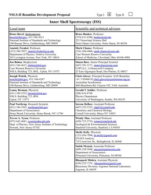
NSLS-II Beamline Development Proposal Type I X Type II<br />
<strong>Inner</strong> <strong>Shell</strong> <strong>Spectroscopy</strong> (<strong>ISS</strong>)<br />
Local team Scientific and technical advisors<br />
Bruce Ravel, Spokesperson<br />
bravel@bnl.gov, 631-344-3613<br />
<strong>National</strong> Institute of Standards and Technology<br />
100 Bureau Drive, Gaithersburg, MD 20899<br />
Anatoly Frenkel, Professor,<br />
(212) 340-7827, anatoly.frenkel@yu.edu<br />
Department of Physics, Yeshiva University<br />
245 Lexington Avenue, New York, NY 10016<br />
Jen Bohon, Biophysicist<br />
(631) 344-4613, jbohon@bnl.gov<br />
Case Western Reserve University<br />
NSLS, Building 725, BNL, Upton, NY 11973<br />
Joseph Woicik, Physicist<br />
woicik@bnl.gov, 631-344-4247<br />
<strong>National</strong> Institute of Standards and Technology<br />
100 Bureau Drive, Gaithersburg, MD 20899<br />
Lonny Berman, Physicist<br />
(631) 344-5333, berman@bnl.gov<br />
NSLS, Building 725, BNL<br />
Upton, NY 11973<br />
Paul Northrup, Research Scientist<br />
(631) 344-3565, northrup@bnl.gov<br />
Department of Geosciences<br />
Stony Brook University, Stony Brook, NY 11794<br />
Trevor A. Tyson, Professor<br />
(973) 642-4681, tyson@adm.njit.edu<br />
Dept. of Physics, New Jersey Institute of Technology<br />
Newark, New Jersey 07102<br />
Bruce Bunker, Professor<br />
(574) 631-6386, bunker@nd.edu<br />
225 Nieuwland Science Hall<br />
Notre Dame University, Notre Dame, IN 46556<br />
Mark Chance, Professor<br />
(216) 368-4406, mark.chance@case.edu<br />
Case Western Reserve University<br />
School of Medicine, Cleveland, Ohio 44106-4988<br />
Simon Bare, Senior Principal Scientist<br />
(847) 391-3171, simon.bare@uop.com<br />
UOP LLC, A Honeywell Company<br />
25 East Algonquin Road, Des Plaines, IL 60017<br />
Chris Glover, Principal Scientist, XAS Beamline<br />
+61 3 85404142,chris.glover@synchrotron.org.au<br />
Australian Synchrotron<br />
800 Blackburn Rd, Clayton VIC 3168, Australia<br />
Gerald T. Seidler, Professor<br />
(206) 616-8746<br />
Physics Department<br />
University of Washington, Seattle, WA 98195<br />
Serena DeBeer, Assistant Professor<br />
(607) 255-2352, sdg63@cornell.edu<br />
Chemistry and Chemical Biology<br />
Cornell University, Ithaca NY 14853<br />
Wendy Mao, Assistant Professor<br />
(650) 723-3718, wmao@stanford.edu<br />
Geological & Environmental Sciences<br />
Stanford University, Stanford, CA 94305<br />
<strong>Shell</strong>y Kelly, Physicist<br />
(123) 456-7890, dr.skelly@gmail.com<br />
EXAFS Analysis<br />
719 Crestview Dr., Bolingbrook, IL 60440<br />
Satish Myneni, Associate Professor<br />
(609) 258-5848, smyneni@princeton.edu<br />
Department of Geosciences<br />
Princeton University, Princeton, NJ 08544<br />
Bhoopesh Mishra, Assistant Physicist<br />
(630) 252-7376 , bhoopeshm@gmail.com<br />
Bioscience Division, Argonne <strong>National</strong> <strong>Laboratory</strong><br />
Argonne, IL 60439
Scientific justification for the <strong>ISS</strong> beamline<br />
The unprecedented brightness and flux of NSLS-II will enable measurements with the high spatial,<br />
energy, and time resolution necessary to fully characterize … complex systems. Advanced<br />
capabilities will include … application of new experimental techniques, such as high-resolution xray<br />
emission spectroscopy and x-ray Raman scattering, to provide new spectroscopic information;<br />
and the use of combinatorial methods for large scale screening of novel materials.<br />
NSLS-II CD-0 document<br />
Absorption of an X-ray by an atom is one of the fundamental interactions of light with matter and the<br />
measurement of absorption is one of the core competencies of any synchrotron. X-ray absorption spectroscopy<br />
(XAS) has a long history at NSLS – many of the first beamlines on the NSLS X-ray ring were for XAS and the<br />
XAS community has remained large and productive for the entire 25 year history of NSLS. In that time, the use<br />
of XAS has become commonplace in a very wide variety of academic and industrial disciplines ranging from<br />
the life and environmental sciences to materials physics and chemistry, engineering materials, geophysics, and<br />
more. The XAS community at NSLS, however, operates within certain constraints.<br />
The dilution of an absorber, the speed at which an XAS spectrum may be collected, and the effective use of high<br />
energy resolution spectrometers are the boundaries within which the NSLS XAS community currently operates.<br />
Each of these boundaries can be pushed back in significant ways by a high-flux source. This proposal is for a<br />
wiggler-based beamline dedicated to XAS and other inner shell spectroscopies. The exceptional flux provided<br />
by a wiggler enables measurement of absorber concentrations at environmentally or technologically relevant<br />
levels impractical to measure at dipole beamlines. Although a companion proposal (the TRS beamline) focuses<br />
on sub-second, time-resolved XAS, this beamline allows collection of high-quality XAS spectra in well under a<br />
minute and is an important part of the strategy for mitigating sample damage under the elevated flux. Finally,<br />
the high flux from the wiggler offers the use of point-to-point focusing and wavelength dispersive<br />
spectrometers, enabling collection of high-resolution XANES spectra, X-ray emission (XES) spectra, and the<br />
measurement of low-energy absorption edges via X-ray energy loss spectroscopy (XELS), all of which are<br />
shown schematically in Fig. 1.<br />
Hard X-ray XES and XELS remain underused by the spectroscopy community not for lack of need or lack of<br />
interest but because of the complexity of the instrumentation and scarcity of beamlines at which such<br />
spectrometers are a routine part of the user program. In recent years, much progress 1,2 has been made in both<br />
spectrometer design and integration into beamline experimental programs. The science examples shown below<br />
are a glimpse at the vast sweep of science that uses inner shell spectroscopy and which benefit by the<br />
Figure 1: Schematic comparing the XAS, XES, and XELS measurements. In XAS, a deep core electronic excitation, the Mn K edge in<br />
this case, is measured by directly absorbing an incident photon. In XES, an electron fills the hole vacated in the XAS process and a<br />
photon is emitted. Shown here is the Mn Kβ emission from a 2p (or possibly some other high-lying) level following the Mn K edge<br />
XAS. In XELS, a photon is scattered inelastically with the energy lost used to promote a deep core electron. Shown here, an oxygen K<br />
edge (1s electron) is measured by XELS. The spectrometer in this example is tuned to 10 keV and the incident photon energy is<br />
scanned through the O K edge energy + the incident energy.<br />
June 21, 2010 1 <strong>ISS</strong> Beamline : NSLS-II BDP 2010
availability of the full complement of inner shell techniques. <strong>ISS</strong> combines the exceptional flux provided by an<br />
NSLS-II wiggler source, the highest quality conventional XAS, and the next generation of XES and XELS<br />
spectrometers into a world-class facility for X-ray spectroscopy as promised in the quote above taken from the<br />
founding document of the NSLS-II project.<br />
Science case: XAS with very low absorber concentration<br />
Biological science: Physiologically relevant concentrations of biomolecules are typically in the sub μM range,<br />
below the sensitivity of a standard XAS beamline. Thus these molecules are generally purified and concentrated<br />
for XAS measurement. Many biological samples cannot be forced into higher concentrations as they precipitate<br />
or form aggregates that are non-biologically relevant or inactive. In order to measure metalloprotein samples in<br />
biologically relevant concentrations, high flux is required, along with highly sensitive fluorescence detection.<br />
Rapid collection of data required for high-quality EXAFS of low concentration absorbers is of immense benefit<br />
to the life science community.<br />
The use of high flux requires damage mitigation<br />
strategies. One such strategy uses continuous-flow<br />
regeneration of fresh sample in solution state, requiring<br />
significant volumes of sample but accommodating<br />
standard fluorescence detection. This strategy also<br />
allows for rapid mixing and stopped- or continuousflow<br />
time-resolved experiments which can reach as<br />
low as the sub-millisecond time regime when full<br />
mixing of reactants can be achieved on this time scale. 3<br />
Cooling of the fluid to near freezing can aid in the<br />
reduction of reaction rates, increasing the number of<br />
relevant measurable reactions. The mixing time<br />
depends on the speed at which the sample is flowed<br />
and the distance of the incident beam from the mixing<br />
point, as depicted in the inset to Fig. 2 while the time<br />
resolution is defined by the size of the beam.<br />
A significant number of enzymatic reactions critical for<br />
biological function occur on the ms to second time<br />
Figure 2: Figure X1. Time-resolved XAS measurements<br />
of TACE (a Zn-binding signal transduction control<br />
enzyme) during enzymatic catalysis using freeze-quench<br />
technology. 4 Changes in Zn coordination and charge<br />
state were observable to 88 ms. The inset shows a<br />
schematic of a micro-fluidic mixer.<br />
scales, particularly when cooled to near freezing temperatures. In general, these are reactions requiring<br />
conformational changes in a protein rather than those which need only perform electron transfer. Freeze-quench<br />
experiments 4 have been used to probe the metal active site chemistry (Fig. 2) of those biomolecules which could<br />
be successfully concentrated without perturbation of function. The <strong>ISS</strong> beamline allows similar measurements<br />
under physiologically relevant concentrations, significantly increasing the number of systems amenable to<br />
investigation.<br />
Environmental science: In a recent XAS experiment at APS (10ID), the adsorption of Hg to Bacillus subtiliis<br />
and Shewanella oneidensis MR-1 biomass was investigated to understand the interaction of Hg with bacterial<br />
cell surfaces. A wide range of Hg 2+ concentration (120 nM to 350 µM) was measured at a fixed bacterial cell<br />
density (2g/L of wet mass) and pH (5.5 ± 0.2). The measurements were performed using a tapered undulator<br />
delivering ~2·10 12 ph/sec to the sample.<br />
The Hg L(III) edge XAS analysis showed that Hg complexes entirely with sulfhydryl groups at the nanomolar<br />
and low micromolar concentrations, and with carboxyl sites at high micromolar concentrations (Fig. 3). Since<br />
Hg-cysteine complexes in aqueous solutions are known to exert strong influence on Hg-methylation 5 , cell<br />
surface bound Hg-(cysteine)3 complexes at environmentally relevant Hg-biomass ratios are likely the key<br />
bottleneck in controlling the rate and extent of Hg-methylation. These results provide first ever insight on the<br />
mechanisms of the transfer of Hg to the cell cytoplasm through the cell membrane for intracellular processes<br />
like methylation 6 . At Hg concentrations above 15 µM, which required hours of measurements at an undulator<br />
June 21, 2010 2 <strong>ISS</strong> Beamline : NSLS-II BDP 2010
Figure 3: (left) Hg LIII edge XANES data show systematic loss of preedge<br />
feature with decreasing Hg concentration (right) Fourier<br />
transformed magnitude of EXAFS data for Hg adsorption to<br />
Shewanella oneidensis MR-1 as a function of adsorbed Hg concentration at pH 5.5 (± 0.2). The red and blue lines in the Fourier<br />
transform magnitude of EXAFS data correspond to 2.02 and 2.51 Å (phase corrected), respectively. A systematic change in the<br />
binding of Hg from Hg-S3, Hg-S to Hg-carboxyl complex was observed with increasing Hg concentration in EXAFS spectra, a trend<br />
that was observed for all bacterial species examined. Cell density in this study was 10 10 cell/L.<br />
beamline with the flux of ~2·10 12 and would have been impossible at dipole beamlines, low abundance<br />
sulfhydryl sites are saturated and masked by high abundance low affinity carboxyl sites which are not relevant<br />
to the intracellular biochemical processes. With the superior flux of <strong>ISS</strong>, measurement of environmentally<br />
relevant, low concentration samples will be routine for environmental contaminants across the periodic table.<br />
Materials science: Thin films are not getting thicker, and dopants are not getting more concentrated. In the<br />
multi-billion dollar semiconductor industry, the “Grand Challenge” is to develop an alternative to the SiO2 gate<br />
dielectric that has enabled Moore's Law scaling of the density of transistors in integrated circuit devices for the<br />
past 40 years. Higher speed with lower power consumption is no longer attainable with ultrathin (
Science case: XES and high resolution XANES<br />
XAS is commonly used for the speciation of valence and chemical<br />
state. This analysis is based on the chemical-dependent edge<br />
shift as well as subtle differences in the features within a few eV<br />
of the edge. The energy resolution of the XAS experiment is<br />
determined both by the bandpass of the monochromator and by<br />
the intrinsic Lorentzian width of the element-specific, finite,<br />
core-hole lifetime. There is a crossover that occurs in the transition<br />
metals where the lifetime broadening of the core-hole<br />
exceeds the monochromator width 8 . Often the ability to distinguish<br />
subtle features of an XAS experiment is limited by the<br />
natural width of the absorbing element rather than by the resolution<br />
of the monochromator. This effect can be reduced by better<br />
defining the observed final-state energy of the electron-core hole<br />
pair decay channel. 9,10,11 A spectrometer like the instrument in<br />
Ref. 11 is used along with the high flux of the <strong>ISS</strong> beamline for<br />
high-resolution XANES measurements, significantly improving the sensitivity to slight differences in chemical<br />
composition and local structure around absorbing atoms.<br />
The same spectrometer is be used to energy analyze the emission spectrum, revealing information about the<br />
electronic state of the absorber species that is complementary to what is obtained with XAS. Fig. 6 shows the<br />
very rich resonant emission spectrum through the absorber edge in CaF3. With the high flux of <strong>ISS</strong> and the<br />
miniXS spectrometer described in Appendix C, high quality 2-D RXES maps like the one shown can be<br />
achieved in ~5 minutes at <strong>ISS</strong> with individual exposures of 1-2 sec.<br />
XES in industrial catalysis: In industrial research,<br />
throughput and efficiency is essential. Heterogeneous<br />
catalysts are regularly probed with a fluorescence microprobe,<br />
with μXAS measured at points of interest. With<br />
<strong>ISS</strong>, non-resonant X-ray emission spectroscopy (XES)<br />
measurement using the miniXS instrument (described in<br />
Appendix C) are feasible over an entire 2D region.<br />
The performance of a Co catalyst used to produce ultralow<br />
sulfur gasoline is positively correlated to the cobalt<br />
sulfidation. Mapping the location of the sulfided and<br />
oxidic Co within an extrudate is relevant to the development<br />
of these catalysts for industrial use. In Fig. 7, the<br />
XES spectra show a shift in the Kβ1,3 peak depending on<br />
the ionicity/covalency of the Co bond. A sulfide bond<br />
has its Kβ1,3 maximum at 2 eV lower in emission energy<br />
than an oxidized bond. At APS 20ID and with the<br />
current generation of miniXS detector, it took 30 seconds<br />
to acquire each XES spectrum in Fig. 7. A single image<br />
from the areal detector is shown in the inset. The x-ray<br />
emission intensities at each pixel are binned in energy to<br />
Figure 5: A conventional Mn K edge XAFS spectrum<br />
of MnO compared to a high-resolution spectrum from<br />
a high-resolution photon spectrometer. 9<br />
Figure 6: A 2-D RXES study for the Ce La emission of CeF3<br />
using the miniXS spectrometer at APS 20-ID. Note the strong<br />
splitting of the pre-edge resonance at ~5719 eV incident<br />
photon energy. The spectral characteristics in this energy<br />
range are useful indicators of the nature of the f-orbital<br />
ground states in such systems. Data courtesy R. Gordon (Univ.<br />
Washington).<br />
give the Kβ emission spectrum. MiniXS uses a focused beam of ~50 μm to resolve the XES spectrum, which is<br />
a relevant length scale in these catalysts. With the high flux of <strong>ISS</strong>, the XES spectra shown in Fig. 7 take under<br />
a second. Thus the spatial distribution of sulfided/oxidized Co within a large, heterogeneous, thin section of<br />
catalyst material can be measured in hours, making measurement of real systems under real conditions quite<br />
feasible.<br />
June 21, 2010 4 <strong>ISS</strong> Beamline : NSLS-II BDP 2010
Figure 7: Non-resonant XES showing the shift in the<br />
Kβ1,3 maximum x-ray emission energy depending on<br />
the sulfided or oxidized Co bonding environment. The<br />
inset shows the XES spectrum collected from in a<br />
single snapshot from the miniXS for one of the<br />
samples. The refection from six crystals is shown.<br />
XES in Biochemistry: Heme and non-heme enzymes carry out<br />
a diverse array of metabolic transformations requiring the<br />
binding and activation of dioxygen. These include proteins such<br />
as methane monooxygenase (MMO) and others capable of C-H<br />
hydroxylations. These enzymes have generated intense interest<br />
aimed at understanding the mechanisms and nature of the active<br />
oxidizing species and the potential for translation to synthetic<br />
catalysts. In both model and enzyme systems, high-valent iron<br />
species are invoked as reactive intermediates. However, due the<br />
inherent reactivity of the synthetic and biological intermediates,<br />
the direct structural characterization is often elusive and spectroscopic<br />
identification has presented significant challenges. <strong>Inner</strong><br />
shell spectroscopies are uniquely suited to address many questions<br />
of oxidation state, local geometry, and spin state of the<br />
active intermediates.<br />
The active site of MMO is an example. The hydroxylation of<br />
methane is carried out by MMO found in methantrophic<br />
bacteria. The soluble form of MMO uses a diiron active site<br />
which reacts with dioxygen to generate first a μ-peroxo-Fe(III)Fe(III) species (MMO-P), then a putative<br />
Fe(IV)Fe(IV) species (MMO-Q), which is responsible for oxidizing methane. Despite intense experimental and<br />
theoretical studies, MMO-Q and MMO-P have eluded structural characterization, and many questions about the<br />
nature of these intermediates remain. The experimental data for MMO-P have been used to argue for a cis-μ-<br />
1,2-peroxo bridging mode, while computational studies favor a µ-η 2 :η 2 -O2 core. For MMO-Q the 2.5 Å Fe-Fe<br />
distance from EXAFS favors a bis-µ-oxo Fe(IV)-Fe(IV) diamond core structure. However, no vibrational data<br />
exist to support any of the postulated core structures and the mechanism for conversion of MMO-P to MMO-Q<br />
is unknown. There are many inherent challenges in studying these enzyme systems – relatively low<br />
concentrations (
Science case: XELS – Soft X-ray edges measured with hard X-rays<br />
X-ray energy loss spectroscopy (XELS) offers an enticing alternative to conventional soft X-ray spectroscopy.<br />
As shown in Fig. 1, the XELS measurement uses high energy incident X-rays and a spectrometer tuned to some<br />
high energy value. The incident beam is scanned through an energy range above the spectrometer tuning energy<br />
such that the difference between the incident and tuned energies passes through an absorption edge. In this way,<br />
the K edges of light elements, L and M edges of transition metals, and more exotic edges such as actinide N and<br />
O are measured. As the incident beam is highly penetrating, XELS is compatible with in situ, operando, and<br />
other sample environments as well as with wet samples and samples exposed to atmosphere. Furthermore, the<br />
XELS measurement is both bulk sensitive and unaffected by the self-absorption attenuation that can be<br />
problematic for soft x-ray spectra measured in fluorescence yield. As a result, XELS is an attractive<br />
complement to any soft x-ray spectroscopy measurement in any of the many scientific disciplines which use<br />
such measurements.<br />
As on example, battery materials have long been the subject of study by XAS, including many lithiated<br />
compounds such as LiFePO4 and Li(Ni,Co)O2. To date, most spectroscopic studies have concentrated on the<br />
measurements of the transition metal K edge, including its measure in an operando environment, or ex situ<br />
measurements of the low-Z K edges or transition metal L edges. An XELS measurement offers the possibility of<br />
a more complete spectroscopic study of materials. Recent work at Argonne 12 has applied XELS to O K and<br />
transmission metal L and M edge spectra in lithiated transition metals. All these ex situ experiments are readily<br />
transferred to the operando environment. XELS measurements of the various low energy edges in the battery<br />
system combined with the superb XAS capabilities of <strong>ISS</strong> provide a more complete spectroscopic picture of the<br />
behavior of the system. Oxides, nitrides, and carbides of transition and heavier metals are of interest to every<br />
spectroscopy-using discipline.<br />
Beamline Concept<br />
The <strong>ISS</strong> beamline is a wiggler-based beamline for <strong>Inner</strong> <strong>Shell</strong> Spectroscopies: XAS, XES, and XELS. This<br />
beamline delivers flux approaching 10 14 photons/second, superior to any existing spectroscopy beamline and enabling<br />
science that is time-prohibitive or simply impossible on dipole sources.<br />
This beamline uses the on-axis portion of a wiggler source with the following optical elements:<br />
• vertical collimating mirror (VCM),<br />
• double crystal monochromator (DCM)<br />
• energy refining monochromator (ERM)<br />
• toroidal focusing mirror (TFM).<br />
The DCM operates in the range from 5 keV to 40 keV, delivers energy resolution of ΔE/E≈10 -4 (typical for a<br />
Si(111) monochromator), and offers scan-to-scan variation in energy calibration under 0.05 eV. The ERM is a<br />
channel-cut Si(311) monochromator which can be placed in the beam to refine the energy resolution coming<br />
from the DCM for experiments which need it. The position of the beam delivered to the experimental hutch is<br />
stable to about 5 μm for proper operation of optics and spectrometers. In this sense, <strong>ISS</strong> benefits from the stability<br />
of the NSLS-II ring as well as from top-off operations. The TFM delivers a spot size of about 1 mm×0.3 mm<br />
at 9 keV.<br />
The VCM and DCM are high heat load elements, requiring careful consideration of cooling strategies. Along<br />
with special design considerations (discussed in detail in Appendix A) for these components, use of <strong>ISS</strong> requires<br />
a variety of operational configurations (mirror angle and filter settings) designed to distribute heatload appropriately<br />
among the various elements when operating in different energy ranges. The configuration modes outlined<br />
in Appendix B serve as the model for operations at <strong>ISS</strong>.<br />
The experimental hutch is a large end station with three experimental installations:<br />
June 21, 2010 6 <strong>ISS</strong> Beamline : NSLS-II BDP 2010
1. An upstream optical table for conventional XAS and the XES spectrometers, as described in Appendix<br />
CC, with energy discriminating detectors and ample room for additional equipment. A set of<br />
KB mirrors focuses to a
espect to the current design of the NSLS-II DW, the concept in that Appendix offers an actionable approach for<br />
<strong>ISS</strong>. The earlier proposal would have delivered flux of over 10 13 ph/sec, competitive with any spectroscopy<br />
beamline in the world. (See Fig. 12 on page 18.) This is the baseline, the lower bound of performance we expect<br />
for <strong>ISS</strong>. Appendix A outlines an approach requiring R&D to delivering truly world-leading flux from the DW<br />
source.<br />
Alternately, a variable gap source could tune the critical energy and power distribution, thus minimizing some<br />
of the challenges associated with the DW source. Of course, any wiggler capable of producing the high flux required<br />
for a world-class spectroscopy beamline will require design of optical elements capable of supporting<br />
very high heat loads.<br />
Regardless of choice of wiggler, this proposal benefits from work already undertaken by the NSLS-II project.<br />
The XPD beamline is developing windows and filters capable of absorbing significant heat load. <strong>ISS</strong> will use<br />
these developments. Calculations commissioned by NSLS-II from Accel show that a directly cooled glidcop<br />
mirror can support up to 7 kW incident heat, deforming with a slope error which remains approximately linear<br />
over the entire active surface of the mirror. (See Fig. 9 in Appendix A.) Although significant, this can be corrected<br />
dynamically, particularly given that top-off operations will assure that the heat load remains constant over<br />
time for a given mirror setting.<br />
The wiggler XAS beamline at the Australian Synchrotron easily supports a 700 W load on a directly-cooled,<br />
Si(111) crystal. At SSRL, power as high as 1.1 kW with minimal thermal distortion to the first crystal have been<br />
demonstrated. 15 As discussed further in Appendix A, this is a somewhat conservative approach to a high heatload<br />
monochromator. Recent calculation show that a directly-cooled first crystal can support significantly higher<br />
heat-loads with manageable thermal distortion to the crystal. Additional R&D is required to understand the<br />
full impact of this thermal distortion on beam performance in terms of impact on flux, resolution, and positional<br />
spread.<br />
Ultimately, it is the responsibility of the NSLS-II project to determine the optimal wiggler design for the <strong>ISS</strong><br />
beamline. We on the proposal team recommend that the use of an existing DW source be considered as the first<br />
option. Strategies for mitigating many aspects of the heat load already exist. Other aspects, most notably the<br />
design of the DCM first crystal, are areas that will require R&D. At the least, a beamline that is competitive<br />
with the best spectroscopy beamlines in the world is clearly tenable. The prospect of delivering truly worldleading<br />
flux to the experimental station merits the technical risk associated with an R&D effort.<br />
Detectors<br />
Energy discriminating detection: Recent work 16 demonstrates that the silicon drift detector (SDD) handles<br />
count rates up to 4·10 5 ph/sec/element with an energy resolution of around 220 eV at 6500 eV using analog signal<br />
chains with 0.1 μsec shaping time. A multi-element SDD is the workhorse fluorescence detector for XAS.<br />
For high energy edges such as 4d metal K edges, the SDD is inefficient due to the limited stopping power of the<br />
Si detection element. A large-area germanium detector (like the Canberra instruments at the CLS HXMA or<br />
Australian XAS beamlines) or the germanium drift detector being developed by the BNL Instrumentation Division<br />
is required.<br />
Wavelength dispersive detection: The miniXS instrument, shown in Fig. 17 on page 22, uses an array of crystals<br />
scattering dispersively onto an area detector to measure a bandpass wide enough to cover entire fluorescence<br />
lines. This short working distance XES spectrometer requires an assortment of crystal carriages to cover<br />
different fluorescence energy ranges, as described in Appendix C. This spectrometer also requires a low background<br />
noise area detector. The large area Pilatus 300K by Dectris combines with the high flux of <strong>ISS</strong> to<br />
provide world-leading throughput for this non-resonant XES system.<br />
The long working distance spectrometer is a proven technology, having been implemented at NSLS, SSRL,<br />
ESRF, CHESS, and elsewhere. It consists of an array of scattering elements on Rowland circles and focused<br />
onto a point or areal detector. A model for this instrument is the one in use at ESRF ID26, 17 although with up to<br />
June 21, 2010 8 <strong>ISS</strong> Beamline : NSLS-II BDP 2010
10 14 ph/sec on the sample, <strong>ISS</strong> outperforms ID26 by an order of magnitude. About 20% of this flux is preserved<br />
through the focusing optics for use by the spectrometers. A large array of spherically bent crystal analyzers are<br />
used for XELS measurements, as described in detail in Appendix D. An energy resolution of about 0.5 eV in<br />
the range of transition metal Kβ fluorescence is the target for this instrument, requiring the use of the ERM.<br />
Without the ERM, the energy resolution will be closer to 1.1 eV. This is a somewhat larger target than for similar<br />
instruments being proposed for NSLS-II or built elsewhere, but it is a wise target. 0.5 eV is sufficient resolution<br />
for both XES and XELS and that relatively large bandpass is appropriate for a high throughput instrument.<br />
Mitigating radiation damage to the sample<br />
With significant power (~100 mW) deposited onto the sample, radiation damage occurs quickly for many<br />
samples, especially especially organic or water-containing samples. Strategies for mitigating this problem are<br />
implemented deeply into the beamline concept. Low temperature sample containment is used for many<br />
experiments and all sample stages and spectrometers are designed to accommodate cryostats. More<br />
significantly, measurement strategies that minimize the exposure of the sample to the beam are adopted. The<br />
step scan conventionally used at XAS beamlines is problematic in that time spent stepping and settling motors<br />
is time spent exposing the sample to the beam without actually collecting data. At <strong>ISS</strong>, the standard mode of<br />
operation for XAS experiments, then, is the slew scan in which the mono is driven continuously in increasing<br />
energy and data is streamed into time-delimited bins. The shutter is closed as the mono rewinds and is opened<br />
for the subsequent scan. Given sufficiently large samples, the sample is periodically rastered to a new location,<br />
avoiding excessive exposure at any one spot. Individual EXAFS scans are typically be measured in about 20<br />
seconds while XANES scans can be as short as 5 seconds.<br />
Required Technical Advances<br />
1. Optimization of optics and beamline configurations.<br />
Management of the high heat load from the wiggler<br />
source is the area most in need of R&D attention, particularly for the DW source. The range of<br />
configuration options and their consequence on each optical element must be explored to optimize<br />
performance over the entire operational range of the beamline.<br />
a) Development of a high heat load VCM – likely to be made of Glidcop, directly cooled, and<br />
coated with Pt and Rh – capable of supporting as much as 7 kW incident heat load. This mirror<br />
will need to dynamically compensate for heat-induced figure error, which is calculated 18 to be<br />
linear over the active surface of the mirror.<br />
b) Development of a high heat load DCM. At the SSRL, a directly cooled Si(111) crystal was<br />
found15 to deform negligibly with heat loads as high as 1.1 kW. This should be the minimum<br />
target of the DCM R&D effort. As outlined in Appendix A, a directly cooled DCM can support<br />
very high heat load, although the effect of the thermal distortion on flux and other performance<br />
attributes must be explored. One particularly important area is stability of LN2 flow which is<br />
known to be a serious source of systematic noise for cryo-cooled monochromators.<br />
c) Development of an ERM. A secondary Si(311) mono to refine the energy resolution requires<br />
development of feedback and tracking system for effective, high-throughput operation.<br />
d) Other high heat load components. <strong>ISS</strong> benefits by work done for the XPD Project Beamline.<br />
e) Beamline configuration management software. Any array of beamline configurations as extensive<br />
as those listed in Appendix B will create substantial complexity of beamline operation. Optical<br />
configuration software, combining database look-up with optimization algorithms, will be<br />
required to make effective use of <strong>ISS</strong> with high user throughput.<br />
2. Spectrometers compatible with many sample environments must be designed to meet the goal of<br />
applying the full suite of inner shell spectroscopies to the broadest possible range of user experiments.<br />
For the miniXS and XELS instruments, work on this has already begun in the group of G.T. Seidler<br />
June 21, 2010 9 <strong>ISS</strong> Beamline : NSLS-II BDP 2010
from University of Washington. The long working distance spectrometer is adapted from a design like<br />
those in use at ESRF, SSRL, CHESS, and elsewhere. See Appendices E and F.<br />
User Community and Demands<br />
By any measure, XAS accounts for around 1/6 of the NSLS user community. XAS and related techniques are<br />
routinely performed at 12 of NSLS' 65 beamlines. In 2006, nearly 20% of on-site visitors to NSLS worked at<br />
XAS beamlines, over 22% of all NSLS users worked at an XAS beamline., and about 15% of all publications<br />
resulting from work at NSLS reported on XAS data. In the period from 2008-2009, users of the beamlines<br />
devoted to XAS and related techniques accounted for 20.4% of the total community of ~2200 NSLS users.<br />
Subscription rates 19 at XAS beamlines are mostly in excess of 1 and the aggregate subscription rate in that<br />
period is nearly 2. NSLS turns XAS users away. XAS beamlines at the other DOE synchrotrons also report<br />
subscription rates above 1. The users NSLS XAS beamlines are all potential users of the <strong>ISS</strong> beamline. The<br />
user base for this beamline is enormous.<br />
NSLS XAS users are actively engaged in the development of spectroscopy at NSLS-II. A technique-based<br />
workshop in 2008 had over 50 participants. The June 1, 2010 XAS beamline development workshop had ~40<br />
participants. Access to a high-performance inner shell spectroscopy beamline was identified as a requirement in<br />
four of the 2008 NSLS-II Scientific Strategic Planning whitepapers.<br />
Proposal Team Expertise and Experience<br />
Four team members (BR, JB, JW, PN) are beamline scientists at NSLS XAS beamlines. One (AF) is the co-PI<br />
of the NSLS Synchrotron Catalysis Consortium. One (LB) has extensive experience in all aspects of optics and<br />
beamline design during a distinguished career at NSLS. One (TT) was part of a team that developed an XES<br />
spectrometer here at NSLS back in the 90s. Three of the advisory team (BB, MC, SB) are senior members of the<br />
synchrotron community and serve on scientific advisory panels for NSLS, BNL, and DOE. One (CG) is the<br />
principal scientist at the wiggler-based XAS beamline in Australia. One (GS) is an innovative designer of X-ray<br />
spectrometers. Two (SD and WM) are faculty at top-tier universities and outstanding synchrotron scientists. One<br />
(SK) is a renowned expert in XAS and author of a recent, important review article on the practice and analysis<br />
of XAS. Two (SM and BM) are experts in the application of synchrotron radiation in the field of<br />
biogeochemistry. Together we represent the breadth and depth of the spectroscopy community. A one page bio<br />
of each Proposal Team member appears at the end of this proposal.<br />
Suggestions for BAT Membership<br />
All local team members as well as Jerry Seidler and Serena DeBeer would be excellent candidates for the BAT.<br />
June 21, 2010 10 <strong>ISS</strong> Beamline : NSLS-II BDP 2010
Appendix A: High heat load beamline optics<br />
The unprecedented brightness and flux of NSLS-II in combination with anticipated developments in<br />
optics, detectors, and computing power will lead to many advanced experimental capabilities that<br />
are not possible today. Access to these new capabilities and the unique infrastructure envisioned<br />
for this new facility will have profound impact on a wide range of scientific disciplines and initiatives<br />
and lead to many exciting discoveries in the coming decades.<br />
NSLS-II CD-0 Document<br />
The NSLS-II damping wiggler (DW) offers extraordinary promise in terms of the broad-band, incoherent flux<br />
required for inner shell spectroscopy, but also extraordinary challenge in terms of design and development of<br />
optics that can accommodate the very high heat load. As we show in this appendix, an ultimate flux of 10 14<br />
ph/sec in the range of 5 keV to 25 keV with the energy resolution required of an XAS experiment is possible<br />
using this source. This ambitious target is an order of magnitude higher than the advertised performance of the<br />
world's current highest flux spectroscopy beamline and fully two orders of magnitude higher than most of the<br />
world's high-performance spectroscopy beamlines. The promise of NSLS-II has always been to provide new<br />
science by advancing synchrotron technology. Here we begin an exploration of how the heat load might be<br />
managed to deliver the full potential flux of the DW source.<br />
To deliver the full flux of an NSLS-II wiggler source, beamline optics capable of handling a very high heat load<br />
must be developed. This includes windows, filters, mirror, and monochromator. As a baseline for consideration<br />
of how this might be done, we can start with the plan developed two years ago for the proposal for an XAS<br />
Project Beamline. The heat load management plan developed at that time is presented for reference as Appendix<br />
B. Using that plan, we demonstrated how to provide in excess of 10 13 ph/sec into the experimental hutch while<br />
presenting only modest technical and cost risks.<br />
The current situation is somewhat different from the assumptions of two years ago. Most significantly, we had<br />
assumed working with canted 3.5 m DW sources. The current DW spec does not allow for canting. However,<br />
considering the longer source, a different filtration strategy, and mirror configurations that allow for a larger<br />
vertical acceptance, we can, in principle, increase the delivered flux by almost an order of magnitude. This<br />
requires several improvements upon what is presented in Appendix B.<br />
1. Remove the first beryllium window. The difficulty of transferring heat adequately out of a thin Be<br />
window makes for one of the most serious challenges of designing a high heat load beamline.<br />
2. Design variable thickness filters which can be inserted into or removed from the beam depending on<br />
experiment energy and first mirror setting. Filter design is an issue already being pursued by NSLS-II<br />
for the XPD Project Beamline. <strong>ISS</strong> will benefit from these developments.<br />
3. Model and design a high heat load collimating mirror.<br />
In 2007, NSLS-II commissioned a study from Accel<br />
into the performance of a mirror under high heat load.<br />
Those calculations showed that a directly cooled<br />
glidcop mirror will certainly suffer large peak slope<br />
errors of ~40 μrad. However, the figure error under<br />
heat loads as high as 7 kW remain approximately<br />
linear over the entire active surface of the mirror.<br />
Although this slope error is substantial, it should be<br />
correctable dynamically. Substantial modeling will be<br />
required to fully characterize the performance of this<br />
high heat load mirror and its impact on beam<br />
properties.<br />
Figure 9: Slope error and temperature on a directly<br />
cooled glidcop mirror (red lines) at 7 kW, as calculated<br />
by Accel for NSLS-II, August 2007.<br />
4. Model and design a high heat load DCM. This is the area that will require the greatest R&D. As a<br />
starting point, we can consider work from SSRL. Their design 15 for a directly cooled, Si(111)<br />
June 21, 2010 11 <strong>ISS</strong> Beamline : NSLS-II BDP 2010
monochromator has been shown to handle up to a 1.1 kW load without significant attenuation of the<br />
theoretical transmission. Refinement of the cooling design is certainly a possibility, although experience<br />
at the XAS beamline as the Australian synchrotron stresses the importance of designing a low vibration<br />
cooling system.<br />
Given that work on filter design is already under way for the XPD beamline and that the 2007 work by Accel<br />
suggests an avenue forward for mirror design, we will concentrate here on the issue of DCM design, with an<br />
eye towards what is required of the optical configuration to deliver the target flux of 10 14 ph/sec.<br />
Starting from two existing monochromator crystal designs, the SSRL model 15 which has cooling liquid passing<br />
through the body of Si block and the so-called hockey puck which has cooling fins which extend into the<br />
cooling liquid, Viswanath Ravindranath from NSLS-II has performed a series of FEA studies of Si(111) under<br />
heat load from the DW source. To begin the FEA analysis, beam characteristics for the DW source are<br />
computed using Ruben Reinenger's software for the 7 m DW source through a 1 mrad x 0.27 mrad aperture.<br />
With the first mirror placed at about 30 m from the source, this aperture collects just over half of the vertical<br />
swath and a horizontal swath that will fill a mirror of normal width.<br />
Figure 10: (Left) Photograph of the monochromator from Ref. 15. Note the channels into<br />
which LN2 flow cartridges are inserted. (Right) The so-called hockey-puck design. The<br />
diffracting surface is at the bottom in this photo. In operation, the fins extend into a<br />
flowing LN2 bath, providing a large surface area for heat transfer.<br />
This aperture passes 10.5 kW of the total 62.5 kW output of the DW source. This beam is filtered by a 100 μm<br />
thick graphite filter, which absorbs 1.6 kW while transmitting >70% of the flux at 5 keV. A 1.2 m long Pt coated<br />
mirror is placed at an angle of 7 mrad, with a critical energy of ~9.7 keV. This absorbs 6.5 kW of power,<br />
leaving 2.8 kW incident upon the first crystal of the monochromator. This configuration results in a flux out of<br />
the monochromator of 10 14 ph/sec. This, then is the initial condition of the FEA analysis, which was performed<br />
for incident angle corresponding to 5 keV and 9 keV operations.<br />
FEA calculations are made using the FEA models created for the two monochromator configurations shown in<br />
Fig. 10, 2.8 kW incident power, and the same LN2 convection cooling model as in Ref. 15. The calculations are<br />
made for the cases of 5 keV and 9 keV. These energies are chosen as representative of the range of use of the<br />
beamline as as difficult test cases for heat load management. Indeed, the lower energy, 5 keV, represents the<br />
most difficult case to be considered for <strong>ISS</strong>. The peak power densities are 4.25 W/mm 2 at 5 keV and<br />
2.41 W/mm 2 at 9 keV.<br />
The SSRL design is promising. For the 5 keV case, the peak temperature is 172 K and the peak meridional and<br />
sagittal slope errors are 40 μrad and 36 μrad. At that energy, the Si(111) Darwin width is 60 μrad. These are<br />
large slope errors, but not so large that further consideration is unwarranted. By increasing the number of flow<br />
channels and increasing the LN2 flow rate, the peak slope errors are reduced to 21 μrad and 18 μrad,<br />
respecitvely.<br />
At 9 keV, the situation is improved. For the SSRL design, the peak slope errors are 7 μrad and 9 μrad, compared<br />
to a Darwin width of about 30 μrad. The peak temperature is137 K. The factor-of-2 improvement afforded by<br />
the increase in number of channels and flow rate reduces the slope error to around 15% of the Darwin width.<br />
June 21, 2010 12 <strong>ISS</strong> Beamline : NSLS-II BDP 2010
The situation improves substantially upon considering the hockey puck design. With the large surface afforded<br />
by the cooling fins, this crystal suffers meridional and sagittal slope errors of 18 μrad and 15 μrad at 5 keV. At<br />
that energy, the peak temperature is 153 K. At 9 keV, the peak slope errors are only 1 μrad each with a peak<br />
temperature of 125 K.<br />
Figure 11: (Top left) Temperature plot of the modeled crystal. Maximum temperature is 153 K. (Bottom left) Displacement<br />
plot. (Top right) Meridional displacement and slope error. Peak meridional slope error is ±18 μrad. (Bottom right) Sagittal<br />
displacement and slope error. Peak sagittal slope error is ±15 μrad.<br />
The results at 5 keV for the hockey puck are shown in Fig. 11. Even with this level of slope error, the Si(111)<br />
crystal is highly transmitting, although there are significant effects of energy and positional spread of the beam<br />
that would have an impact on use of the beam. By 9 keV, the slope errors are quite small even despite the high<br />
heat load of 2.8 kW.<br />
These results are extremely encouraging although clearly this is an incomplete analysis. This FEA analysis<br />
suggests that the directly cooled hockey puck crystal can support the 2.8 kW load required to deliver a flux of<br />
10 14 ph/sec. The situation is somewhat worse at the lower energy. But even there, these results are encouraging.<br />
At the very least, the heat load can be reduced by additional filtration and flux well in excess of 10 13 ph/sec is<br />
possible. Ray tracing to determine the effect of the slope error on the performance of the beamline is warranted.<br />
Given the modest requirements of <strong>ISS</strong> on source brightness, the impact of the slope errors at lower energy might<br />
be tolerable.<br />
June 21, 2010 13 <strong>ISS</strong> Beamline : NSLS-II BDP 2010
Appendix B: Heat Load Management at the Damping-Wiggler XAS<br />
Beamline<br />
Paul Northrup (March 7, 2008)<br />
[Note: This section was prepared for presentation to the March 2008 meeting of the NSLS-II EFAC and was<br />
submitted as supporting documentation for the XAS Project Beamline proposal. It is presented here verbatim<br />
except for two sections which are not relevant to the current proposal. Although some details of the damping<br />
wiggler design, most importantly the ability to cant 3.5 m source, have changed in the intervening years, this<br />
remains useful as a template for designing a beamline to work on the current DW design.]<br />
Summary: This document was prepared as a supplement to the Preliminary Design Report (PDR) in response<br />
to reviewer concerns of the manageability of the high power delivered by the NSLS-II Damping Wiggler (DW)<br />
insertion device. The DW was chosen as the best source for the Project X-ray absorption spectroscopy (XAS)<br />
beamline based on its properties of high flux, broad continuous energy range, and non-coherent nature. A<br />
divide-and-conquer approach was employed to distribute heatload over different beamline components. Initial<br />
considerations of heatload assumed worst-case configurations in an effort to make the most robust design<br />
possible. However, it soon became clear that several components could not tolerate the full brunt of unfiltered<br />
power being considered for a 7m-long DW.<br />
Therefore the calculations presented in this document were undertaken to determine realistic heat loads on<br />
beamline components in the configurations expected under actual use. As a result, heatloads on individual<br />
components are shown to be well within the desired tolerances. Further, it will not be necessary to compromise<br />
beamline performance or flux in order to manage heat load.<br />
Beamline requirements, parameter limits and assumptions: This document supplements the PDR, last<br />
revised January 2008. The goal of the XAS beamline is to deliver very high flux over a wide continuous energy<br />
range, while maintaining excellent stability and repeatability of beam position and energy calibration. Energy<br />
range is limited on the high end by the insertion device and on the low end by the Be window and necessary<br />
filtration. The DW source is assumed to be a 3.5m-long DW90 device (37 poles, K=15.2) in the upstream<br />
canted position. This follows the design decision to employ a canted geometry from day one rather than two inline<br />
segments that would be canted at a later time.<br />
The acceptance angles for beamline optics determine the maximum fraction of the DW fan that will impact<br />
components. Vertical acceptance is calculated for a collimating mirror of variable angle (depending on<br />
configuration as described below) assuming a length of 1.5 m (1.45 m reflective surface) and a position of 33.65<br />
m from the center of the DW. These positions are based on the shieldwall positions as of January 2008 PDR<br />
revision, and use of the upstream (outboard) canted wiggler segment allowing 0.5 m for the canting bend.<br />
Horizontal acceptance is limited by the 1.0 mrad design maximum (based largely on front end mask<br />
limitations), and also by effective acceptance of the monochromator. This is a function of the effect of<br />
horizontal divergence on the diffraction angle, relative to the rocking curve of the crystal at a given energy. For<br />
example the maximum vertical angular deviation for 1 mrad of horizontal acceptance is 6 μrad. This becomes a<br />
significant consideration at higher energies and for higher-energy-resolution experiments, where the<br />
monochromator rocking curve is less than 6 μrad.<br />
Carbon foil array filters and the standard 0.25mm Be window are positioned at 31 m and 32 m, respectively, and<br />
the monochromator at 35 m.<br />
Selected configurations: The following operational configurations were considered. These were chosen as the<br />
smallest set of fixed repeatable configurations to best serve experimental needs, cover the energy range,<br />
maintain harmonic-rejection, and distribute heat load. Possible adjustments to filter thicknesses, mirror angles,<br />
and coatings will shift these energy ranges and acceptances, but the current set is also optimized for<br />
experimental needs. If necessary, additional configurations could be included for even closer heatload<br />
management. Note that collimating mirror angle effectively determines maximum vertical acceptance.<br />
June 21, 2010 14 <strong>ISS</strong> Beamline : NSLS-II BDP 2010
Configuration Filters VCM<br />
angle/coating<br />
Energy range Angular acceptance<br />
(horiz x vert)<br />
1 250 μm C 3.15 mrad, Si 5.7-9.8 keV 1.0 x 0.136 mrad<br />
2 500 μm C 2.0 mrad, Si 7-15.2 keV 1.0 x 0.086 mrad<br />
3 5 mm C 3.15 mrad, Pt 14.4-26 keV 1.0 x 0.136 mrad<br />
4 7 mm C 2.0 mrad, Pt 17-35 keV 1.0 x 0.086 mrad<br />
5 5 mm C +<br />
110 μm Ni<br />
Table 1: Optical configurations used in different energy ranges.<br />
1.6 mrad, Pt 35-50 keV 0.3 x 0.069 mrad<br />
Be window and pre-filters: The Be window is required as a vacuum-isolation measure between beamline<br />
components and the front end. A windowless configuration would have much more stringent beamline vacuum<br />
requirements, and is not recommended where the monochromator is direct-cooled with LN2 (a possible design<br />
here) or the collimating mirror is direct-water-cooled (as it is here). The standard Be window is 250 microns<br />
thick, 10 mm high, and as wide as necessary for the maximum fan of beam used.<br />
Protection of the Be window from the lowest-energy radiation requires a graphite pre-filter, in an array of<br />
increasing-thickness foils. This is based on the expected requirement to keep the Be temperature below 100C;<br />
above that temperature the stress of repeated thermal cycling may eventually weaken the window. Interestingly,<br />
this component appears to be the most power-limiting element of the XAS beamline. The combined absorption<br />
of the pre-filter and the Be window effectively set the minimum usable energy for the beamline.<br />
Initial FEA calculations by Accel (provided in report of September 2007 and supplement October 2007)<br />
demonstrated that for a vertical acceptance of up to 0.15 mrad (4.65 mm), a 10 mm high Be window would not<br />
perform adequately using any reasonable pre-filter thickness. However, a 5 mm high window would maintain<br />
an acceptable temperature provided its absorbed heat load was kept below 72 W. Using this approximation, for<br />
what is a complex function of total power, power density, and footprint, the minimum pre-filter thickness was<br />
re-calculated for the selected operational configurations. The result is 250 microns of graphite as a pre-filter,<br />
and a minimum usable photon energy of approximately 5.7 keV. There is still significant flux below that energy,<br />
but the slope of flux vs energy is too great for reasonable normalization. These parameters will continue to be<br />
refined as beamline design and shieldwall configuration evolve. Another round of FEA calculations is required.<br />
Using a window height that is barely larger than the beam footprint (5 mm compared with 4.65 mm) will<br />
require careful alignment as well as measures to protect the window frame and braze from direct beam. This can<br />
be best accomplished by placing the Be window on the downstream end of the in-vacuum Bremsstrahlung<br />
collimator at 30 m and providing a very limited vertical adjustment.<br />
Note that changing Be thickness does not offer any advantages. Thicker Be will conduct heat better, thus<br />
cooling more efficiently, but will absorb approximately an equivalent amount more power. Thinner Be is<br />
similarly indifferent, and is less robust than the standard thickness. The other geometric option is to place the<br />
window at an angle to the beam, by rotating in the horizontal plane, to thus spread the power density across a<br />
greater width of Be. Although this increases absorption, using a thinner window would result in a marginal gain<br />
of 5-10% in heat load capacity. That is not sufficient to justify the added complexity and risk at this phase of<br />
design.<br />
Depending on cooling-water temperatures (current design is considering raising this to 85 F), and new FEA<br />
results, it will likely be necessary to employ a colder cooling loop for just the Be window.<br />
The pre-filter design of choice is similar to that employed at NSLS Beamline X25, and consists of an array of<br />
graphite foils. These are of increasing thickness, from 5 to 100 microns each, and are cooled radiatively. The<br />
power is indirectly captured by water-cooling of the filter holder frames and by surrounding the array with a<br />
June 21, 2010 15 <strong>ISS</strong> Beamline : NSLS-II BDP 2010
cooled shielding enclosure. Further “baffles” between the foils and on each end, having openings just larger<br />
than the beam size, limit thermal radiation and scatter that may otherwise heat other components.<br />
Retractable filters: A sequential array of optional additional filters will be used for configurations above the<br />
lowest energy range (Table Error: Reference source not found). These thicker graphite filters absorb more of the<br />
lower energy power, and bear more of the heatload burden for configurations where the collimating mirror<br />
absorbs less. Thus the heatload delivered to the monochromator remains tolerable.<br />
The filter sequence would start at 250 microns graphite and include up to 7 mm net thickness. For the highest<br />
energy ranges, filters composed of heavier elements such as Si or Ni (freestanding or in composite with<br />
graphite) could be added. Heatloads for operations above 35 keV are well below concern due to the limited<br />
acceptance, but power density remains an important issue.<br />
FEA calculations will be needed for these filters as well, but such filters are not expected to be pushing the<br />
limits of the technology. It may be necessary for the first one in the series to be constructed from more than one<br />
layer, akin to the pre-filter design.<br />
Collimating mirror: The collimating mirror bears most of the heatload at lower energies and corresponding<br />
higher incidence angles. Initial FEA by Accel showed that by using direct cooling one could effectively<br />
dissipate approximately 2.5 kW, and fully correct for the thermal distortion by adjusting the bend. However, a<br />
heatload of 4 kW produced a small slope error (0.1 μrad) even after compensation by adjusting the bend.<br />
Indirect (side) cooling of the mirror was unacceptable even at less than 2.5 kW. Therefore it is reasonable to<br />
make the design decision to use direct cooling, and to set a maximum operational heatload of 2.5 kW.<br />
Further FEA calculations will be needed to more closely quantify thermal distortion both along and across the<br />
reflecting surface, for the appropriate set of configurations. Realistic estimates of surface roughness and coating<br />
thickness will also need to be incorporated. Accel’s calculations considered over-illuminating the reflective<br />
surface so as to avoid boundary effects; in practical application this portion of the mirror (and the fate of its<br />
portion of the reflected beam) must be accounted for.<br />
Monochromator: The most critical component for heatload concerns is clearly the monochromator. Thermal<br />
distortion of the first crystal affects throughput (flux), focusing performance (angular divergence), and stability<br />
(over angular changes with energy). Such distortion is modeled by FEA and usually quantified on the basis of<br />
maximum slope error.<br />
A survey of existing facilities worldwide indicated that first-crystal heatloads of up to 700 W can be effectively<br />
managed using cryo-cooled Si(111). Designs differ: most are direct-cooled, which is more difficult to<br />
implement and to maintain, while some are indirect-cooled (for example the Australian Synchrotron XAS<br />
wiggler beamline). FEA by Accel for the NSLS-II beamline indicated that a direct-cooled monochromator could<br />
handle total heatloads up to 1.3 kW with acceptable slope error of less than 4 μrad. An indirect-cooled design<br />
was shown to handle up to approximately 700 W with similar distortion. These were calculated for 23 degrees<br />
theta (the worst-case geometry, for 5 keV monochromatic beam), although they used pre-filter thicknesses<br />
which were not realistic for actual operation. Crystal geometry used for the indirect- cooled model was a large<br />
rectangular block as described in the Accel monochromator design document (and employed at the Australian<br />
Synchrotron); the same geometry was used for the direct-cooled model but a modified set of thermal contact<br />
parameters was employed.<br />
In-house preliminary FEA (conducted by V. Ravindranath) used the direct-cooled “hockey puck” design<br />
employed at several beamlines. These calculations did not yet include any filters or mirrors, but optimized<br />
crystal dimensions and considered the benefits of under-cooling the LN2. Results indicated that for 0.15 x 0.25<br />
mrad acceptance (1.8 kW from an unfiltered 7m DW100 source) the maximum slope error is 23 μrad.<br />
Extrapolating from these results indicated that about 1.2 kW would be the maximum tolerable heatload for a<br />
0.15 x 1.0 mrad footprint.<br />
Based on these early results, the current design includes provisions for direct cooling of the first crystal, with<br />
the expectation that indirect cooling would be the preferred option if more detailed study showed it feasible. A<br />
June 21, 2010 16 <strong>ISS</strong> Beamline : NSLS-II BDP 2010
maximum operational heatload is set at 700 W. This is achievable without compromising beamline performance<br />
based on operational configurations in Table 1 and a canted 3.5 m DW90 source. Further FEA is under way<br />
utilizing more appropriate filter conditions and crystal geometries. These constraints are, however, only<br />
qualitative boundaries: greater thermal distortion may be acceptable for some applications where performance<br />
(energy resolution, focus) is less critical, and even stricter requirements may be applicable to the most<br />
demanding of experiments.<br />
Slope error is only one aspect of thermal distortion to be considered. A rigorous analysis will combine FEA and<br />
ray-tracing in a 3D treatment of the monochromator. Thermal distortion will include energy distribution across<br />
the beam footprint, energy-dependent penetration depth and power absorbed in the 3D solid, 2D slope error<br />
across the beam footprint, and lattice (d-spacing) change throughout the solid. Ray-tracing will utilize the fullycharacterized<br />
solid with respect to the 3D diffracting volume, including such aspects as angular mis-orientation,<br />
surface distortion, d-spacing mismatch, tune between crystals, and rocking-curve width.<br />
Predicted maximum heat loads: The following table shows realistic expected heat loads on the various<br />
components in the five configurations described above (Table 1). Filters absorb a higher fraction when working<br />
at higher energies, while the collimating mirror absorbs a larger share at lower energies. The effect of reduced<br />
angular acceptance at higher energies is shown in the total power column. All values are below the maximum<br />
allowed heatloads as described in the sections above, and represent the highest heatloads theoretically possible<br />
for each configuration.<br />
Configuration Total power Filters Be window VCM Mono<br />
1 (5.7-9.8 keV) 3624 W 693 W 69 W 2363 W 499 W, Si(111)<br />
2 (7-15.2 keV) 2416 W 599 W 33 W 1139 W 643 W, Si(111)<br />
3 (14.4-26 keV) 3624 W 2222 W 15 W 728 W 661 W, Si(111)<br />
4 (17-35 keV) 2416 W 1606 W 8 W 189 W 613 W, Si(111)<br />
5 (35-50 keV) 607 W 529 W 1 W 18 W 57 W, Si(333)<br />
Maximum<br />
allowed load<br />
72 W 2500 W 700 W<br />
Table 2: Heatloads on various heat-bearing optical components in the various configurations.<br />
Considerations for achieving higher energy resolution: For applications requiring higher energy resolution,<br />
typical strategy elsewhere is to use an alternate higher-resolution monochromator crystal pair. The strategy<br />
developed here, in light of the large heatload issue, is unique. Rather than relying on an alternate crystal set (e.g.<br />
Si311) in the high heatload monochromator, the DW XAS beamline design employs a Si(111) primary<br />
monochromator to bear the brunt of the heat load and a synchronized second monochromator in series to refine<br />
the energy to higher resolution. In that way, greater thermal distortion is tolerated in the wider-bandpass Si(111)<br />
crystals, while greater throughput is achieved with the undistorted high-resolution crystals. This also allows<br />
selection of appropriate high-resolution crystal materials without regard to their thermal tolerance.<br />
Crystals with narrower bandpass naturally have lower tolerance for thermal distortion. Performance calculations<br />
were made comparing throughput of Si(311) in the high-heatload monochromator versus Si(111) followed by<br />
Si(311). Taking into account a reasonable approximation of thermal distortion (as discussed above), these<br />
calculations showed that the two-monochromator design produced approximately 40 times higher throughput<br />
than would Si(311) alone.<br />
Scatter shielding: For such a high-power beamline, even scattered radiation may transfer significant heat to<br />
components not in the direct beampath. This requires the addition of water-cooled shielding, baffles, and masks<br />
around and between white beam components. Such shielding will surround the pre-filters, retractable filter<br />
assemblies, and Be window. An additional design feature of the collimating mirror will be a shielding shroud<br />
June 21, 2010 17 <strong>ISS</strong> Beamline : NSLS-II BDP 2010
over the sides and above the face of the mirror (dubbed the “Conestoga wagon”), to keep scattered radiation<br />
from heating the enclosure and the positioning and bending mechanism components.<br />
Particular attention will be paid to scatter-shielding within the monochromator, both for thermal load and for<br />
monochromatic beam quality. Both crystals will be LN2-cooled, to minimize thermal effects from scatter, while<br />
water-cooled shielding will protect other components and mechanisms. The feasibility of a “tracking mask”<br />
between the first and second crystals will be investigated.<br />
Changes in configuration: Since these calculations were initiated, there have been some changes to shieldwall<br />
position and beamline component locations. These refinements are expected to continue, especially at this early<br />
design stage, and will impact such parameters as power density and angular acceptance. Design changes will be<br />
monitored, and heatloads recalculated periodically, but the values presented here should remain substantially<br />
correct (within 10%). In addition, as the larger design issues become settled, beamline component design can<br />
then be optimized to deliver the best possible performance. This will include such aspects as filter thicknesses,<br />
mirror angles, and component cooling schemes.<br />
Figure 12: Flux using the various configurations described in this appendix and using the canted 3.5 m DW sources that<br />
were assumed for the 2008 XAS Project Beamline proposal. The margins represent a spread of delivered flux based on<br />
optimistic and pessimistic assumptions about thermal distortions on the VCM and DCM.<br />
June 21, 2010 18 <strong>ISS</strong> Beamline : NSLS-II BDP 2010
Appendix C: High energy resolution inner shell spectrometry<br />
A typical XAS measurement made using an energy discriminating solid state detector is capable of resolving the<br />
α fluorescence line of transition metal and heavier atoms from the β line. However the energy resolution of any<br />
energy discriminating detector is inadequate to resolve any structure within those fluorescence lines. This is<br />
shown for Mn in Fig. 13. The ultimate energy resolution of an XAS experiment is determined by the bandpass<br />
of the x-ray monochromator and the natural Lorentzian width that results from finite lifetime of the core-hole.<br />
With a silicon monochromator and in the energy range of hard X-ray XAS beamline, the lifetime broadening of<br />
the core-hole exceeds the monochromator bandpass. 8 The natural linewidth can be reduced by better defining<br />
the observed final-state energy of the electron-core hole pair decay channel. 10,11 We are proposing the<br />
implementation of two spectrometers at the <strong>ISS</strong> beamline.<br />
Figure 13: (Left) The Mn Kα and Kβ fluorescence lines measured with a silicon drift detector with ~220 eV<br />
energy resolution. (Right) The Mn Kβ line measured with a crystal analyzer with ~0.5 eV resolution. The Kβ1,3<br />
and Kβ' lines are clearly resolved.<br />
Long working distance XES spectrometer<br />
The long working distance XES spectrometer 20 works by placing one or more bent crystal analyzers on<br />
Rowland circles. With more than one analyzer, the Rowland circles are made to intersect at two points, as<br />
shown in the inset of Fig. 14. The sample sits at one intersection and a point detector sits at the other. The<br />
working distance – the separation between sample and analyzer crystals – is typically around 1 m. A<br />
spectrometer of this sort is the principal instrument for the XAS/XES program at ESRF ID26. Other such<br />
spectrometers are in use at SSRL 6-2, CHESS Station C1, and elsewhere. One of the earliest such instruments<br />
was developed here at NSLS. 9 The very high flux of <strong>ISS</strong> offers the enticing promise of making XES and highresolution<br />
XANES measurements quick and routine, particularly if combined with a spectrometer with a far<br />
larger number of analyzer elements.<br />
The XES spectrum is measured by fixing the incident beam energy and scanning the spectrometer through a<br />
bandpass wide enough to cover an entire fluorescence line, typically a few 10s of eV as shown in Fig. 13. This<br />
energy scan is accomplished by translating the crystal elements and the detector vertically, thus changing the<br />
wavelength for which the Rowland circles shown in Fig. 14 overlap. A high-resolution XANES scan is then<br />
measured by placing the spectrometer at some feature of the XES spectrum, for instance the peak of the Kβ 1,3<br />
peak, and varying the incident energy as in a normal XAS scan. In this way, the broadening due to the core-hole<br />
lifetime is reduced, resulting in spectra like the one shown in Fig. 5 on page 4 on the main body of the proposal.<br />
June 21, 2010 19 <strong>ISS</strong> Beamline : NSLS-II BDP 2010
Figure 14: Experimental set up of the long working distance spectrometer. The arrows indicate the motion of the<br />
components during an XES spectrum. The inset shows the orientation of the Rowland circles for the four analyzer crystals<br />
shown in the schematic. Figure from Ref. 20.<br />
High-resolution XANES is applicable to virtually every XAS experiment in every field that uses XAS. As<br />
stated in the main body of this proposal, a common use of XAS is to determine valence and chemical state via<br />
the near-edge structure. Because of the core-hole broadening, this is often challenging. For example, the<br />
various forms of iron oxide and iron oxyhydroxide are quite similar in their XANES. In a mixed phase system,<br />
quantification of phases by a linear combination analysis is often uncertain due to the core-hole broadening.<br />
Higher resolution XANES spectra, in many cases, removes ambiguity from this analysis, thus improving<br />
quantification of phases. For higher energy edegs, uranium L3 for example, the core-hole broadening is quite<br />
large relative to the valence shift observed between oxidized and partially reduced forms of uranium, thus<br />
impeding the ability to quantify a partially reduced system. Again, high resolution XANES is a great benefit.<br />
Although the effect of reducing the broadening is most pronounced near the edge, this spectrometer can be put<br />
to very good use for a variety of exotic EXAFS measurements. In Ref. 26, the authors show a EXAFS<br />
spectrum for the mixed-spin, iron cyanide Prussian Blue. By tuning the spectrometer to carefully chosen points<br />
in the XES, XAS spectra dominated by one spin state or the other can be measured. The fine resolution of<br />
spectrometer allows measurement of specific fluorescence lines even when other elements in the sample<br />
fluoresce at nearby energies. In this way, EXAFS can be extended 21 beyond an intervening edge. The XES<br />
spectrometer can also be used in the manner of the typical energy dispersive detector to measure XAS spectrum<br />
on a minority element can be measured in the presence of majority component with similar fluorescence energy,<br />
albeit with the ability to reject the majority signal far more efficiently than with the energy dispersive detector.<br />
Long working distance spectrometer used for XELS<br />
The XELS spectrometer is described in detail in Appendix D and operates similarly to the long working<br />
distance XES spectrometer. Instead of tuning the analyzer crystals to an energy associated with a particular<br />
fluorescence line, they are tuned to some high energy. In the schematic of the XELS process shown in Fig. 1,<br />
the analyzers are tuned to 10 keV. A soft X-ray edge is thus measured by scanning the incident beam through an<br />
energy range above the tuning energy of the fixed analyzer crystals. On top a large Compton background, the<br />
energy loss features associated with soft X-ray edges are seen. This is seen in Fig. 15 which shows the XELS as<br />
measured by the LERIX spectrometer 22 at APS beamline 20ID. This spectrometer is similar to the long working<br />
distance spectrometer proposed for <strong>ISS</strong> except that the analyzer crystals are arranged in an arc around the<br />
sample such that XELS is measured for a large range of momentum transfer. In Fig. 15, the Compton peak is<br />
seen dispersing in energy as the momentum transfer is increased. On top of that, the much smaller XELS<br />
features are seen. In this experiment, 23 a titanium-bearing pyrochlore was measured by making fine scans<br />
through the regions indicated in Fig. 15. The data for the O K, Ti L2,3, and Ti M are shown in Fig. 16.<br />
June 21, 2010 20 <strong>ISS</strong> Beamline : NSLS-II BDP 2010
Figure 15: Momentum transfer dependent XELS measured on a<br />
titanium pyrochlore with the LERIX 22 instrument. Note the broad<br />
Compton peak dispersing in energy as the momentum transfer is<br />
changed. The soft X-ray edge regions are highlighted.<br />
With an estimated flux on the sample of about 5x10 12 ph/sec, these<br />
two figures represent about a day of beamtime at APS 20ID. With an<br />
order of magnitude high flux and a spectrometer with a larger number<br />
of scattering elements, <strong>ISS</strong> makes this sort of measurement much<br />
more routine.<br />
The applicability of XELS extends anywhere that soft X-ray<br />
spectroscopy is currently performed. Because it uses a hard X-ray<br />
incident beam, it is an unambiguously bulk-sensitive probe, making it a useful complement to conventional soft<br />
X-ray spectroscopy. The hard x-ray probe also makes this measurement compatible with any sample<br />
environment designed for a conventional XAS experiment, making it possible to combine XAS and XELS in<br />
any kind of in situ or operando experiment.<br />
Short working distance XES spectrometer (miniXS)<br />
Figure 16: Bulk absorption spectra<br />
measured by XELS for O K, Ti<br />
L2,3, and Ti M for the titanium<br />
pyrochlore.<br />
The aspect of the long working distance spectrometer that limits the data collection rate are the total collection<br />
solid angle (for weak XES) or the motor motion times (for strong signals). The use of a position sensitive<br />
detector has led to interesting trade-offs in this field, with vastly improved measurement times for very highresolution<br />
studies 24 and interesting options to convert focusing optics to wider-band dispersive optics by<br />
working off-circle. 25 Another variant which leads to a high-throughput dispersive spectrometer is the short<br />
working distance, or miniXS, spectrometers. 1 In the latest iteration of these instruments (see Fig. 17), which will<br />
soon be a standard part of the 20-ID XAS microprobe endstation at 20-ID, several flat dispersing elements are<br />
placed onto a Rowland circle geometry with only ~8 cm diameter. The active region of each crystal subtends a<br />
collection solid angle comparable to one or two typical spherically bent crystal analyzers, resulting in net<br />
collection solid angles comparable to that for large arrays of spherically bent crystal analyzers in an inexpensive<br />
and versatile framework. Due to the Rowland-like configuration, each diffracting element sends the same<br />
analyzed energy bandwidth through an appropriately placed exit aperture. When the crystals are properly<br />
placed, as per solution of the relevant inverse problem, the resulting dispersed spectrum from each crystal will<br />
perfectly fill the areal detector with no overlaps (see Fig. 18, which comes from a high-pressure DAC study<br />
using a similar instrument).<br />
June 21, 2010 21 <strong>ISS</strong> Beamline : NSLS-II BDP 2010
Figure 17: The miniXS spectrometer. Photo courtesy<br />
of Jerry Seidler, University of Washington.<br />
In Fig. 17, which shows the miniXS instrument configured to perform<br />
U M-edge XES, the beam is incident from the back of the<br />
spectrometer. The emitted radiation which strikes the small (~ 1<br />
cm) crystals on the right hand side of the white, plastic optics<br />
holder is Bragg-scattered and analyzed, resulting in several copies<br />
of the same dispersed spectrum on the Pilatus 100K camera,<br />
placed ~14cm above the exit aperture for the instrument. The<br />
sample is mounted behind the yellow polyimide window, which<br />
allows passage of x-rays for conventional XAS. An example of<br />
Figure 18: XES for Fe2O3 as measured using the miniXS<br />
instrument. The bright stripes are the Fe Kβ1,3 peak as imaged<br />
by each crystal. The greenish bands to the right of each bright<br />
stripe are the weaker, lower energy Kβ' lines.<br />
resulting XES image is shown in Fig. 18. Note that the separation<br />
between the sample and the crystal carriage is quite large enough<br />
to accommodate a specialized sample environment for temperature<br />
controlled or in situ experiments.<br />
Specialized image processing is required to convert these images to XES spectra. First elastic scattering is<br />
exposed to the areal detector through the energy range of the fluorescence lines. This defines the association of<br />
each pixel in the detector to emitted energy. Then the signals<br />
from each pixel associated with the same energy are binned to<br />
yield spectra like those shown in Fig. 19. With a relatively<br />
modest flux of 10 11 ph/sec from APS 16ID, a decent XES<br />
spectrum can be collected in 1 sec. After about two minutes, the<br />
statistics are good enough to resolve the very small Kβ' peak.<br />
With the much superior flux of the <strong>ISS</strong> beamline, this counting<br />
time is substantially reduced for concentrated samples and<br />
dilute samples become easier.<br />
Figure 19: XES spectra from stainless steel after<br />
processing as described in the text.<br />
With efficiencies gained by measuring both dispersively and<br />
redundantly and by using the larger Pilatus 300K, the miniXS<br />
instrument opens up a range of experimental possibilities. The<br />
XES measurement is fast enough to consider μXES mapping of<br />
a heterogeneous sample. The full RXES 26 plane, that is the<br />
measure of XES as a function of incident energy through an<br />
absorption edge, can be measured efficiently. Finally, the fast XES measurement allows for time-resolved XES,<br />
as described in the main body of the proposal.<br />
June 21, 2010 22 <strong>ISS</strong> Beamline : NSLS-II BDP 2010
Appendix D: XELS end station at the <strong>ISS</strong> beamline<br />
Gerald T. Seidler (University of Washington)<br />
Given the rapidly diversifying application of x-ray Raman scattering (i.e., nonresonant inelastic x-ray scattering<br />
from semi-core and core level initial states), we propose to construct a high throughput X-ray Scattering (XRS)<br />
spectrometer to serve the users of NSLS-II. This technique is especially well-suited to NSLS-II, thanks to the<br />
anticipated extremely high brilliance of the new light source: XRS is a photon-starved technique in all but the<br />
simplest experiments. Either of two directions may then be reasonably pursued. First, one my aim at the<br />
absolute highest throughput while retaining the option of ‘good enough’ energy resolution to preserve much, but<br />
admittedly not all, of the information in the pre- and near-edge region. This would mean, for example, having<br />
net energy resolution (for monochromator plus spectrometer) of ~0.5 eV for studies of the near-edge region but<br />
of only ~1.5 - 2 eV for studies of the extended region. Second, one may instead focus exclusively on the<br />
problem of achieving near-edge spectroscopy limited essentially by the core-hole lifetimes of the target species.<br />
For the 1s shell of light elements and the 2p shells of the 3d transition metals, this means an energy resolution of<br />
~0.1-0.2 eV.<br />
While it is possible to combine these two options in the same spectrometer 27 , to do so runs into some<br />
fundamental limitations which compromise the performance of the lower-resolution, high-throughput option.<br />
Most obviously, the low-resolution and high-resolution instruments are best sited at, respectively, wiggler and<br />
undulator sources; the wiggler provides optimal flux but insufficient collimation for effective use of the<br />
monochromators needed for high-resolution studies, and conversely for the undulator source. Also, the highresolution<br />
instrument requires the use of ‘dispersion compensation’ with x-ray sensitive area detectors 24,28 to<br />
reach the desired energy resolution with a 1-m diameter Rowland circle instrument. The resulting coupled<br />
constraints on the area detector location(s) and the density and arrangement of large arrays of spherically bent<br />
crystal analyzers provide a substantial barrier to achieving the highest throughput for the lower-energy<br />
resolution (i.e., 0.5 eV – 1.5 eV) option. We admit that an additional complication arises for applications<br />
requiring extremely small spot sizes, mainly Mbar-pressure DAC studies: such studies would benefit from a<br />
wiggler source for flux, but require an undulator to have fine enough collimation for the needed focus. Hence,<br />
it is clearly the case that one instrument, and in fact one beamline, cannot optimally satisfy both useful goals.<br />
In the present case, where we are operating a wiggler beamline, we have chosen to pursue only the lowerresolution,<br />
higher-throughput direction. We believe that such an instrument at NSLS-2 will effectively serve<br />
numerous scientific communities, as we have summarized in the scientific portion of this BDP. To this end, we<br />
propose to build a massively multi-analyzer IXS spectrometer, which we have named the X-ray Energy Loss<br />
Spectrometer (XELS), to emphasize the conceptual similarities between nonresonant IXS and EELS, and also<br />
to indicate that the spectrometer’s applications will span valence, semicore, and core level initial states – i.e., an<br />
XRS spectrometer geared for spectroscopic studies.<br />
We now outline the projected spectrometer design and performance targets. We say ‘projected’ due to long<br />
timeline until proposed completion of the <strong>ISS</strong> beamline together with the lessons which can be learned in the<br />
interim from the several ongoing projects to modify or commission large XRS spectrometer at the APS<br />
(LERIX-1B and LERIX-2), the ESRF (ID-16 and the planned ID-16 upgrade), and SSRL (Bergmann’s ~100<br />
analyzer system). Members of the <strong>ISS</strong> team are involved in the APS efforts and will remain in close contact<br />
with the relevant scientists at the other facilities as the various instruments are designed, constructed, and<br />
commissioned. We do not doubt that unforeseen challenges, and opportunities, will occur in the construction of<br />
such instruments in the next few years; the <strong>ISS</strong> team will be well-placed to learn from these positive and<br />
negative experiences, and to incorporate that information into the final XELS design.<br />
That being said, our starting point is as follows:<br />
A. Spherically bent crystal analyzers: Si 660 analyzers will be used. These will be strain relieved 10 cm<br />
diameter optics with 1-m radius of curvature. As shown in figure X, such optics have been successfully<br />
deployed at ID-16 of the ESRF, and have demonstrated an 0.41 eV energy bandpass (the resolution in<br />
June 21, 2010 23 <strong>ISS</strong> Beamline : NSLS-II BDP 2010
that figure is broadened by the 200 meV monochromator bandpass). When combined with the energy<br />
refining monochromator at 10 keV the resulting net energy resolution will be 0.5 eV.<br />
B. Analyzer alignment modules: Following the ID16 ESRF experience, the methods of the SSRL<br />
instrument, and the plans for the LERIX-1B and LERIX-2 instruments, each spherically bent crystal<br />
analyzer (SBCA) will be controlled on two tilt axes and one linear translator (toward and away from the<br />
center of the Rowland circle). This allows the maximum freedom for distributing the analyzer<br />
reflections between different detection channels (i.e., overlapping or non-overlapping images on area<br />
detectors) or for modifying the overall instrument ray-tracing to allow larger clearances near the sample<br />
location for special sample environmental apparatus.<br />
C. Spectrometer superstructure: There are several different approaches to the placement of analyzer<br />
modules so as to obtain high coverage while retaining flexibility in analyzer positioning – in fact, the<br />
several LERIX instruments, the ESRF ID16 instrument and planned upgrade, and the SSRL instrument<br />
all take reasonable but different approaches to this problem. Given that the main goal of the XELS<br />
instrument at <strong>ISS</strong> are optimum throughput and given that the analyzers, being relatively low energy<br />
resolution (i.e., wide rocking curves), will be fairly easy to align as long as the wafer mis-cut transferred<br />
to the final analyzer is well-characterized, we favor a brute-force approach. Specifically, we will work<br />
along the lines of the planned LERIX-2 instrument at the Advanced Photon Source. A partial 2.5 m<br />
diameter geodesic-like dome will be constructed with numerous fittings for high-tolerance kinematic<br />
mounting of units of three to 14 analyzer modules within ±30 degrees of the vertical scattering plane<br />
when at 2θ = 90 deg and farther out of the scattering plane when near the forward and backward<br />
scattering geometries.<br />
D. Detectors: Barring significant new developments in low-noise x-ray area detectors, three to four of the<br />
Pilatus100k detectors (or a custom assembly thereof) will be used. These will be augmented by a<br />
multiple stages of spatial filtering to minimize stray scatter, such as is required in any such instrument.<br />
While the spatial degrees of freedom may, in some cases, be used for filtering of the ‘imaged’ scattering<br />
from sample chamber walls, more often we will define ROI’s on the face of the area detector and then<br />
effectively simplify the data processing down to an array of ‘one-pixel’ detectors. The targeted energy<br />
resolution does not require the use of dispersion compensation.<br />
E. Overall ray-tracing: Some details are still under development. Briefly, neighboring analyzers at similar<br />
q will typically share a small detector region, and will often, but not always, share the same Rowland<br />
circle. The instrument will be configured to occasionally move between two different Bragg angles:<br />
88.5 deg for ‘typical’ operation and 87 deg as a ‘maximum clearance’ mode when required by several<br />
users with environmental chambers which must be long in the direction perpendicular to the vertical<br />
scattering plane.<br />
F. Collection solid angle: We target a final coverage of 20% of the upper 2π steradian half-sphere; given<br />
the polarization dependence of XRS, this is closer to 40% of the useful scattered radiation at typical<br />
values of q. Such coverage requires ~200 spherically bent crystal analyzers. Note that XELS is<br />
inherently a modular system. Consequently, the instrument can be built and commissioned with a much<br />
smaller number of analyzers, i.e., ~50 SBCA, in assemblies of ~10 SBCA which can be re-positioned<br />
and re-tuned as needed. Final scale-up to fill the spectrometer frame can then follow, building on the<br />
results of the beta-version spectrometer.<br />
G. Primary and secondary monochromator, and net energy resolution: The heat-bearing principal<br />
monochromator combined with the energy refining monochromator provide adequate energy resolution<br />
for calibration and operation of the XELS end station.<br />
H. Focusing optics: A secondary source aperture and Kirkpatrick-Baez mirrors will be used to focus to a<br />
~50 μm spot. If required, several analyzers at the upstream end of the XELS instrument can be removed<br />
to accommodate the KB optics.<br />
June 21, 2010 24 <strong>ISS</strong> Beamline : NSLS-II BDP 2010
I. User-ready support equipment: Custom cryostats, furnaces, and He-filled or vacuum-compatible<br />
sample chambers will be available for users. A motorized 2-axis goniometer with high-tolerance XYZpositioning<br />
will be integral with all sample chambers.<br />
J. Additional user support: The LERIX-helper package (from Seidler’s group at UW) is now in betatesting<br />
at the Advanced Photon Source. LERIX-helper is a GUI-driven package for calibration and data<br />
processing (i.e., re-gridding and statistically-optimal re-binning) for multi-analyzer instruments working<br />
in the inverse-scanning mode. This package will be scaled-up and slightly modified for use with<br />
LERIX-2, and will then transfer easily to use with XELS. This package allows users to view final<br />
energy loss spectra, averaged over selected q, immediately upon completion of energy scanning. Rough,<br />
interim spectra (as a function of incident photon energy) will also be computed in real-time during data<br />
collection using standard beamline support software.<br />
XELS: Measurement Times for O K-edge Extended XRS Studies<br />
1-analyzer rate 1-analyzer 30 analyzer<br />
sample (counts/sec) estimated int. time for 0.3% total time<br />
I0 = 1*10^14/sec bground/step statistics (min) for study (hrs)<br />
TiO2 (bare) 2000 1.7 5.5 0.5<br />
LiTiO2 (in situ) 2000 4 18.8 1.6<br />
LiCoO2 (in situ) 640 4 58.6 4.9<br />
CO2 (100 atm) 4000 2 3.4 0.3<br />
UO2 320 5 168.8 14.1<br />
Table 3: Estimated measurement times for several different O K extended XELS studies, all at q = 8 Å -1 with<br />
1 eV incident bandwidth. The calculations are based on a combination of theoretical values for the O 1s edge<br />
step in the dynamic structure factor, 1×10 14 ph/sec incident flux at ~10 keV, detection bandwidth and integral<br />
reflectivity appropriate for stress-relaxed Si 660 analyzers, and calculated penetration lengths and/or known<br />
experimental constraints on scattering lengths. The column “1-analyzer rate …” gives the anticipate O 1s<br />
edge-step height in counts per second as measured by a single Si 660 analyzer. The column “1-analyzer int.<br />
time….” gives the needed integration time in minutes for a single analyzer to achieve Poisson statistics,<br />
including the dominant Compton background from all sample constituents, at the level of 0.3% of the O K edgestep.<br />
In other words, the resulting spectra are targeted to have 300:1 signal to noise after subtraction of the<br />
Compton tail. We assume that ~200 individual energies must be measured for a completed study of the extended<br />
XELS from an O K-edge. The longer measurement time for in situ battery studies are due to the added<br />
Compton scattering signal from the polymer electrolyte matrix which holds the TMO particles. The in situ<br />
battery studies would have to be supplemented with measurement of the ~20% O K-edge signal of the polymer<br />
electrolyte and (ideally) matching ex situ studies for additional validation. The estimated count rates, above,<br />
are also in reasonable agreement with prior experience at the APS LERIX-1 instrument, after scaling for<br />
differences in beam and analyzer characteristics. Near-edge studies, also at 1 eV incident bandwidth, would<br />
typically require only ~70 energy steps and also would often place much less stringent requirement on signal to<br />
noise, so that such measurements would be ~1 order of magnitude faster.<br />
June 21, 2010 25 <strong>ISS</strong> Beamline : NSLS-II BDP 2010
1 B. Dickinson, et al. Rev. Sci. Instrum. 79, 123112 (2008)<br />
2 XAS-XES beamline ID26 at ESRF: http://www.esrf.eu/UsersAndScience/Experiments/DynExtrCond/ID26/<br />
3 Kane et al. (2008) Anal. Chem. 80, 9534<br />
4 Solomon et al. (2007) PNAS 104, 4931<br />
5 S. Jeffra and M. Francois, Nature Geoscience, 2 (2009)<br />
6 B. Mishra, J. Fein, N. Yee, T. Beveridge, S. Myneni, Nature Geoscience, (In review)<br />
7 J.C. Woicik, et al. , Appl. Phys. Lett. 73, 1269 (1998).<br />
8 M.O. Krause and J.H. Oliver, J. Phys. Chem. Ref. Data 8, 329 (1976).<br />
9 K. Hämäläinen, et al. Phys. Rev. B 46 14274 (1992).<br />
10 E.Jensen, et al., Phys. Rev. Lett. 62, 71 (1989)<br />
11 K. Hämäläinen, et al., Phys. Rev. Lett. 67, 2850 (1991).<br />
12 M. Balasubramanian and G. Seidler, private communication<br />
13 A 1 micron change in gap on the NSLS-II U20 corresponds to a 5 volt change in peak energy at 17 keV. At that energy, a<br />
0.5 eV step is commonly used in a XANES scan. A repeatable 100 nanometer step in gap height is unrealistic.<br />
14 G. Bunker, Introduction to XAFS: A Practical Guide to X-ray Absorption Fine Structure <strong>Spectroscopy</strong>, (2010)<br />
Cambridge University Press, ISBN 978-0521767750<br />
15 M. Rowan, J.W. Peck and T. Rabedeau, Nucl. Instrum. and Methods, A 467, p. 400 (2001)<br />
16 J. Woicik et al. J. Synchrotron Rad. 17 (2010) 409-413 doi:10.1107/S0909049510009064<br />
17 P. Glatzel, et al. Catalysis Today 145 (2009) 294–299 doi:10.1016/j.cattod.2008.10.049<br />
18 Source Power Load Consideration, Accel presentation to NSLS-II, August 2007<br />
19 NSLS User Administration Office, private communication<br />
20 P. Glatzel and U. Bergmann, Coordination Chemistry Reviews 249 (2005) 65–95<br />
21 J. Yano et al, J. Am. Chem. Soc. 127 (2005) pp 14974–14975<br />
22 T.T Fister et al., Rev. S ci. Instrum. 77, 063901 (2006), doi: 10.1063/1.2204581<br />
23 Unpublished work by the spokesperson and collaborators from University of Sheffield.<br />
24 S. Huotari et al, Rev. Sci. Instrum. 77 053102 (2006)<br />
25 P. Glatzel, private communication<br />
26 P. Glatzel, et al. J. Chem Soc. Am. 126 (2004) 9946<br />
27 R. Vereni, et al, J. Synchrotron Rad. 16 469 (2009); doi:10.1107/S090904950901886X<br />
28 S. Huotari, et al, J. Synchrotron Rad. 12 467 (2005); doi:10.1107/S0909049505010630<br />
June 21, 2010 26 <strong>ISS</strong> Beamline : NSLS-II BDP 2010
Name: Bruce Ravel (<strong>ISS</strong> Spokesperson)<br />
Education/Training:<br />
Institution and Location Degree Year Field of Study<br />
University of Washington Ph.D 1997 Physics<br />
Wesleyan University B.A. 1989 Physics (magna cum laude)<br />
Research and Professional Experience:<br />
<strong>National</strong> Institute of Standards and Technology<br />
Scientist, Synchrotron Measurement Group Nov. 2007-present<br />
<strong>National</strong> Research Council Postdoctoral Fellow 1997-1999<br />
Argonne <strong>National</strong> <strong>Laboratory</strong><br />
Physicist, Biology Division 2005-2007<br />
Naval Research <strong>Laboratory</strong><br />
Physicist, Chemistry Division 2001-2005<br />
Centre <strong>National</strong>e de la Recherche Scientifique<br />
Visiting scientist 2000-2001<br />
Professional Activities:<br />
• Beamline experience<br />
◦ Local contact, beamline X23A2 2007-present<br />
◦ Staff member, APS Sector 10, 2005-2007<br />
• Committees<br />
◦ Executive committee member, International X-ray Absorption Society, 2005-present<br />
◦ Chair, NSLS User Executive Committee, 2009-2010<br />
• XAS software and education<br />
◦ Organizer/instructor XAS Schools: NSLS (2003-2005, 2008, 2009), APS (2006-2009), SLS (2007,2009),<br />
Thai Synchrotron (2009,2010), Univ. Alberta (2005), Polish Academy of Science (2006), Univ. Ghent (2010)<br />
◦ Co-author of the IFEFFIT package, a widely used package for XAS data analysis, 2001-present<br />
◦ Contributing author of FEFF8, a widely used XAS theory program<br />
• Review Panels<br />
◦ Proposal Review Panel, <strong>National</strong> Synchrotron Light Source, 2007-present<br />
◦ Proposal Review Panel, Advanced Photon Source, 2006-present<br />
◦ Proposal Review Panel, Canadian Light Source, 2006-present<br />
Selected Publications:<br />
1. ATHENA, ARTEMIS, HEPHAESTUS: data analysis for X-ray absorption spectroscopy using IFEFFIT, B. Ravel<br />
and M. Newville, J. Synchrotron Rad. 12, pp. 537-541 (2005), doi:10.1107/S0909049505012719<br />
At 706 citations, this is the most frequently cited article from The Journal of Synchrotron Radiation.<br />
2. Real Space Multiple Scattering Calculation and Interpretation of X-Ray Absorption Near Edge Structure, A.L.<br />
Ankudinov, B. Ravel, J.J. Rehr, S. Conradson, Phys. Rev B58:12, 7565, (1998), doi:10.1103/PhysRevB.58.7565<br />
3. Simultaneous XAFS measurements of multiple samples, B. Ravel, C. Scorzato, D.P. Siddons, S.D. Kelly, S.R Bare,<br />
J. Sycnhrotron Radiat., 17 (2010) 380-385, doi:10.1107/S0909049510006230<br />
4. Performance of a four-element Si drift detector for X-ray absorption fine-structure spectroscopy:<br />
resolution, maximum count rate, and dead-time correction with incorporation into the ATHENA data analysis<br />
software, J.C. Woicik, B. Ravel, D.A. Fischer, W.J. Newburgh, J. Sycnhrotron Radiat., 17 (2010) 409-413,<br />
doi:10.1107/S0909049510009064<br />
5. The structure of ion beam amorphised zirconolite studied by grazing angle X-ray absorption spectroscopy, D.P.<br />
Reid, M.C. Stennett, B. Ravel, J.C. Woicik, N. Peng, E.R. Maddrell and N.C. Hyatt, Nuclear Instruments and<br />
Methods in Physics Research B 268:11-12, (2010) 1847-1852; doi:10.1016/j.nimb.2010.02.026<br />
6. Condensed Matter Astrophysics: A Prescription for Determining the Species-specific Composition and Quantity of<br />
Interstellar Dust Using X-rays, J.C. Lee, J. Xiang, B. Ravel, J. Kortright K. Flanagan, The Astrophysical Journal<br />
702:2 (2009) 970; doi: 10.1088/0004-637X/702/2/970<br />
7. Protein Oxidation Implicated as the Primary Determinant of Bacterial Radioresistance M. J. Daly,<br />
E. K. Gaidamakova, V. Y. Matrosova, A. Vasilenko, M. Zhai, R. D. Leapman, B. Lai, B. Ravel, S.-M. W. Li,<br />
K. M. Kemner, J. K. Fredrickson PLOS Biology 5:4, (2007), doi:10.1371/journal.pbio.0050092
Anatoly I. Frenkel<br />
Biographical Sketch<br />
Education and Training<br />
Undergraduate, graduate: University of St. Petersburg, Russia, M.S, Physics, 1981-1987<br />
Graduate: Tel-Aviv University, Israel, Ph.D., Physics, 1990-1995<br />
Postdoctoral: University of Washington, Seattle, 1995 – 1996. Postdoctoral advisor: E. A. Stern<br />
Research and Professional Experience<br />
Yeshiva University, Department of Physics. Professor: 2007, Tenure: 2004, Assoc. Professor: 2001<br />
<strong>Brookhaven</strong> <strong>National</strong> Lab., NSLS. Visiting Scientist, sabbatical appointment: Jan-May, 2009<br />
U. of Illinois, Urbana-Champaign. Research Scientist at XAFS beamline (X16C), 1996 – 2001<br />
Synergistic activities<br />
• Synchrotron Catalysis Consortium (SCC). Principal Investigator and the Spokesperson.<br />
• Co-organizer, International Operando-IV Congress, <strong>Brookhaven</strong> <strong>National</strong> <strong>Laboratory</strong>, 2012.<br />
• Research interests: In situ x-ray studies of nanomaterials; x-ray studies of reactivity; metal-insulator<br />
transitions; polarity of thin films; development of synchrotron instrumentation and data analysis<br />
methods.<br />
• Guest editor of two special issues of Synchrotron Radiation News focusing on “Catalysis with Hard<br />
X-rays”, published in 2009.<br />
• Organizer and instructor in synchrotron summer schools and short courses in 2001-2009.<br />
• I have been a member of the NSLS Users Executive Committee, NSLS, APS and SSRL Proposal<br />
Study Panels, APS AMOC science reviewer, DOE and NSF proposal reviewer.<br />
• Chair of the Division of Natural Sciences and Mathematics at Yeshiva University<br />
Representative publications (total number 155).<br />
• S. Khalid, W. Caliebe, P. Siddons, I. So, B. Clay, T. Lenhard, J. C. Hanson, Q. Wang, A. I.<br />
Frenkel, N. Marinkovic, N. Hould, G. Landrot, D. L. Sparks, A. Ganjoo “QEXAFS instrument with<br />
milli-second time scale optimized for in situ applications” Rev. Sci. Instrum. 81, 015105 (2010).<br />
• A. Yevick, A. I. Frenkel “Effects of surface disorder on EXAFS modeling of metallic clusters”<br />
Phys. Rev. B 81, 115451-7 (2010)<br />
• A. Kowal, M. Li, M. Shao, K. Sasaki, M. B. Vukmirovic, J. Zhang, N. S. Marinkovic, P. Liu, A.<br />
I. Frenkel, R. R. Adzic “Ternary Pt/Rh/SnO2 Electrocatalysts for Oxidizing Ethanol to CO2”, Nature<br />
Materials, 2009, 8, 325-330.<br />
• E. Poverenov, I. Efremenko, A. I. Frenkel, Y. Ben-David, L. J. W. Shimon, G. Leitus, J. M. L.<br />
Marin, D. Milstein, “A terminal Pt(IV)-oxo complex bearing no stabilizing electron withdrawing<br />
ligands and exhibiting diverse reactivity”, Nature, 2008, 255, 1093-1096.<br />
• Q. Wang, J. C. Hanson, A. I. Frenkel “Solving the structure of reaction intermediates by timeresolved<br />
synchrotron x-ray absorption fine structure spectroscopy” J. Chem. Phys. 2008, 129, 234502<br />
• A. I. Frenkel “Solving the 3D structure of metal nanoparticles”, Zeitschrift fur Krystallographie,<br />
2007, 222, 605-611. Invited article for the special issue "Structure Studies of Nanocrystals."<br />
• A. I. Frenkel, D. M. Pease, J. Budnick, P. Metcalf, E. A. Stern, P. Shanthakumar, T. Huang<br />
”Strain-induced bond buckling and its role in insulating properties of Cr-doped V2O3”<br />
Phys. Rev. Lett. 2006, 97, 195502.<br />
• A. I. Frenkel, A. V. Kolobov, I. K. Robinson, J. O. Cross, Y. Maeda, and C. E. Bouldin, “Direct<br />
separation of SRO in intermixed nanocryst. and amorph. phases” Phys. Rev. Lett. 2002, 89, 285503.
NAME<br />
Jen Bohon<br />
EDUCATION/TRAINING<br />
BIOGRAPHICAL SKETCH<br />
POSITION TITLE<br />
Instructor<br />
INSTITUTION AND LOCATION DEGREE YEARS FIELD OF STUDY<br />
Johns Hopkins University, Baltimore, MD B.A. 1992-1996 Biology<br />
Stony Brook University, Stony Brook, NY Ph.D. 1996-2004 Physiology & Biophysics<br />
<strong>Brookhaven</strong> <strong>National</strong> <strong>Laboratory</strong>, Upton, NY Postdoctoral 2004-2005 Biophysics<br />
Case Western Reserve U., Cleveland, OH Postdoctoral 2005-2009 Biophysics<br />
A. Professional Positions and Honors<br />
Positions and Employment<br />
1996-2004 Research Assistant, Stony Brook University, Stony Brook, NY<br />
2004-2005 Postdoctoral Research Associate, <strong>Brookhaven</strong> <strong>National</strong> <strong>Laboratory</strong>, Upton, NY<br />
2005-2009 Postdoctoral Fellow and Beamline Scientist, Case Western Reserve University, Cleveland, OH<br />
2009-2010 Senior Research Associate and Beamline Scientist, CWRU, Cleveland, OH<br />
2010- Instructor and Beamline Scientist, CWRU, Cleveland, OH<br />
Honors<br />
2007-2008 Ruth L. Kirschstein <strong>National</strong> Service Award, Cell Physiology<br />
2008-2009 Ruth L. Kirschstein <strong>National</strong> Service Award, Heart and Lung Physiology<br />
2009- General Member, NSLS User Executive Committee, Elected May 2009<br />
B. Selected Peer Reviewed Publications<br />
1. Bohon, J. and de los Santos, C.R. (2003) Structural effect of the anti-cancer agent 6-thioguanine on<br />
duplex DNA. Nucleic Acids Research, 31, 1331-1338.<br />
2. Bohon, J. and de los Santos, C.R. (2005) Effect of 6-thioguanine on the stability of duplex DNA.<br />
Nucleic Acids Research, 33, 2880-2886.<br />
3. Sullivan MR, Rekhi S, Bohon J, Gupta S, Abel D, Toomey J, Chance MR (2008) Installation and testing<br />
of a focusing mirror at beamline X28C for high flux x-ray radiolysis of biological macromolecules. Review of<br />
scientific instruments, 79(2 Pt 1), 025101.<br />
4. Bohon, J., Jennings, L.D., Phillips, C.M., Licht, S., Chance, M.R. (2008) Synchrotron Protein<br />
Footprinting Supports Substrate Translocation by ClpA via ATP-Induced Movements of the D2 Loop.<br />
Structure, 16, 1157-1165.<br />
5. Jennings, L.D., Bohon, J., Chance, M.R., Licht, S. (2008) The ClpP N-terminus Coordinates Substrate<br />
Access with Protease Active Site Reactivity. Biochemistry 47, 11031-11040.<br />
6. Smedley, J., Bohon, J., Wu, Q., Rao, T. (2009) Laser patterning of diamond. Part I. Characterization of<br />
surface morphology, Journal of Applied Physics 105, 123107.<br />
7. Smedley, J., Jaye, C., Bohon, J., Rao, T., Fischer, D. (2009) Laser patterning of diamond. Part II.<br />
Surface nondiamond carbon formation and its removal, Journal of Applied Physics 105, 123108.<br />
8. Bohon, J., Smedley, J., Muller, E., Keister, J. W., Development of Diamond-Based X-ray Detection for<br />
High Flux Beamline Diagnostics, in Diamond Electronics and Bioelectronics — Fundamentals to<br />
Applications III, edited by P. Bergonzo, J.E. Butler, R.B. Jackman, K.P. Loh, M. Nesladek (Mater. Res. Soc.<br />
Symp. Proc. Volume 1203, Warrendale, PA, 2010), 1203-J19-03.<br />
9. Smedley, J., Keister, J. W., Muller, E., Jordan-Sweet, J., Bohon, J., Distel, J., Dong, B. Diamond<br />
Photodiodes for X-ray Applications, in Diamond Electronics and Bioelectronics — Fundamentals to<br />
Applications III, edited by P. Bergonzo, J.E. Butler, R.B. Jackman, K.P. Loh, M. Nesladek (Mater. Res. Soc.<br />
Symp. Proc. Volume 1203, Warrendale, PA, 2010), 1203-J17-21.<br />
10. Shi, W., Bohon, J., Han, D., Habte, H., Qin, Y., Cho, M., Chance, M. R. (2010) Structural<br />
Characterization of HIV GP41 with the Membrane Proximal External Region, J. Biol. Chem., in press.<br />
11. Bohon, J. Muller, E., Smedley, J. (2010) Development of Diamond-Based X-ray Detection for High<br />
Flux Beamline Diagnostics, J Synchrotron Rad., submitted.
Name: Joseph C. Woicik<br />
Education/Training:<br />
Institution and Location Degree Year Field of Study<br />
Stanford University, Stanford CA Ph.D. and M.S. 1989 Applied Physics<br />
Cornell University, Ithaca NY B.S. 1983 Applied Physics<br />
Research and Professional Experience:<br />
<strong>National</strong> Institute of Standards and Technology<br />
Senior Physicist 1999-present<br />
Physicist 1991-1999<br />
<strong>National</strong> Research Council Post Doctoral Fellow 1989-1991<br />
Professional Activities:<br />
Spokesperson, beamline X2A2 <strong>National</strong> Synchrotron Light Source<br />
Local contact, beamline X24A <strong>National</strong> Synchrotron Light Source<br />
Staff member, Advanced Photon Source Sector 33, 2002-2004<br />
Chair, International Workshop for New Opportunities in Hard X-ray Photoelectron <strong>Spectroscopy</strong>: HAXPES 2009,<br />
<strong>Brookhaven</strong> <strong>National</strong> <strong>Laboratory</strong>, 5/20/09 – 5/22/09<br />
Chair, Advanced Photon Source <strong>Spectroscopy</strong> Review Panel, 2003-2005<br />
Member Advanced Photon Source Beamline Review Panel, 2006<br />
Member Department of Energy Instrumentation Review Panel, 2007<br />
Recent Awards:<br />
Department of Commerce Bronze Metal 1998<br />
Selected Publications:<br />
1. Performance of a 4-element Si drift detector for x-ray absorption fine-structure spectroscopy: Resolution, maximum count<br />
rate, dead-time correction, and incorporation into the Athena data analysis software, J.C. Woicik, B. Ravel, D.A. Fischer, and<br />
W.J. Newburgh, J. Synch. Rad. 17, 409 (2010).<br />
2*. Oxygen vacancies in N doped anatase TiO2: Experiment and first-principles calculations, A.K. Rumaiz, J.C. Woicik, E.<br />
Cockayne, H.Y. Lin, G.H. Jaffari, and S.I. Shah, Appl. Phys. Lett. 95, 262111 (2009).<br />
3**.A ferroelectric oxide made directly on silicon, M.P. Warusawithana, C. Cen, C.R. Sleasman, J.C. Woicik, Y. Li, L.F.<br />
Kourkoutis, J.A. Klug, H. Li, P. Ryan, L.-P. Wang, M. Bedzyk, D.A. Muller, L.-Q. Chen, J. Levy, and D.G. Schlom, Science<br />
324, 367 (2009).<br />
4*. Phase identification of self-forming Cu-Mn based diffusion barriers on p-SiOC:H and SiO2 dielectrics using x-ray absorption<br />
fine structure, J.M. Ablett, J.C. Woicik, Zs. Tokei, S. List, and E. Dimasi, Appl. Phys. Lett. 94, 042112 (2009).<br />
5. Strain-induced change in local structure and its effect on the ferromagnetic properties of La0.5Sr0.5CoO3 thin films, C.K. Xie,<br />
J.I. Budnick, W.A. Hines, B.O. Wells, and J.C. Woicik, Appl. Phys. Lett. 93, 182507 (2008).<br />
6. Ferroelectric distortion in SrTiO3 thin films on Si(001) by x-ray absorption fine structure spectroscopy: Experiment and firstprinciples<br />
calculations, J.C. Woicik, E.L. Shirley, C.S. Hellberg, K.E. Andersen, S. Sambasivan, D.A. Fischer, B.D.<br />
Chapman, E.A. Stern, P. Ryan, D.L. Ederer, and H. Li, Phys. Rev. B 75, Rapid Communications, 140103 (2007).<br />
7. Site-specific valence x-ray photoelectron spectroscopy, J.C. Woicik, Synchrotron Radiation News 17, 48 (2004).<br />
8*. Hybridization and bond-orbital components in site-specific x-ray photoelectron spectra of rutile TiO2, J.C. Woicik, E.J.<br />
Nelson, L. Kronik, M. Jain, J.R. Chelikowsky, D. Heskett, L.E. Berman, and G.S. Herman, Phys. Rev. Lett. 89, 077401<br />
(2002).<br />
9. Direct measurement of valence-charge asymmetry by x-ray standing waves, J.C. Woicik, E.J. Nelson, and P. Pianetta, Phys.<br />
Rev. Lett. 84, 773 (2000).<br />
10*.Bond-length distortions in strained-semiconductor alloys, J.C. Woicik, J.G. Pellegrino, B. Steiner, K.E. Miyano, S.G.<br />
Bompadre, L.B. Sorensen, T.-L. Lee, and S. Khalid, Phys. Rev. Lett. 79, 5026 (1997).<br />
11. Diffraction anomalous fine structure: A new x-ray structural technique, H. Stragier, J.O. Cross, J.J. Rehr, Larry B. Sorensen,<br />
C.E. Bouldin, and J.C. Woicik, Phys. Rev. Lett. 23, 3064 (1992).<br />
12. Studies of Si - Ge interfaces with Surface EXAFS and photoemission, J.C. Woicik and P. Pianetta, in Synchrotron Radiation<br />
Research: Advances in Surface and Interface Science: Issues and Technology, Ed. R.Z. Bachrach, 1992 Plenum Press, New<br />
York.<br />
* NSLS/BNL Highlight<br />
** APS/ANL Highlight
NAME: Lonny E. Berman POSITION TITLE: Physicist<br />
EDUCATION INSTITUTION AND LOCATION DEGREE YEAR FIELD OF STUDY<br />
The College of Arts and Sciences, Cornell University A.B. (Honor) 1981 Physics<br />
The Graduate School, Cornell University M.S. 1983 Applied Physics and Materials Science<br />
The Graduate School, Cornell University Ph.D. 1988 Applied Physics and Materials Science<br />
Post-Graduate Experience:<br />
1987 - 1989 Assistant Scientist, <strong>National</strong> Synchrotron Light Source (NSLS), <strong>Brookhaven</strong> <strong>National</strong> <strong>Laboratory</strong><br />
1989 - 1991 Associate Scientist, NSLS, <strong>Brookhaven</strong> <strong>National</strong> <strong>Laboratory</strong><br />
1991 - Present Scientist (Physicist) (Tenure Appt. April 1994), NSLS, <strong>Brookhaven</strong> <strong>National</strong> <strong>Laboratory</strong><br />
2007 - Present Head, Beamline R&D Section, NSLS, <strong>Brookhaven</strong> <strong>National</strong> <strong>Laboratory</strong><br />
Award:<br />
2000 BSA Science & Technology Award Co-Recipient, <strong>Brookhaven</strong> <strong>National</strong> <strong>Laboratory</strong><br />
Relevant Panels:<br />
1996 - 1997; 1999 - 0 External Review Board, Industrial Macromolecular Crystallographic Association, Argonne Nat Lab<br />
1997 - 99; 2001 - 2 United States <strong>National</strong> Committee for Crystallography, <strong>National</strong> Research Council<br />
1998 - 1999 Technical Advisory Committee, Structural Biology Center Principal Users Group, Argonne Nat Lab<br />
1998 - 2002 Scientific Advisory Board, Southeast Regional Collaborative Access Team, Argonne <strong>National</strong> Lab<br />
1999 - Present Synchrotron Advisory Board, <strong>National</strong> Institute of General Medical Sciences, <strong>National</strong> Inst of Health<br />
Editorships:<br />
2002 - Present Co-Editor, Journal of Synchrotron Radiation, published by the Int Union of Crystallog, Chester, UK<br />
Selected Recent Publications:<br />
1. C.-Y. Kim, M.J. Bedzyk, E.J. Nelson, J.C. Woicik, and L.E. Berman, "Site-Specific Valence-Band Photoemission Study<br />
of α-Fe203", Phys. Rev. B 66, 085115 (2002).<br />
2. J.C. Woicik, E.J. Nelson, L. Kronik, M. Jain, J.R. Chelikowsky, D. Heskett, L.E. Berman, and G.S. Herman,<br />
"Hybridization and Bond-Orbital Components in Site-Specific X-Ray Photoelectron Spectra of Rutile TiO2", Phys. Rev.<br />
Lett. 89, 077401 (2002).<br />
3. L.E. Berman, D.P. Siddons, P.A. Montanez, A. Lenhard, and Z. Yin, "A Cryogenically Cooled Channel-Cut Crystal<br />
Monochromator Using a Helium Refrigerator and Heat Exchanger", Rev. Sci. Instrum. 73, 1481-1484 (2002).<br />
4. L.E. Berman, Q. Shen, K.D. Finkelstein, P. Doing, Z. Yin, and G. Pan, "Characterization of a Diamond Crystal X-Ray<br />
Phase Retarder", Rev. Sci. Instrum. 73, 1502-1504 (2002).<br />
5. J.M. Ablett, L.E. Berman, C.-C. Kao, G. Rakowsky, and D. Lynch, "Small-Gap Insertion Device Development at the<br />
<strong>National</strong> Synchrotron Light Source -- Performance of the New X13 Mini-Gap Undulator", J. Synchrotron Radiation 11,<br />
129-131 (2004).<br />
6. W. Shi, H. Robinson, M. Sullivan, D. Abel, J. Toomey, L.E. Berman, D. Lynch, G. Rosenbaum, G. Rakowsky, L. Rock,<br />
W. Nolan, G. Shea-McCarthy, D. Schneider, E. Johnson, R.M. Sweet, and M.R. Chance, “Beamline X29: A Novel<br />
Undulator Source for X-Ray Crystallography”, J. Synchrotron Rad. 13, 365-372 (2006).<br />
7. R.F. Fischetti, D.W. Yoder, S. Xu, S. Stepanov, O. Makarov, R. Benn, S. Corcoran, W. Diete, M. Schwoerer-Boehing,<br />
R. Signorato, L. Schroeder, L.E. Berman, P.J. Viccaro, and J.L. Smith, “Optical Performance of the GM/CA-CAT<br />
Canted Undulator Beamlines for Protein Crystallography”, Proceedings of the Ninth International Conference on<br />
Synchrotron Radiation Instrumentation, AIP Conf. Proc. 879, 754–757 (2006).<br />
8. T. Tanabe, J.M. Ablett, L.E. Berman, D.A. Harder, S. Hulbert, M. Lehecka, G. Rakowsky, J. Skaritka, A. Deyhim, E.<br />
Johnson, J. Kulesza, and D. Waterman, “X25 Cryo-Ready In-Vacuum Undulator at the NSLS”, Proceedings of the Ninth<br />
International Conference on Synchrotron Radiation Instrumentation, AIP Conf. Proc. 879, 283-286 (2006).<br />
9. J.M. Ablett and L.E. Berman, “Spectral Measurements and Synchrotron Radiation Calculation Comparisons of the New<br />
X25 Mini-Gap Undulator”, Proceedings of the Fourteenth <strong>National</strong> Conference on Synchrotron Radiation<br />
Instrumentation, Nucl. Instrum. Methods Phys. Res., Sect. A 582, 37-39 (2007).<br />
10. T. Shaftan, S. Hulbert, and L. Berman, “Comparison of calculated brightness and flux of radiation from a longperiod<br />
wiggler and a short-period undulator”, J. Synch. Rad. 15, 335-340 (2008).<br />
11. E.M. Dufresne, S.B. Dierker, Z. Yin, and L.E. Berman, “Development of New Apertures for Coherent X-ray<br />
Experiments”, J. Synch. Rad. 16, 358-367 (2009).<br />
12. J. Dvorak, L.E. Berman, S.L. Hulbert, D.P. Siddons, and K. Wallwork, “Simple Tools for Characterization of<br />
Synchrotron Beam Flux, Energy Resolution, and Stability”, Proceedings of the Tenth International Conference on<br />
Synchrotron Radiation Instrumentation, AIP Conf. Proc. 1234, 639-642 (2009).<br />
13. O. Chubar, J. Bengtsson, L.E. Berman, A. Broadbent, Y.Q. Cai, S. Hulbert, Q. Shen, and T. Tanabe, “Parametric<br />
Optimization of Undulators for NSLS-II Project Beamlines”, Proceedings of the Tenth International Conference on<br />
Synchrotron Radiation Instrumentation, AIP Conf. Proc. 1234, 37-40 (2009).
Paul Northrup<br />
Department of Geosciences, Stony Brook University<br />
Education:<br />
Dowling College Biology B.A., 1989<br />
Stony Brook University Earth and Space Sciences M.S., 1992<br />
Stony Brook University Geology Ph.D., 1996<br />
Bell Laboratories Post-Doctoral Associate 1996-1998<br />
Appointments:<br />
2009-present Lecturer and Research Scientist, Department of Geosciences, Stony Brook University<br />
2004-2009 Assistant, Associate Environmental Scientist, Environmental Sciences Department, BNL<br />
2003-2004 Research Scientist, Department of Geosciences, Stony Brook University<br />
1998-2003 Physicist, Member of Technical Staff, Bell Laboratories, Lucent Technologies<br />
NSLS:<br />
2003-present Spokesperson and Program Director, Beamline X15B<br />
1996-present Beamline Scientist, Participating Research Team (PRT), X15B<br />
2004-2009 Contributing User Group Beamline Scientist, X27A Microprobe<br />
2004-2009 PRT member, X11A and X11B EXAFS beamlines<br />
2005-2008 Scientist, Center for Environmental Molecular Science,<br />
an NSF/DOE Center at BNL and Stony Brook<br />
NSLS-II Project:<br />
2009-present Beamline Development Scientist (25%),<br />
NSLS-II Submicron Resolution X-ray (SRX) Beamline<br />
2008-2009: Group Leader (interim), SRX Beamline<br />
2008-present Beamline Advisory Team member, SRX Beamline<br />
2007-2008 Group Leader, NSLS-II Project Beamline for XAS<br />
Selected Relevant Publications:<br />
P. Northrup, “X-ray Absorption <strong>Spectroscopy</strong> Beamline”, Part 2, Chapter 5, in NSLS-II Preliminary Design Report<br />
(PDR), NSLS-II, 2008.<br />
P. Northrup and R. Reeder, “Evidence for the importance of growth-surface structure to trace element incorporation in<br />
topaz” Am. Min., 79, 1994.<br />
F. Einsiedl, T. Schäfer, and P. Northrup, “Combined sulfur K-edge XANES spectroscopy and stable isotope analyses of<br />
fulvic acids and groundwater sulfate identify sulfur cycling in a karstic catchment area” Chemical Geology 238,<br />
268-276, 2007.<br />
P. Bingham, A. Connelly, R. Hand, N. Hyatt, P. Northrup, “Incorporation and Speciation of Sulphur in Glasses for Waste<br />
Immobilization” Glass Technol., 50(3), 135-138, 2009.<br />
P. Northrup, A. Lanzirotti, A. Celestian, “Growth of Environmental Science at the NSLS” Synch. Rad. News, 20:3, 2007.<br />
P. Voyles, P. Citrin, D. Chadi, D. Muller, J. Grazul, P. Northrup, H.-J. Gossmann, “Evidence for a New Class of Defects<br />
in Highly n-doped Si: Donor-pair-vacancy-interstitials” Phys. Rev. Lett., 91, 125505, 2003.<br />
P. Citrin, P. Northrup, R. Birkhahn, A. Steckl, “Local Structure and Bonding of Er in GaN: A Contrast with Er in Si”<br />
Appl. Phys. Lett., 76: 2865, 1999.<br />
Synergistic activities:<br />
1. Beamline design: a) current X15B Helium-atmosphere endstation, design, construction and commissioning,<br />
b) NSLS-II beamline for bulk/microbeam XAS, full preliminary design, c) NSLS-II SRX Beamline,<br />
collaborator on initial BAT design, adaptation to NSLS-II Project constraints, ongoing design effort.<br />
2. Beamline management and program development: X15B.<br />
3. User community: NSLS User Executive Committee representative for imaging. Workshops, short courses<br />
and seminars.<br />
4. Facility strategic planning: Participation in NSLS/NSLS-II scientific and facility strategic planning<br />
workshops, and development of white papers.
Trevor Anthony Tyson’s Short Curriculum Vitae<br />
Department of Physics Tel. #: (973) 642-4681<br />
New Jersey Institute of Technology FAX #: (973) 596-5794<br />
Newark, New Jersey 07102-1982 E-mail: tyson@adm.njit.edu<br />
Professional Preparation<br />
Professor (physics) New Jersey Institute of Technology 2003-<br />
Associate Professor (physics) New Jersey Institute of Technology 2000-2003<br />
Member of Graduate Faculty (physics) Rutgers Univ., Newark Campus 1996-<br />
Assistant Professor (physics) New Jersey Institute of Technology 1996-2000<br />
Postdoctoral Position Los Alamos <strong>National</strong> <strong>Laboratory</strong> 1994<br />
Postdoctoral Position - Atomic Physics and <strong>Spectroscopy</strong> 1991<br />
Stanford University School of Humanities and Sciences Postdoc. Fellow<br />
INFN Frascati Italy - Stanford University<br />
Ph. D. (Applied Physics) Stanford University 1991<br />
B. S., Honors (Physics with Mathematics Minor) Andrews University1983<br />
Related Publications<br />
P. Gao, H. Chen, T. A. Tyson, Z. Liu, and S.-W. Cheong, “Infrared and x-ray<br />
absorption measurements of anomalous phonon effects in orthorhombic LuMnO3: Evidence<br />
for an electronically driven ferroelectric state”, submitted to Phys. Rev. Lett.<br />
S. Sumit aand T. A. Tyson, "New Insights in Electronic Structure and<br />
Antiaromatic Instabilities of BoronNanoribbons", Physical Review Letters, in press.<br />
V. V. Poltavets,K. A. Lokshin, H. Nevidomskyy, M. Croft, T. A. Tyson, J.<br />
Hadermann, G. Van Tendeloo, T. Egami, G. Kotliar , N. ApRoberts-Warren, A. P. Dioguardi,<br />
N. J. Curro, and M. Greenblatt, "Bulk Magnetic Order in a Two-Dimensional Ni1þ=Ni2þ<br />
(d9=d8) Nickelate, Isoelectronic with Superconducting Cuprates", Physical Review Letters,<br />
104, 206403 (2010).<br />
T. A. Tyson, Z. Chen, Q. Jie, Q. Li and J. J. Tu, “Local Spin-Coupled Distortions in<br />
Hexagonal Multiferroic HoMnO3”, Phys. Rev. B 81, 054101 (2010).<br />
T. A. Tyson, Z. Chen, Q. Jie, Q. Li, and J. J. Tu, “Local structure of thermoelectric<br />
Ca3Co4O9”, Phys. Rev. B: Condens. Matter Mater. Phys. 79, 024109/1 (2009).<br />
Q. Qian, T. A. Tyson, M. Deleon, C. C. Kao, J. Bai, and A. I. Frenkel,<br />
"Influence of strain on the atomic and electronic structure of manganite films",<br />
Journal of Physics and Chemistry of Solids 68, 458 (2007).<br />
Q. Qian, T. A. Tyson, C. Dubourdieu, A. Bossak, J. P. Sénateur, M. Deleon, J.<br />
Bai, and G. Bonfait, “Structural, Magnetic and Transport Studies of La0.8MnO3<br />
Films, J.Appl. Phys. 92, 4518 (2002).<br />
Q. Qian, T. A. Tyson, C. Dubourdieu, A. Bossak, J. P. Sénateur, M. Deleon, J. Bai,<br />
and G. Bonfait, “Structural studies of annealed ultrathin La0.8MnO3 films”, Appl. Phys. Lett.<br />
80, 2663 (2002).<br />
Funding (Active)<br />
[1] T. A. Tyson, “Understanding the Spin-Lattice Coupling in Multiferroic Oxides”<br />
DOE-BES ($640,000, 8/2007 to 7/2011)<br />
[2] T. A. Tyson (NJIT), P. Siddons (BNL), J.Bai (UT), Gianluigi De Geronimo, (BNL), C.-<br />
C. Kao (BNL) “Development of a Silicon Detector for Synchrotron Based X-Ray<br />
<strong>Spectroscopy</strong>, X-Ray Holography and Materials Education”, NSF MRI ($550,000, 8/2007<br />
to 7/2010)<br />
[3] T. A Tyson, T. Zhou, M. Xanthos, Z. Iqbal and N. M. Ravindra, “MRI: Acquisition of<br />
a Properties Measurement System for Education and Research in Energy Related Materials”,<br />
NSF MRI ($300,000, 9/2009 to 9/2010)<br />
Ph. D Thesis Advisors: K. O. Hodgson (Stanford University, principal advisor)<br />
S. Doniach (Stanford University)
Name: Bruce A. Bunker<br />
Education/Training:<br />
Institution and Location Degree Year Field of Study<br />
University of Washington Ph.D 1974 Physics<br />
University of Washington B.A. 1980 Physics<br />
Research and Professional Experience:<br />
University of Notre Dame<br />
Department Chair, Department of Physics Aug. 1998 – Aug. 2003.<br />
Professor, Department of Physics Aug. 1994 - present.<br />
Associate Professor, Deparment of Physics Aug. 1987 – Aug. 1994.<br />
Assistant Professor, Department of Physics Sep. 1983 – Aug. 1987.<br />
Professional Activities:<br />
• Director, Materials Research Collaborative Access Team, Advanced Photon Source, Argonne <strong>National</strong> <strong>Laboratory</strong>.<br />
• Vice Chair and Chair Elect, Executive Committee, International X-ray Absorption Society<br />
• Panel Chair, <strong>National</strong> Science Foundation Review Panel, MIT CUBIX Science and Technology Center, Oct. 11-<br />
13, 2009.<br />
• Member, Partner User Council, Advanced Photon Source<br />
• Member, General User Program Advisory Committee, Advanced Photon Source.<br />
• Member, Canadian Light Source Peer Review Committee, 2006-2008.<br />
• Member, <strong>National</strong> Science Foundation Review Panel, Cornell High-Energy Synchrotron Source, October 15-16<br />
2007, and April 27-29 2009.<br />
• Co-Organizer, XAFS X, The 10th International Conference on X-ray Absorption Fine Structure (XAFS X)<br />
(Chicago, Illinois, USA, August 10-14, 1998).<br />
Selected Publications:<br />
1. Mishra, B., Boyanov, M., Bunker, B. A., Kelly, S. D., Kemner K. M., Nerenberg, R., Read-Daily, B. L., Fein, J.<br />
B.. “X-ray Absorption <strong>Spectroscopy</strong> Study of Cd Adsorption Onto Bacterial Consortia”, Geochimica et<br />
Cosmochimica Acta 73 4311–4325, (2009).<br />
2. Mishra, B., Haack, E. A., Maurice, P. A., and Bunker, B. A. (2009). “Effects of the Microbial Siderophore DFO-B<br />
on Pb and Cd Speciation in Aqueous Solution”, Environ. Sci. Technol., 43 (1), pp 94–100, (2009).<br />
3. Vasconcelos, I. F., Haack, E. A., Maurice, P. A., Bunker, B. A., “EXAFS Analysis of Cadmium(II) Adsorption to<br />
Kaolinite”, Chem. Geol. 249/3-4, 237-250, (2008).<br />
4. Vasconcelos, I. F., Bunker, B. A., and Cygan, R. T., "Molecular Dynamics Modeling of Ion Adsorption to the<br />
Basal Surfaces of Kaolinite", J. Phys. Chem. C 111(18) pp 6753 – 6762 (2007) .<br />
5. Mishra, B.., Kelly, S. D., Fein, J. B., Boyanov, M., Kemner , K. M. and Bunker, B. A.. "Cd adsorption onto<br />
Bacillus subtilis bacterial cell walls: Integrating isotherm and EXAFS studies" Geochimica Et Cosmochimica Acta<br />
69 A675-a675, (2005)<br />
6. Robel , I. Girishkumar, G., Bunker, B. A., Kamat, P. V., Vinodgopal, K. (2006). “Structural changes and catalytic<br />
activity of platinum nanoparticles supported on C60 and carbon nanotube films during the operation of direct<br />
methanol fuel cells”, App. Phys. Lett. 88, 073113-1-3.<br />
7. Lahiri, D., Subramania, V., Bunker, B. A., and Kamat, P. V. (2006). “Probing Photochemical Transformations at<br />
TiO2/Pt and TiO2/Ir interfaces using X-ray Absorption <strong>Spectroscopy</strong>”, J. Chem. Phys. 124 (20), 204720-1-<br />
204720-7.<br />
8. Robel, I., Bunker, B. A., Kamat , P. V. and Kuno , M. (2006). "Exciton recombination dynamics in CdSe<br />
nanowires: Bimolecular to three-carrier Auger kinetics" Nano Letters 6(7): 1344-1349 (2006).<br />
9. Lahiri, D., Bunker, B. A., Mishra, B., Zhang, Z., Meisel, D., Doudna, C. M., Bertino, M. F., Blum, F. D.,<br />
Tokuhiro, A. T., , S. Chattopadhyay, Shibata, T., Terry, J. (2005). “Bimetallic Pt–Ag and Pd–Ag nanoparticles”, J.<br />
Applied Phys., 97, 094304.<br />
10. Robel, I., Bunker, B. A., and Kamat, P. V.. "SWCNT-CdS nanocomposite as light harvesting assembly: Photoinduced<br />
charge transfer interactions", Adv. Mater., 17, 2458-2463 (2005).
NAME<br />
Mark R. Chance<br />
BIOGRAPHICAL SKETCH<br />
POSITION TITLE<br />
Director and Professor<br />
EDUCATION/TRAINING (Begin with baccalaureate or other initial professional education, such as nursing, and include postdoctoral training.)<br />
INSTITUTION AND LOCATION DEGREE YEAR(s) FIELD OF STUDY<br />
Wesleyan University, Middletown, CT B.A. 1980 Biology<br />
MIT, Boston, MA 1980-1984 Biochemistry<br />
University of Pennsylvania, Philadelphia, PA Ph.D. 1986 Biophysics<br />
AT&T Bell Labs, Murray Hill, NJ Post-Doc 1986-1988 Biophysics<br />
A. Professional Positions<br />
Positions and Employment<br />
1995- Director, Center for Synchrotron Biosciences, NSLS, <strong>Brookhaven</strong> Labs (BNL)<br />
2005- Director, Case Center for Proteomics and Cleveland Foundation Center for Proteomics<br />
2005- Professor of Physiology & Biophysics, Case Western Reserve Medical School, Cleveland OH<br />
2007- Director, Translational Technologies Core, Cleveland Center for Translational Science<br />
<strong>National</strong> and International Committees<br />
2003- Scientific Advisory Committee, Protein Crystallography Research Resource, BNL, Chair (2007)<br />
2007-2010 Experimental Facilities Advisory Committee, <strong>National</strong> Synchrotron Light Source-II, BNL<br />
2006-2010 Biophysics Proposal Review Panel, Advanced Light Source, Lawrence Berkeley <strong>National</strong> Lab.<br />
2009- Editorial Board Member, Cancer Genomics and Proteomics<br />
2009 Reviewer, Research Grants Council, Hong Kong, China<br />
B. Selected Recent Publications<br />
1. Angel, T., Chance, M.R., and Palczewski, K. Conserved waters mediate structural and functional activation of family A<br />
(rhodopsin-like) G protein-coupled receptors, Proc. Nat. Acad. Sci., 106(21):8555-60, 2009. PMCID: PMC2688986<br />
2. Angel, T.E., Gupta, S., Jastrzebska, B., Palczewski, K., Chance, M.R. “Structural waters define a functional channel<br />
mediating activation of the GPCR, rhodopsin”, Proc. Nat. Acad. Sci, 106(34):14367-72, 2009. PMCID: PMC2732891<br />
3. Kiser, P., Golczak, M., Lodowski, D., Chance, M.R., Palczewski, K., “Crystal Structure of Native RPE65, the Retinoid<br />
Isomerase of the Visual Cycle”, Proc. Nat. Acad. Sci, 106(41):17325-30, 2009. PMCID: PMC2765077<br />
4. Orban, T., Gupta, S., Palczewski, K., Chance, M.R. “Visualizing Water Molecules in Transmembrane Proteins Using<br />
Radiolytic Labeling Methods,” Biochemistry, 49(5):827-34, 2010. PMCID: PMC2819031<br />
5. Orban, T., Bereta, G., Miyagi, M., Wang, B., Chance, M.R., Sousa, M.C., Palczewski, K. “Conformational Changes in<br />
Guanylate Cyclase-Activating Protein 1 Induced by Ca 2+ and N-Terminal Fatty Acid Acylation,” Structure, 18(1):116-<br />
26, 2010. PMCID: PMC2822722<br />
6. Gupta, S., Bavro, V.N., D’Mello, R., Tucker, S.J., Venien-Bryan, C., Chance, M.R. “Conformational Changes During<br />
the Gating of a Potassium Channel Revealed by Structural Mass Spectrometry,” Structure, in press, 2010.<br />
C. Research Support<br />
Ongoing Research Support<br />
P30-EB-009998 (Chance) 9/09-8/14<br />
NIH<br />
Case Center for Synchrotron Biosciences<br />
P20-DA-026133 (Chance) 4/09-3/12<br />
NIH<br />
Case Center for Proteomics in HIV/AIDS and Drug Abuse<br />
P30-EB009998 (Chance) 9/09-8/14<br />
NIH<br />
Case Center for Synchrotron Biosciences<br />
P01-AI-074286 (Cho) 4/08-3/11<br />
NIH<br />
Development of a Subunit Envelope Vaccine<br />
Mark Chance – co-PI- Project 2-Structural Evaluation of Antigens<br />
P01-DE-019759 (Weinberg) 3/09-2/14<br />
NIH<br />
Oral Mucosal Immunity in Vulnerable HIV Infected Populations<br />
UL1-RR-024989 (Davis) 9/07-8/12<br />
NIH<br />
Case Western Reserve University/Cleveland Clinic CTSA<br />
Mark Chance - PI of Translational Technology Core
Simon R. Bare<br />
Biographical Sketch<br />
Education and Training<br />
Undergraduate: University of Liverpool, UK, B.Sc. (Hons), 1979<br />
Graduate: University of Liverpool, UK, Ph.D., Chemistry, 1982<br />
Postdoctoral: Cornell University, Ithaca, NY, 1982-1984. Postdoctoral advisor: Wilson Ho<br />
Postdoctoral: Lawrence Berkeley <strong>National</strong> <strong>Laboratory</strong>, Berkeley, CA, 1984-1986. Postdoctoral<br />
advisor: Gabor A. Somorjai<br />
Research and Professional Experience<br />
UOP LLC, a Honeywell Company, Des Plaines, IL<br />
Senior Principal Scientist, 2003–present, Research & Development Associate 1996-2003<br />
The Dow Chemical Company, Midland, MI<br />
Research Leader, 1993-1996, Project Leader 1989-1993, Senior Research Chemist 1986-1989<br />
Synergistic Activities:<br />
• Synchrotron Catalysis Consortium (SCC). Industrial member<br />
• Member of Basic Energy Sciences Advisory Committee (BESAC)<br />
• Member of BNL Light Sources Directorate Scientific Advisory Committee<br />
• Co-organizer, International Operando-IV Congress, <strong>Brookhaven</strong> <strong>National</strong> <strong>Laboratory</strong>, 2012.<br />
• Chair of DMSE COV, panel lead SUFD COV<br />
• Former member of the NSLS Users Executive Committee (including Chair), NSLS and APS<br />
Proposal Study Panels, DOE and NSF proposal reviewer.<br />
• Initiator of inaugural course on EXAFS Data Collection & Analysis, 2003. Co-organized<br />
similar course at NSLS and APS. Lectured at course (2003-5, 2007)<br />
• Research interests: Characterization of catalysts, with focus on in situ characterization.<br />
Fundamental surface chemistry of catalysts. Synchrotron-radiation based X-ray methods for<br />
probing the structure of catalysts. Developing structure-activity relationships in catalysis.<br />
Representative Publications (total number 70+):<br />
• "Characterization of catalysts in reactive atmospheres by X-ray absorption spectroscopy",<br />
S.R. Bare and T. Ressler, Advances in Catalysis, (2009) 52 339-465.<br />
• “Structural Characterization of Ni-W hydrocracking catalyst using in situ EXAFS and<br />
HRTEM”, S.D. Kelly, N. Yang, G.E. Mickelson, N. Greenlay, E. Karapetrova, W. Sinkler,<br />
S.R. Bare. J. Catalysis (2009) 263 16-33.<br />
• “Design and operation of a high pressure reaction cell for in situ x-ray absorption<br />
spectroscopy”, S.R. Bare, N. Yang, S.D. Kelly, G.E. Mickelson, and F.S. Modica, Catalysis<br />
Today (2007) 126 18-26<br />
• "Uniform catalytic site in Sn-β zeolite determined using x-ray absorption fine structure", S.R.<br />
Bare, S.D. Kelly, W. Sinkler, J.J. Low, F.S. Modica, S. Valencia, A. Corma, L.T. Nemeth, J.<br />
Am. Chem. Soc. 127 (2005) 12924-12932.<br />
• “Effect of hydrogen adsorption on the X-ray absorption spectra of small Pt clusters”, A.L.<br />
Ankudinov, J.J. Rehr, J.J Low, S.R. Bare, Phys. Rev. Lett., 86 (2001) 1642-1645.<br />
• "The kinetic significance of V 5+ in n-butane oxidation catalyzed by vanadium phosphates",<br />
G.W. Coulston, S.R. Bare, H.H. Kung, K.E. Birkeland, G. Bethke, R. Harlow, N. Herron,<br />
P.L. Lee, Science, 275 (1997) 191-193.<br />
• “Simple flow-through reaction cells for in situ transmission and fluorescence X-ray<br />
absorption spectroscopy of heterogeneous catalysts”, S.R. Bare, G.E. Mickelson, F.S.<br />
Modica, A.Z. Ringwelski, and N. Yang, Rev. Sci. Instrum., (2006) 77 023105/1-023105/6.
Name: Gerald T. Seidler<br />
Education/Training:<br />
Institution and Location Degree Year Field of Study<br />
University of Chicago Ph.D 1993 Physics<br />
University of Chicago M.Sc. 1991 Physics<br />
University of Florida B.Sc. 1988 Physics<br />
Research and Professional Experience:<br />
University of Washington<br />
Associate Professor, Department of Physics 2002-present<br />
Assistant Professor, Department of Physics 1996-2002<br />
NEC Research Institute, Princeton NJ<br />
Research Scientist 1993-1996<br />
Professional Activities:<br />
• Applications and spectrometer development for high-throughput nonresonant inelastic x-ray scattering from<br />
valence- and core-level initial states. This includes the design and construction of the LERIX spectrometer at the<br />
Advanced Photon Source.<br />
• Applications and spectrometer development for high-throughput resonant and nonresonant x-ray emission<br />
spectroscopy. This includes the development of the miniXS spectrometers for rapid XES studies, such as at the 20-<br />
ID XAS microprobe endstation at the Advanced Photon Source.<br />
• Energy sciences research, involving actinide materials, energy storage systems, and polymeric photovoltaics<br />
Selected Publications:<br />
1. A Comparative Study of the Valence Electronic Excitations of N2 by Inelastic X-ray and Electron Scattering, J.A.<br />
Bradley, G.T. Seidler, G. Cooper, M. Vos, A.P. Hitchcock, A.P. Sorini, C. Schlimmer, K.P. Nagle, submitted, Phys<br />
Rev Lett (2010).<br />
2. NaReO4, An Intensity Standard for O K-edge Covalency Studies Using XAS and NRIXS <strong>Spectroscopy</strong>, J.A. Bradley,<br />
E.R. Batista, K.S. Boland, C.J. Burns, D.L. Clark, S.D. Conradson, S.A. Kozimor, R.L. Martin, G.T. Seidler, D.K.<br />
Shuh, T. Tyliszezak, M.P. Wilkerson, L.E. Wolfsberg, Ping Yang, accepted, J Amer Chem Soc (2010).<br />
3. Probing Electronic Correlations in Actinide Materials Using Multipolar Transitions, J.A. Bradley, S. Sen Gupta,<br />
G.T. Seidler, K.T. Moore, M.W. Haverkort, G.A. Sawatzky, S.D. Conradson, D.L. Clark, S.A. Kozimor, and K.S.<br />
Boland, Phys Rev B81:19 193104 (2010).<br />
4. Momentum-resolved Resonant and Nonresonant Inelastic X-ray Scattering at the Advanced Photon Source , T.<br />
Gog, G.T. Seidler, D.M. Casa, et al., Synchrotron Radiation News 22:6 12 (2009).<br />
5. Intermediate-Range Order in Water Ices: Non-resonant inelastic x-ray scattering measurements and real-space full<br />
multiple scattering calculations, T. T. Fister, K. P. Nagle, F. D. Vila, G. T. Seidler, J. J. Rehr, J. O. Cross, and C.<br />
Hamner, Phys Rev B79 174117 (2009).<br />
6. A Short Working Distance Multiple Crystal X-ray Spectrometer. B. Dickinson, G.T. Seidler, Z.W. Webb, J.A.<br />
Bradley, K.P. Nagle, S.M. Heald, R.A. Gordon, and I.M. Chou, Rev Sci Instrum 79, 123112 (2008).<br />
7. The local electronic structure of α-Li3N, T. T. Fister, G. T. Seidler, E. L. Shirley, F. D. Vila, J. J. Rehr, K. P. Nagle,<br />
J. C. Linehan, and J. O. Cross, J Chem Phys 129 044702 (2008).<br />
8. An extraction algorithm for core level excitations in non-resonant inelastic x-ray scattering spectra, H.<br />
Sternemann, C. Sternemann, G. T. Seidler, T. T. Fister, and M. Tolan, J Synch Rad 15:2 162 (2008).<br />
9. The Local Electronic Structure of Dicarba-closo-dodecarboranes C2B10H12, T.T. Fister, F.D. Vila, G.T. Seidler, L.<br />
Svec, J.C. Linehan, J.O. Cross, J Amer Chem Soc 130 925 (2008).<br />
10. High multipole transitions in NIXS: valence and hybridization in 4f systems, R.A. Gordon, G.T. Seidler, T.T. Fister,<br />
M.W. Haverkort, G.A. Sawatzky, A. Tanaka, and T.K. Sham, EPL 81 26004 (2008).<br />
11. Fine Structure and Chemical Shifts in Non-resonant Inelastic x-ray Scattering from Li-intercalated Graphite, M.<br />
Balasubramanian, C.S. Johnson, J.O. Cross, G.T. Seidler, T.T. Fister, E.A. Stern , C. Hamner, and S.O. Mariager,<br />
Appl Phys Lett 91 031904 (2007).<br />
12. Deconvolving Instrumental and Intrinsic Energy Resolution in Excited- State X-ray Spectroscopies, T.T. Fister,<br />
G.T. Seidler, J.J. Rehr, J.J. Kas, W.T. Elam, J.O. Cross, and K.P. Nagle, Phys Rev B75 174106 (2007).<br />
13. A multielement spectrometer for efficient measurement of the momentum transfer dependence of inelastic x-ray<br />
scattering, T.T. Fister, G.T. Seidler, L. Wharton, et al., Rev Sci Instrum 77 063901 (2006).
Serena DeBeer<br />
Education/Training:<br />
Institution and Location Degree Year Field of Study<br />
Stanford University Ph.D. 2002 Inorganic Chemistry<br />
Southwestern University B.S. 1995 Chemistry (with honors)<br />
Research and Professional Experience:<br />
2009-present Assistant Professor, Cornell University<br />
2003-2009 Staff Scientist, Stanford Synchrotron Radiation <strong>Laboratory</strong><br />
2001-2003 Beam line Scientist, Stanford Synchrotron Radiation <strong>Laboratory</strong><br />
1995-2001 Graduate Research Assistant, Department of Chemistry, Stanford University<br />
1996-1999 Advanced Teaching Assistant, Department of Chemistry, Stanford University<br />
1995-1996 Teaching Assistant, Department of Chemistry, Stanford University<br />
Professional Activities:<br />
Co-chair, Structural Molecular Biology Synchrotron Summer School, SSRL (2003-2007).<br />
Lecturer, Berkeley-Stanford Summer School on Synchrotron Radiation, 2006<br />
Lecturer, Advanced Photon Source XAFS School, 2009<br />
Organizer XAS Short Course, SSRL 2007<br />
Co-chair of a STXM workshop for applications in geological, environmental and biological sciences,<br />
SSRL, 2007<br />
Co-chair of a workshop on XANES spectroscopy at the SSRL, 2007<br />
Proposal Review Panel, Advanced Photon Source, 2008-2009<br />
Guest editor of a special issue of Inorganica Chimica Acta<br />
Lecturer, Synchrotron School, <strong>National</strong> Synchrotron Radiation <strong>Laboratory</strong>, Thailand, 2005-2007<br />
Selected Publications:<br />
1. 1. “Probing Valence Orbital Composition with Iron Kβ X-ray Emission <strong>Spectroscopy</strong>”N. Lee, T.<br />
Petrenko, U. Bergmann, F. Neese, S. DeBeer, J. Am. Chem. Soc., in press.<br />
2. 2. “Calibration of Scalar Relativistic Density Functional Theory for the Calculation of Sulfur K-Edge<br />
Absorption Spectra” S. DeBeer George, F. Neese, Inorg. Chem., 2010, 49, 1849-1853.<br />
3. 3. “Type zero copper proteins”, K. M. Lancaster, S. DeBeer George, K. Yokoyama, J. H. Richards, H. B.<br />
Gray, Nature Chem., 2009,1, 711-715.<br />
4. 4. “Polarized X-ray Absorption <strong>Spectroscopy</strong> of Single-crystal Mn(V) Complexes Relevant to the<br />
Oxygen Evolving Complex of Photosystem II” J. Yano, J. Robblee, Y. Pushkar, M. A. Marcus, K. Sauer,<br />
J. Bendix, T. J. Collins, E. I. Solomon, S. DeBeer George, and V. Yachandra, J. Am. Chem. Soc., 2007,<br />
129, 12989-13000.<br />
5. “Characterization of a Genuine Iron(V)Nitrido Species by Nuclear Resonant Vibrational <strong>Spectroscopy</strong><br />
Coupled to Density Functional Calculations” S. DeBeer George, T. Petrenko, N. Aliaga-Alcade, E. Bill,<br />
B. Mienert, W. Sturhahn, Y. Xiao, K. Wieghardt, F. Neese, J. Am. Chem. Soc., 2007, 129, 11053-11060.<br />
6. “An Octahedral Coordination Complex of Iron(VI)” J. F. Berry, E. Bill, E. Bothe, S. DeBeer George, B.<br />
Mienert, F. Neese, K. Wieghardt, Science, 2006, 312, 1937-1941.
Wendy Mao<br />
Education/Training:<br />
Institution and Location Degree Year Field of Study<br />
University of Chicago Ph.D. 2005 Geophysical Sciences<br />
MIT B.S. 1998 Materials Science & Engineering<br />
Research and Professional Experience:<br />
Stanford University<br />
Assistant Professor, Joint appointment in Geological and Environmental Sciences &<br />
Photon Science, SLAC <strong>National</strong> Accelerator <strong>Laboratory</strong> 2007-present<br />
Los Alamos <strong>National</strong> <strong>Laboratory</strong><br />
J. R. Oppenheimer Fellow, Post-Doc 2005-2007<br />
Professional Activities:<br />
• Elected member of COMPRES Facilities Committee<br />
• Elected member of APS Users Organization Steering Committee<br />
• DOE Hydrogen Fuel Initiative Review, Hydrogen Storage Panel Member<br />
• West Coast High Pressure Facilities Review Committee, Advanced Light Source, Lawrence Berkeley <strong>National</strong><br />
<strong>Laboratory</strong><br />
• <strong>National</strong> School on Neutron and X-ray Scattering, Argonne <strong>National</strong> <strong>Laboratory</strong>, Argonne, IL, 2008-present.<br />
Selected Publications:<br />
1. “S. Wang, W. L. Mao, A. P. Sorini, C-C. Chen, T. P. Devereaux, Y. Ding, Y. Xiao, P. Chow, N. Hiraoka, H. Ishii, Y.<br />
Q. Cai, and C-C. Kao, High pressure evolution of Fe2O3 electronic structure revealed by X-ray absorption<br />
spectroscopy, Phys. Rev. Lett., submitted.<br />
2. L. Wang, Y. Ding, W. Yang, W. Liu, Z. Cai, J. Kung, J. Shu, R. J. Hemley, W. L. Mao, and H.-k. Mao, Nanoprobe<br />
measurements of materials at megabar pressures, Proc. Nat. Acad. Sci., doi:10.1073/pnas.1001141107, 2010.<br />
3. Q. Zeng, Y. Ding, W. L. Mao, W. Yang, S. V. Sinogeikin, J. Shu, H-k. Mao, and J.Z. Jiang, Origin of pressureinduced<br />
polyamorphism in Ce75Al25 metallic glass, Phys. Rev. Lett.104, 105702, 2010.<br />
4. M. Baldini, G. Aquilanti, H-k. Mao, W. Yang, G. Shen, S. Pascarelli, and W. L. Mao, High-pressure EXAFS study<br />
of vitreous GeO2 up to 44 GPa, Phys. Rev. B 81, 024201, 2010.<br />
5. T. Yamanaka, W. L. Mao, H-k. Mao, R. J. Hemley, and G. Shen, New structure and spin state of iron-rich<br />
(Mg,Fe)SiO3 post-perovskite, J. Phys.: Conf. Ser. 215 012100, doi: 10.1088/1742-6596/215/1/012099, 2010.<br />
6. Q. Zeng, Y. Ding, W. L. Mao, W. Luo, A. Blomqvist, R. Ahuja, W. Yang, J. Shu, S. V. Sinogeikin, Y. Meng, D.<br />
Brewe, J. Z. Jiang, and H-k. Mao, Substitutional alloy of Ce and Al, Proc. Natl. Acad. Sci.<br />
doi:10.1073/pnas.0813328106, 2009.<br />
7. W. L. Mao, H-k. Mao, Y. Meng, P. J. Eng, M. Y. Hu, P. Chow, J. Shu, and R. J. Hemley, X-ray-induced<br />
dissociation of H2O and formation of an O2-H2 alloy at high pressure, Science 314, 636-638, 2006.<br />
8. W. L. Mao, G. Shen, V. B. Prakapenka, Y. Meng, A. J. Campbell, D. L. Heinz, J. Shu, R. Hemley, and H-k. Mao,<br />
Ferromagnesian postperovskite silicates in the D’’ layer of the Earth, Proc. Nat. Acad. Sci. 101, 15867-15869,<br />
2004.<br />
9. W. L. Mao, H-k. Mao, P. J. Eng, T. P. Trainor, M. Newville, C-c. Kao, D. L. Heinz, J. Shu, Y. Meng, and R.<br />
Hemley, Bonding changes in compressed superhard graphite, Science 302, 425-427, 2003.
Name: <strong>Shell</strong>y D. Kelly<br />
Education/Training:<br />
Institution and Location Degree Year Field of Study<br />
University of Washington Ph.D 1999 Physics<br />
University of Idaho B.A. 1992 Physics<br />
Research and Professional Experience:<br />
EXAFS Analysis<br />
Sole proprietor March 202-present<br />
UOP LLC, a Honeywell Company<br />
Consultant Physicist March 2002-present<br />
Michigan State University<br />
Consultant Physicist Dec 2008-present<br />
Argonne <strong>National</strong> <strong>Laboratory</strong><br />
Postdoc March 1999-April 2003<br />
Assistant Physicist April 2003-March 2008<br />
Physicist March 2008-August 2009<br />
Professional Activities:<br />
• Reviewer of General User Proposals on the Microprobe and Imaging Panel for the APS<br />
• Reviewer of scientific manuscripts for Applied Physics Letters, Geochimica et Cosmochimica Acta, Environmental<br />
Science and Technology, American Mineralogist, Chemical Geology, Contaminant Hydrology, Journal of Synchrotron<br />
Radiation, Adsorption of Metals to Geomedia II, and Journal of Catalysis.<br />
• Reviewer of proposals for Natural Environment Research Council Research (NERC), <strong>National</strong> Science Foundation<br />
(NSF), and Stanford Synchrotron Radiation Light Source (SSRL).<br />
• EXAFS workshop instructor for the past 8 years at the <strong>National</strong> Synchrotron Light Source, 2002, 2003, and 2004<br />
and at the Advanced Photon Source, 2005, 2006, and 2007, 2008, 2009.<br />
• Organizer of “Future Directions in Synchrotron Environmental Science” at the Advance Photon Source Users<br />
Meeting, May 2004.<br />
• Chair of Synchrotron-Based Analytical Techniques for Nuclear and Environmental Sciences session at the American<br />
Chemical Society 225 th Meeting, New Orleans, LA, March 2003.<br />
• Early Career Subcommittee member, Strategic Environmental Initiative, Argonne <strong>National</strong> <strong>Laboratory</strong>, 2002.<br />
• Taught XAFS analysis and data collection at the <strong>National</strong> School on Neutron and X-ray Scattering, Advance<br />
Photon Source, August 12-25, 2001.<br />
Selected Publications:<br />
1. Kelly, S. D. (2010). Uranium Chemistry in Soils and Sediments. Synchrotron-based Techniques in Soil and<br />
Sediment. Grafe, M. and Singh, B., Elsevier B.V. 34: 411-466.<br />
2. Kelly, S. D., Wu, W. M., Yang, F., Criddle, C. S., Marsh, T. L., O'Loughlin, E. J., Ravel, B., Watson, D., Jardine, P.<br />
M. and Kemmer, K. M. (2010). "Uranium Transformation in Static Microcosms." Environ. Sci. & Technol. 44: 236-<br />
242.<br />
3. Kelly, S. D., Yang, N., Mickelson, G. E., Greenlay, N., Karapetrova, E., Sinkler, W. and Bare, S. R. (2009).<br />
"Structural characterization of Ni-W hydrocracking catalysts using in situ EXAFS and HRTEM." Journal of<br />
Catalysis 263(1): 16-33.<br />
4. Kelly, S. D., Hesterberg, D. and Ravel, B. (2008). Analysis of soils and minerals using X-ray absorption<br />
spectroscopy. Methods of soil analysis, Part 5 -Mineralogical methods. Ulery, A. L. and Drees, L. R. Madison, WI,<br />
USA, Soil Science Society of America: 367-463.<br />
5. Kelly, S. D., Kemner, K. M., Carley, J., Criddle, C., Jardine, P. M., Marsh, T. L., Phillips, D., Watson, D. and Wu,<br />
W. M. (2008). "Speciation of uranium in sediments before and after in situ biostimulation." Environmental Science<br />
& Technology 42(5): 1558-1564.<br />
6. Kelly, S. D., Kemner, K. M. and Brooks, S. C. (2007). "X-ray Absorption <strong>Spectroscopy</strong> Identifies Calcium-Uranyl-<br />
Carbonate Complexes at Environmental Concentrations." Geochimica Et Cosmochimica Acta 71(4): 821-834.<br />
7. Kelly, S. D., Rasbury, E. T., Chattopardhyay, S., Kropf, A. J. and Kemnner, K. M. (2006). "Evidence of a Stable<br />
Uranyl Site in Ancient Organic-Rich Calcite." Environmental Science & Technology 40(7): 2262-2268.
Name: Satish C. B. Myneni<br />
Education/Training:<br />
Institution and Location Degree Year Field of Study<br />
The Ohio State University Ph.D. 1995 Environmental Geochemistry<br />
Indian Inst. of Tech., Kharagpur M. Tech. 1991 Applied Geology<br />
Indian Inst. of Tech., Bombay M. Sc. 1989 Applied Geology<br />
Research and Professional Experience:<br />
Princeton University<br />
Associate Professor 2005-present<br />
Assistant Professor 1999-2005<br />
Lawrence Berkeley <strong>National</strong> <strong>Laboratory</strong><br />
Geological Scientist 1998-1999<br />
Postdoctoral Scientist 1995-1998<br />
Professional Activities last 5 years:<br />
Reviewer for grant proposals of USDA, DOE, NSF<br />
Member of the American Chemical Society, Geochemical Society<br />
Review Panels<br />
Proposal Review Panel (2005-present) & Proposal Oversight Panel (2007-present), <strong>National</strong><br />
Synchrotron Light Source<br />
DOE (3 times in the last 5 years)<br />
Significant Awards and Honors<br />
1985-87 Merit scholarship, Indian Institute of Technology, Bombay<br />
1992 Outstanding teaching associate award, The Ohio State University<br />
2001 Best research paper on interfacial processes in the environment, BES, DOE.<br />
Five Selected Publications<br />
1. Tropical dendrochemistry: A novel approach to estimate age and growth from ringless trees.<br />
Poussart P.M., Myneni S.C.B., Lanzirotti A. Geophys. Res. Let. 33 (17) (2006) L17711.<br />
2. Effect of soil fulvic acid on nickel (II) sorption and bonding at the aqueous-boehmite (γ-AlOOH)<br />
interface. Strathmann T.J., Myneni S.C.B. Environ. Sci. Technol. 39: (2005) 4027-4034.<br />
3. Organic aerosol growth mechanisms and their climate forcing implications. Maria S., Russell<br />
L.M., Gilles M.K., Myneni S.C.B. Science 306: (2004) 1921-1924.<br />
4. Formation of stable chlorinated hydrocarbons in weathering plant material. Myneni S.C.B.<br />
Science 295: (2002) 1039-1041<br />
5. Imaging of Humic Substance Macromolecular Structures in Water and Soils. Myneni S.C.B.,<br />
Brown J., Martinez G.A., and Meyer-Ilse. W. Science 286: (1999) 1335-1337.
Name: Bhoopesh Mishra<br />
Education/Training:<br />
Institution and Location Degree Year Field of Study<br />
University of Notre Dame (IN, USA) Ph.D 2006 Physics<br />
Indian Institute of Technology (Bombay, India) M.Sc 2000 Physics<br />
Science College, Patna University (Patna, India) B.Sc 1998 Physics<br />
Research and Professional Experience:<br />
Argonne <strong>National</strong> <strong>Laboratory</strong><br />
Postdoctoral Research Associate, Biosciences Division Aug. 2009 -present<br />
Princeton University<br />
Postdoctoral Research Associate, Department of Geosciences Aug. 2006-Aug. 2009<br />
University of Notre Dame<br />
Research Assistant/Fellow, Department of Physics Aug. 2000- Aug. 2006<br />
Professional Activities:<br />
• Reviewer for Environmental Science and Technology, Water Research, Journal of Environmental Engineering,<br />
Geochimica et Cosmochimica Acta, Biometals, and Chemical Geology.<br />
• Associate Editor of Geochemical News since January 2009<br />
• Guest Editor for the special issue of the journal BioMetals on “Siderophores: From Biogeochemistry to Medical<br />
perspectives”<br />
• Reviewer of <strong>National</strong> Science Foundation (NSF), Geobiology and Low-temperature Geochemistry program<br />
Recent Publications:<br />
1. Bhoopesh Mishra, Jeremy Fein, Nathan Yee, Terry J. Beveridge, Satish C.B. Myneni “Hg-Cysteine Complexation<br />
on Bacterial Cell Walls” (Submitted to Nature Geoscience)<br />
2. Bhoopesh Mishra, Maxim I. Boyanov, Bruce A. Bunker,, <strong>Shell</strong>y D. Kelly, Kenneth M. Kemner, and Jeremy B.<br />
Fein “High- and low-affinity binding sites for Cd on the bacterial cell walls of Bacillus subtilis and Shewanella<br />
oneidensis” Geochimica et Cosmochimica Acta, 2010, (In press, doi:10.1016/j.gca.2010.02.019 )<br />
3. Bhoopesh Mishra, Elizabeth A. Haack, Patricia A. Maurice, Bruce A. Bunker, "Effects of Microbial siderophore<br />
DFO-B on Pb Adsorption to Kaolinite" Chemical Geology, 2010, (In press, doi:10.1016/j.chemgeo.2010.05.009 )<br />
4. Bhoopesh Mishra, Maxim I. Boyanov, Bruce A. Bunker,, <strong>Shell</strong>y D. Kelly, Kenneth M. Kemner, Robert<br />
Nerenberg, Brenda L. Read-Daily, and Jeremy B. Fein “An X-ray Absorption <strong>Spectroscopy</strong> Study of Cd Binding<br />
Onto Bacterial Consortia” Geochimica et Cosmochimica Acta, 73(15), 4311-4325 (2009)<br />
5. Thomas Wichard, Bhoopesh Mishra, Satish C.B. Myneni, Jean-Phillipe Bellenger, Anne M.L. Kraepiel, “Storage<br />
and bioavailability of Mo in soils increased by organic matter complexation” Nature Geoscience, 2, 625-629<br />
(2009)<br />
6. Bhoopesh Mishra, Elizabeth A. Haack, Patricia A. Maurice, Bruce A. Bunker, “Effects of Microbial Siderophore<br />
DFO-B on Pb and Cd Speciation in Aqueous Solution” Environmental Science and Technology, 43(1), 94-100<br />
(2009)<br />
7. (Invited Review) Patricia Maurice, Elizabeth Haack, Bhoopesh Mishra, “Siderophores Sorption to Clays”<br />
BioMetals, 22(4), 649-658 (2009)<br />
8. John Komlos, Bhoopesh Mishra, Antonio Lanzirotti, Satish C.B. Myneni and Peter R. Jaffé, “Real Time<br />
Speciation of Uranium during Active Bioremediation and U(IV) Reoxidation” Journal of Environmental<br />
Engineering, 134(2), 78-86 (2008)<br />
9. Debdutta Lahiri, Bruce Bunker, Bhoopesh Mishra, Zhenyuan Zhang, Dan Meisel, C.M. Doudna, M.F. Bertini,<br />
Frank D. Blum, A.T. Tokuhiro, Soma Chattopadhyay, Tomohiro Shibata, Jeff Terry, “Bimetallic Pt-Ag and Pd-Ag<br />
nanoparticles”, Journal of Applied Physics, 97, (9), 943041-943047 (2005)


