

Inner Shell Spectroscopy (ISS) - Brookhaven National Laboratory
Inner Shell Spectroscopy (ISS) - Brookhaven National Laboratory
Inner Shell Spectroscopy (ISS) - Brookhaven National Laboratory

You also want an ePaper? Increase the reach of your titles
YUMPU automatically turns print PDFs into web optimized ePapers that Google loves.
Appendix C: High energy resolution inner shell spectrometry<br />
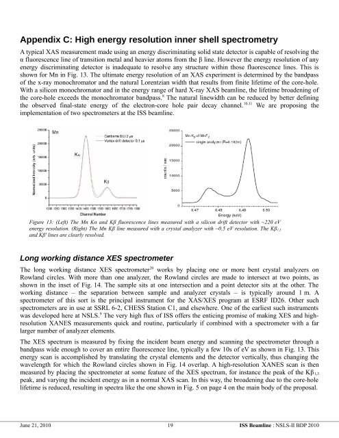
A typical XAS measurement made using an energy discriminating solid state detector is capable of resolving the<br />
α fluorescence line of transition metal and heavier atoms from the β line. However the energy resolution of any<br />
energy discriminating detector is inadequate to resolve any structure within those fluorescence lines. This is<br />
shown for Mn in Fig. 13. The ultimate energy resolution of an XAS experiment is determined by the bandpass<br />
of the x-ray monochromator and the natural Lorentzian width that results from finite lifetime of the core-hole.<br />
With a silicon monochromator and in the energy range of hard X-ray XAS beamline, the lifetime broadening of<br />
the core-hole exceeds the monochromator bandpass. 8 The natural linewidth can be reduced by better defining<br />
the observed final-state energy of the electron-core hole pair decay channel. 10,11 We are proposing the<br />
implementation of two spectrometers at the <strong>ISS</strong> beamline.<br />
Figure 13: (Left) The Mn Kα and Kβ fluorescence lines measured with a silicon drift detector with ~220 eV<br />
energy resolution. (Right) The Mn Kβ line measured with a crystal analyzer with ~0.5 eV resolution. The Kβ1,3<br />
and Kβ' lines are clearly resolved.<br />
Long working distance XES spectrometer<br />
The long working distance XES spectrometer 20 works by placing one or more bent crystal analyzers on<br />
Rowland circles. With more than one analyzer, the Rowland circles are made to intersect at two points, as<br />
shown in the inset of Fig. 14. The sample sits at one intersection and a point detector sits at the other. The<br />
working distance – the separation between sample and analyzer crystals – is typically around 1 m. A<br />
spectrometer of this sort is the principal instrument for the XAS/XES program at ESRF ID26. Other such<br />
spectrometers are in use at SSRL 6-2, CHESS Station C1, and elsewhere. One of the earliest such instruments<br />
was developed here at NSLS. 9 The very high flux of <strong>ISS</strong> offers the enticing promise of making XES and highresolution<br />
XANES measurements quick and routine, particularly if combined with a spectrometer with a far<br />
larger number of analyzer elements.<br />
The XES spectrum is measured by fixing the incident beam energy and scanning the spectrometer through a<br />
bandpass wide enough to cover an entire fluorescence line, typically a few 10s of eV as shown in Fig. 13. This<br />
energy scan is accomplished by translating the crystal elements and the detector vertically, thus changing the<br />
wavelength for which the Rowland circles shown in Fig. 14 overlap. A high-resolution XANES scan is then<br />
measured by placing the spectrometer at some feature of the XES spectrum, for instance the peak of the Kβ 1,3<br />
peak, and varying the incident energy as in a normal XAS scan. In this way, the broadening due to the core-hole<br />
lifetime is reduced, resulting in spectra like the one shown in Fig. 5 on page 4 on the main body of the proposal.<br />
June 21, 2010 19 <strong>ISS</strong> Beamline : NSLS-II BDP 2010


