

Inner Shell Spectroscopy (ISS) - Brookhaven National Laboratory
Inner Shell Spectroscopy (ISS) - Brookhaven National Laboratory
Inner Shell Spectroscopy (ISS) - Brookhaven National Laboratory

You also want an ePaper? Increase the reach of your titles
YUMPU automatically turns print PDFs into web optimized ePapers that Google loves.
Scientific justification for the <strong>ISS</strong> beamline<br />
The unprecedented brightness and flux of NSLS-II will enable measurements with the high spatial,<br />
energy, and time resolution necessary to fully characterize … complex systems. Advanced<br />
capabilities will include … application of new experimental techniques, such as high-resolution xray<br />
emission spectroscopy and x-ray Raman scattering, to provide new spectroscopic information;<br />
and the use of combinatorial methods for large scale screening of novel materials.<br />
NSLS-II CD-0 document<br />
Absorption of an X-ray by an atom is one of the fundamental interactions of light with matter and the<br />
measurement of absorption is one of the core competencies of any synchrotron. X-ray absorption spectroscopy<br />
(XAS) has a long history at NSLS – many of the first beamlines on the NSLS X-ray ring were for XAS and the<br />
XAS community has remained large and productive for the entire 25 year history of NSLS. In that time, the use<br />
of XAS has become commonplace in a very wide variety of academic and industrial disciplines ranging from<br />
the life and environmental sciences to materials physics and chemistry, engineering materials, geophysics, and<br />
more. The XAS community at NSLS, however, operates within certain constraints.<br />
The dilution of an absorber, the speed at which an XAS spectrum may be collected, and the effective use of high<br />
energy resolution spectrometers are the boundaries within which the NSLS XAS community currently operates.<br />
Each of these boundaries can be pushed back in significant ways by a high-flux source. This proposal is for a<br />
wiggler-based beamline dedicated to XAS and other inner shell spectroscopies. The exceptional flux provided<br />
by a wiggler enables measurement of absorber concentrations at environmentally or technologically relevant<br />
levels impractical to measure at dipole beamlines. Although a companion proposal (the TRS beamline) focuses<br />
on sub-second, time-resolved XAS, this beamline allows collection of high-quality XAS spectra in well under a<br />
minute and is an important part of the strategy for mitigating sample damage under the elevated flux. Finally,<br />
the high flux from the wiggler offers the use of point-to-point focusing and wavelength dispersive<br />
spectrometers, enabling collection of high-resolution XANES spectra, X-ray emission (XES) spectra, and the<br />
measurement of low-energy absorption edges via X-ray energy loss spectroscopy (XELS), all of which are<br />
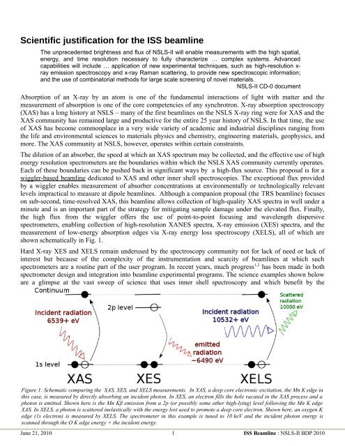
shown schematically in Fig. 1.<br />
Hard X-ray XES and XELS remain underused by the spectroscopy community not for lack of need or lack of<br />
interest but because of the complexity of the instrumentation and scarcity of beamlines at which such<br />
spectrometers are a routine part of the user program. In recent years, much progress 1,2 has been made in both<br />
spectrometer design and integration into beamline experimental programs. The science examples shown below<br />
are a glimpse at the vast sweep of science that uses inner shell spectroscopy and which benefit by the<br />
Figure 1: Schematic comparing the XAS, XES, and XELS measurements. In XAS, a deep core electronic excitation, the Mn K edge in<br />
this case, is measured by directly absorbing an incident photon. In XES, an electron fills the hole vacated in the XAS process and a<br />
photon is emitted. Shown here is the Mn Kβ emission from a 2p (or possibly some other high-lying) level following the Mn K edge<br />
XAS. In XELS, a photon is scattered inelastically with the energy lost used to promote a deep core electron. Shown here, an oxygen K<br />
edge (1s electron) is measured by XELS. The spectrometer in this example is tuned to 10 keV and the incident photon energy is<br />
scanned through the O K edge energy + the incident energy.<br />
June 21, 2010 1 <strong>ISS</strong> Beamline : NSLS-II BDP 2010


