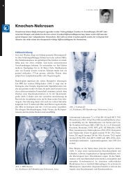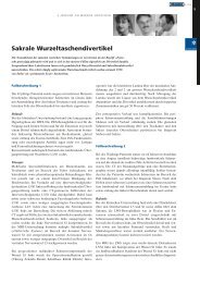
Das UniversitätsSpital Zürich - Rheuma Schweiz
Das UniversitätsSpital Zürich - Rheuma Schweiz
Das UniversitätsSpital Zürich - Rheuma Schweiz

You also want an ePaper? Increase the reach of your titles
YUMPU automatically turns print PDFs into web optimized ePapers that Google loves.
14<br />
expressed in both vimentin and CD68 positive cells. Western blot analysis confirmed<br />
that CXCR7 is more abundant in RA samples than in OA (ratio<br />
CXCR7/·-tubulin: 0.81± 0.57 SEM vs 0.34± 0.17 SEM, n=4 each). After stimulation<br />
of cultured RASF (n=4) with Il-1�, TNF-� and TGF-�, there were no significant<br />
changes in the expression of CXCR7 mRNA and protein in all the<br />
measured timepoints.<br />
Conclusions: The discovery of the alternative receptor CXCR7, opened a new<br />
field in the study of CXCL12 pathways. This is the first time that the chemokine<br />
receptor CXCR7 is described in synovial tissue and fibroblasts of RA patients.<br />
Based on this data, we suggest that the upregulation of CXCR7 in RA is not<br />
driven by cytokines like IL-1�, TNF-� and TGF-�, but possibly by alternative<br />
mechanisms like epigenetic modulations.<br />
09<br />
LONG DISTANCE MIGRATION OF RASF: ROLE OF EXTRACELLULAR<br />
MATRIX<br />
Stephanie Lefèvre 1 , Anette Knedla 1 , Christoph Tennie 1 , Ingo H. Tarner 1 , Henning<br />
Stürz 2 , Jürgen Steinmeyer 3 , Steffen Gay 4 , Ulf Müller-Ladner 1 , Elena Neumann 1 .<br />
1 Internal Med and <strong>Rheuma</strong>tology, Justus-Liebig-University of Gießen, Bad<br />
Nauheim, Germany; 2 Dept Orthopedics and Orthopedic Surgery, Justus-Liebig-<br />
University of Gießen, Gießen, Germany; 3 Dept Orthopedics and Exp Orthopedics,<br />
Justus-Liebig-University of Gießen, Gießen, Germany; 4 Ctr Exp <strong>Rheuma</strong>tology,<br />
USZ, <strong>Zürich</strong>, Switzerland<br />
Background: Key players in rheumatoid arthritis (RA) pathophysiology are<br />
activated synovial fibroblasts (SF) which actively attach to, invade into and<br />
degrade cartilage which can be simulated in the SCID mouse model of RA. In<br />
preliminary experiments, we could show that RASF are able to migrate from a<br />
primary implantation site to a distant one, mainly via the blood stream. Therefore,<br />
the route of migration and the role of the extracellular matrix (ECM) were<br />
further analyzed in this study.<br />
Methods: Healthy human cartilage was implanted into SCID mice together<br />
with RASF. At the contralateral flank, cartilage without cells was implanted. In<br />
addition, RASF were injected intravenously (iv), subcutaneously (sc) or<br />
intraperitoneally (ip) 14 days after cartilage implantation. To evaluate the role<br />
of the ECM towards the migratory behavior of RASF, complete RA synovium,<br />
bovine cartilage or necrotic cartilage, respectively, and contralaterally normal<br />
human cartilage were implanted. After 60 days, implants, organs, blood, murine<br />
ear cartilage and joints were removed. To detect human cells, species-specific<br />
immunohisto- and -cytochemistry were performed.<br />
Results: RASF were not only able to invade and degrade cartilage which was<br />
inserted simultaneously with RASF, they also migrated to and invaded into the<br />
contralateral cartilage (Scores: inv 2.3±0.8 and 1.9±0.9, deg 1.8±0.8 and 1.6±0.6).<br />
Injection of cells led to strong destruction of the implanted cartilage, particularly<br />
after sc and iv application. Interestingly, implantation of complete synovial<br />
tissue, containing ECM and other cell types, led to migration of RASF to the<br />
contralateral cartilage in 5/11 animals (inv 2.0±1.0, deg 2.3±0.4). Single RASF<br />
were able to invade bovine and human necrotic cartilage and they could also be<br />
found in the murine ear and joint, but no invasion and degradation could be<br />
detected at day 60 after RASF application. Conclusions: RASF have the ability<br />
to migrate from a primary implant of cartilage or RA synovium to the contralateral<br />
implantation site via the blood stream and to mediate cartilage degradation.<br />
The cells are also able to cross the peritoneum after ip injection. Cartilage<br />
degradation appears to be independent of chondrocyte viability and of<br />
species background since RASF invade necrotic human as well as bovine cartilage.Therefore,<br />
the exposure to cartilage matrix, e. g. after microinjuries, may be<br />
sufficient for RASF to attach to the cartilage, to initiate destruction and spread<br />
RA from one joint to another.<br />
10<br />
FIBRIN TRIGGERS AN INNATE IMMUNE RESPONSE IN RHEUMA-<br />
TOID ARTHRITIS SYNOVIAL FIBROBLASTS ACTING AS AN ENDOGE-<br />
NOUS LIGAND OF TOLL LIKE RECEPTOR<br />
Olga Sanchez-Pernaute 1 , Fabia Brentano1, Caroline Ospelt 1 , Christoph Kolling 2 ,<br />
Beat A. Michel 1 , Renate E. Gay 1 , Gabriel Herrero-Beaumont 3 , Steffen Gay 1 ,<br />
Michel Neidhart 1 .<br />
1 Center of Experimental <strong>Rheuma</strong>tology, University Hospital and Zurich Center<br />
for Integrative Human Physiology (ZIHP), Zurich, Switzerland;<br />
2 Orthopaedic Surgery, Schulthess Klinic, Zurich, Switzerland; 3 <strong>Rheuma</strong>tology<br />
Section, Fundacion Jimenez Diaz, Madrid, Spain<br />
Purpose: The deposition of fibrin inside joints is followed by its citrullination<br />
and has been associated with the perpetuation of rheumatoid arthritis (RA)<br />
through the development of a specific immune response. In macrophages, fibrin(ogen)<br />
has been shown to be a ligand of Toll-like receptor 4 (TLR4). Since<br />
RA synovial fibroblasts (RASF) express functional TLR4, we investigated the<br />
role of fibrin in the activation of RASF as well as the effect of citrullination on<br />
the cellular response.<br />
Methods: Isolated RASF obtained from surgical explants were used for the<br />
experiments. Fibrin was polymerized in situ by the addition of 0.7 U/ml thrombin<br />
to 0.8 mg/ml fibrinogen. Citrullination was performed by adding 2.84 U/ml<br />
peptidylarginine deiminase 2 to the components of the clot. Differential gene<br />
expression was analysed using cDNA arrays (Affymetrix GeneChip ® ) and<br />
mRNA isolated from RASF, alone or with fibrin, native or citrullinated, after<br />
an incubation period of 18h. The transcripts induced by native or citrullinated<br />
fibrin were compared with the untreated RASF, establishing a bidirectional<br />
2-fold filter and merging results from two experiments. Quantitative real time<br />
PCR using SYBR green was employed to confirm the up-regulation of selected<br />
genes by native or citrullinated fibrin in additional cultures of RASF.<br />
Results: The cDNA arrays showed that the exposure of RASF to fibrin resulted<br />
in the up-regulation of genes participating in the innate response and relevant<br />
to the pathogenesis of RA, including the pro-inflammatory cytokine IL-6,<br />
the chemokines IL-8, CXCL1 and CCL2, the TNF· induced peptide 3 and the<br />
adhesion molecule ICAM-1. These inductions were observed with both native<br />
and citrullinated fibrin. Quantitative real time PCR confirmed the up-regulation,<br />
after incubation with fibrin, of IL-8 (959 ± 418 fold), CXCL1 (98 ± 37<br />
fold), IL-6 (22 ± 11 fold), COX-2 (21 ± 6 fold) and CCL2 (14 ± 3 fold) (n = 3<br />
RASF, mean ± SEM). Interestingly, compared to native fibrin, citrullinated fibrin<br />
revealed an even stronger effect on the induction of IL-8.The up-regulation<br />
of COX-2, CXCL1 and IL-8 induced by fibrin was suppressed by 70% after 2h<br />
preincubation of RASF with TLR4 blocking monoclonal antibodies (HTA125,<br />
10 mg/ml, Abcam).<br />
Conclusion: Fibrin and in particular its citrullinated form can trigger an innate<br />
immune response in RASF through a TLR4 dependent pathway. Based on<br />
these data, it can be concluded that fibrin contributes significantly to sustain<br />
the inflammatory process in RA.<br />
11<br />
DIFFERENTIAL INDUCTION OF IL-23 SUBUNITS BY TLR LIGANDS<br />
IN RHEUMATOID ARTHRITIS SYNOVIAL FIBROBLASTS AND<br />
MONOCYTES<br />
Fabia Brentano 1 , Caroline Ospelt 1 , Joanna Stanczyk 1 , Renate E. Gay 1 , Christoph<br />
Kolling 2 , Steffen Gay 1 , Diego Kyburz 1 .<br />
1 Center of Experimental <strong>Rheuma</strong>tology and Zurich Center of Integrative<br />
Human Physiology (ZIHP), University Hospital of Zurich, Switzerland;<br />
2 Schulthess Clinic, Zurich, Switzerland<br />
Purpose: IL-23 was found to have key roles in autoimmune diseases. We analyzed<br />
the expression of IL-23 in rheumatoid arthritis (RA) and osteoarthritis<br />
(OA) synovial tissues. Furthermore we studied the induction of both subunits<br />
of IL-23, p19 and p40, in cultured RA synovial fibroblasts (RASF) and monocytes<br />
after the activation of TLR2, 3 and 4.<br />
Methods: Expression of p19 and p40 was analyzed in synovial tissues by in situ<br />
hybridization (n=3) and immunohistochemistry (n=6). RASF and blood monocytes<br />
from healthy donors were stimulated with bacterial lipoprotein (bLP),<br />
poly(I-C) (PIC) and lipopolysaccharide (LPS) for 24h (n=4). The expression of<br />
p19 and p40 mRNA was analyzed by RT-PCR as well as by Real-time PCR. To<br />
detect IL-23 in supernatants of RASF and monocytes a bioassay was performed:<br />
MACS sorted CD8+ T-cells were preactivated with anti-CD3 and anti-<br />
CD28 antibodies and stimulated with supernatants of TLR-ligand stimulated<br />
RASF and monocytes or rhIL-23 as a positive control. Subsequently IL-23<br />
dependent IL-17 production was determined by ELISA.<br />
Results: p19 mRNA and protein were abundantly expressed in RA synovial<br />
tissues. Specifically p19 was expressed in the sublining and lining layer as well<br />
as at sites of invasion. Double staining with cell type specific markers revealed<br />
that p19 positive cells expressed the fibroblast marker vimentin or the<br />
macrophage marker CD68. In addition we found that not all p19 positive cells<br />
were double positive for p40. In OA synovial tissues only low levels of p19 were<br />
expressed.<br />
By in vitro stimulation of RASF with the TLR2 ligand bLP, the TLR3 ligand<br />
PIC and the TLR4 ligand LPS we found p19 mRNA to be induced most<br />
markedly by PIC (PIC: 24.4±4; bLP: 9.3±3.5; LPS: 5.8±2.8 fold upregulation relative<br />
to control). However, in RASF the expression of the p40 subunit could<br />
not be induced by any of the TLR ligands. In monocytes both subunits for IL