Cloning Protocols and Applications Guide-A4 format - Promega
Cloning Protocols and Applications Guide-A4 format - Promega
Cloning Protocols and Applications Guide-A4 format - Promega

You also want an ePaper? Increase the reach of your titles
YUMPU automatically turns print PDFs into web optimized ePapers that Google loves.
||||||||||||| 13<strong>Cloning</strong><br />
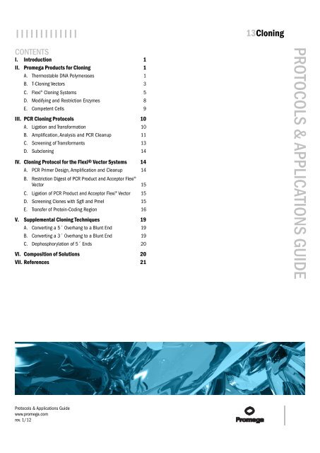
CONTENTS<br />
I. Introduction 1<br />
II. <strong>Promega</strong> Products for <strong>Cloning</strong> 1<br />
A. Thermostable DNA Polymerases 1<br />
B. T-<strong>Cloning</strong> Vectors 3<br />
C. Flexi® <strong>Cloning</strong> Systems 5<br />
D. Modifying <strong>and</strong> Restriction Enzymes 8<br />
E. Competent Cells 9<br />
III. PCR <strong>Cloning</strong> <strong>Protocols</strong> 10<br />
A. Ligation <strong>and</strong> Trans<strong>format</strong>ion 10<br />
B. Amplification, Analysis <strong>and</strong> PCR Cleanup 11<br />
C. Screening of Transformants 13<br />
D. Subcloning 14<br />
IV. <strong>Cloning</strong> Protocol for the Flexi® Vector Systems 14<br />
A. PCR Primer Design, Amplification <strong>and</strong> Cleanup 14<br />
B. Restriction Digest of PCR Product <strong>and</strong> Acceptor Flexi®<br />
Vector 15<br />
C. Ligation of PCR Product <strong>and</strong> Acceptor Flexi® Vector 15<br />
D. Screening Clones with SgfI <strong>and</strong> PmeI 15<br />
E. Transfer of Protein-Coding Region 16<br />
V. Supplemental <strong>Cloning</strong> Techniques 19<br />
A. Converting a 5´ Overhang to a Blunt End 19<br />
B. Converting a 3´ Overhang to a Blunt End 19<br />
C. Dephosphorylation of 5´ Ends 20<br />
VI. Composition of Solutions 20<br />
VII. References 21<br />
<strong>Protocols</strong> & <strong>Applications</strong> <strong>Guide</strong><br />
www.promega.com<br />
rev. 1/12<br />
PROTOCOLS & APPLICATIONS GUIDE
||||||||||||| 13<strong>Cloning</strong><br />
I. Introduction<br />
The cloning of genes, gene fragments <strong>and</strong> other DNA<br />
sequences is a fundamental part of molecular biology. To<br />
study the function of a particular DNA sequence, you must<br />
be able to manipulate that sequence. There are two main<br />
ways to achieve this: the polymerase chain reaction (PCR)<br />
<strong>and</strong> the more traditional use of restriction enzymes <strong>and</strong><br />
modifying enzymes to “cut <strong>and</strong> paste” the desired DNA<br />
fragments into cloning vectors, which can then be replicated<br />
using live cells, most commonly E. coli. The use of PCR has<br />
an advantage in that it gives you the option to re-amplify<br />
the target DNA each time your DNA supplies dwindle<br />
without ligation into a vector or trans<strong>format</strong>ion into E. coli.<br />
Alternatively, PCR products can be ligated into a suitable<br />
vector, which can then be transformed into <strong>and</strong> replicated<br />
by E. coli. This chapter covers the basics of cloning using<br />
PCR <strong>and</strong> restriction enzymes, including DNA cleanup prior<br />
to ligation, ligation, trans<strong>format</strong>ion <strong>and</strong> screening to<br />
identify recombinant clones.<br />
The PCR process is a useful tool to quickly <strong>and</strong> easily<br />
amplify the desired sequences. With the successful<br />
sequencing of whole <strong>and</strong> partial genomes of organisms<br />
across all biological kingdoms, DNA cloning by PCR is an<br />
easily attainable option. Public DNA databases allow<br />
researchers to design primers to amplify their DNA<br />
fragment of interest directly from the genomic DNA of the<br />
desired organism. With the simple addition of a reverse<br />
transcription step prior to PCR, RNA sequences can be<br />
converted to cDNA, which can then be cloned into a<br />
suitable vector. For additional in<strong>format</strong>ion about<br />
amplification of DNA <strong>and</strong> RNA sequences using PCR, see<br />
the PCR Amplification chapter of the <strong>Protocols</strong> <strong>and</strong><br />
<strong>Applications</strong> <strong>Guide</strong>.<br />
PCR products generated using a nonproofreading DNA<br />
polymerase such as Taq DNA polymerase, which lacks<br />
3´→5´ exonuclease activity, have a single templateindependent<br />
nucleotide at the 3´ end of each DNA str<strong>and</strong><br />
(Clark, 1988; Newton <strong>and</strong> Graham, 1994). This<br />
single-nucleotide overhang, which is most commonly an<br />
A residue, allows hybridization with <strong>and</strong> cloning into T<br />
vectors, which have a complementary 3´ single T overhang.<br />
PCR products generated using a proofreading DNA<br />
polymerase, such as Pfu DNA polymerase, have blunt ends<br />
<strong>and</strong> must be cloned into a blunt-ended vector or need a<br />
single 3´A overhang added to ligate into a T vector (Knoche<br />
<strong>and</strong> Kephart, 1999).<br />
If PCR amplification of the desired DNA fragment is not<br />
possible or desirable, restriction enzyme digestion of the<br />
target DNA can be employed. The desired fragment may<br />
need to be separated from other DNA fragments in the<br />
reaction, so the size of the desired DNA fragment should<br />
be known. Once isolated, the fragment is cloned into a<br />
vector with compatible ends. If the vector ends are capable<br />
of religating (e.g., the vector has blunt ends or is cut with<br />
a single restriction enzyme), the vector is often treated with<br />
alkaline phosphatase to discourage recircularization <strong>and</strong><br />
maximize ligation between the insert <strong>and</strong> vector.<br />
<strong>Protocols</strong> & <strong>Applications</strong> <strong>Guide</strong><br />
www.promega.com<br />
rev. 1/12<br />
Following trans<strong>format</strong>ion into E. coli, the resulting bacterial<br />
colonies are screened by PCR for the correct recombinant<br />
vector using primers to amplify the insert. Alternatively,<br />
the recombinant vector can be identified by performing a<br />
restriction enzyme digestion to determine the presence of<br />
the correct insert. Screening is often simplified by using<br />
vectors that contain an antibiotic-resistance gene, so cells<br />
containing the vector will survive on medium<br />
supplemented with the appropriate antibiotic. Screening<br />
can be further simplified by choosing a vector <strong>and</strong> E. coli<br />
strain that are compatible with blue/white screening, which<br />
takes advantage of intracistronic α-complementation to<br />
regenerate β-galactosidase activity. Many E. coli strains<br />
used for cloning <strong>and</strong> propagation of plasmids contain a<br />
chromosomal deletion of the lac operon but carry an F´<br />
episome that provides the remaining coding sequence of<br />
the lacZ gene. The functional lacZ gene product,<br />
β-galactosidase, is produced when the lacZ coding<br />
in<strong>format</strong>ion missing on the F´ episome is provided by the<br />
plasmid. This activity is detected by plating bacteria<br />
transformed by plasmids on plates containing isopropyl<br />
β-D-thiogalactopyranoside (IPTG; an inducer of the lac<br />
promoter) <strong>and</strong> 5-bromo-4-chloro-3-indolyl-β-D-galactoside<br />
(X-Gal; a dye that produces a blue color when hydrolyzed<br />
by β-galactosidase). When the reading frame of the<br />
α peptide is disrupted by insertion of a foreign DNA<br />
fragment or deletion of vector sequences,<br />
α-complementation does not occur, <strong>and</strong> the bacterial<br />
colonies remain white or occasionally light blue.<br />
II. <strong>Promega</strong> Products for <strong>Cloning</strong><br />
A. Thermostable DNA Polymerases<br />
The use of amplification enzymes is the first step in cloning<br />
by PCR. Most people use PCR for cloning, taking advantage<br />
of the single nucleotide A overhang left after amplification<br />
with a nonproofreading DNA polymerase to ligate the<br />
amplimer to a vector containing T overhangs. However,<br />
products will be blunt-ended if the DNA polymerase has<br />
3′→5′ exonuclease activity, also known as proofreading<br />
activity. Alternatively, PCR primers can add sequences for<br />
restriction enzyme sites, <strong>and</strong> the resulting products can be<br />
digested <strong>and</strong> ligated into a vector with compatible ends.<br />
<strong>Promega</strong> provides several thermostable DNA polymerases.<br />
These include the GoTaq® Amplification Family, Tfl DNA<br />
Polymerase <strong>and</strong> proofreading polymerases. A detailed list<br />
of the various enzymes for use in PCR can be found in the<br />
Protocol <strong>and</strong> <strong>Applications</strong> <strong>Guide</strong> chapter on PCR<br />
Amplification, in the section "Thermostable DNA<br />
Polymerases". The GoTaq® Amplification Family of<br />
products is highlighted in the following section.<br />
GoTaq® Amplification Family<br />
GoTaq® DNA Polymerase is available in various<br />
formulations to suit your needs: the st<strong>and</strong>ard GoTaq® DNA<br />
Polymerase, which is supplied with a 1X reaction buffer<br />
that contains 1.5mM MgCl2; GoTaq® Flexi DNA<br />
Polymerase, which allows a range of MgCl2 to be used for<br />
PROTOCOLS & APPLICATIONS GUIDE 13-1
||||||||||||| 13<strong>Cloning</strong><br />
PCR; GoTaq® Green Master Mix, which is a premixed,<br />
ready-to-use solution containing GoTaq® DNA Polymerase,<br />
dNTPs, MgCl2 <strong>and</strong> reaction buffer at optimal concentrations<br />
for efficient amplification of DNA templates by PCR;<br />
GoTaq® Hot Start Polymerase, which includes a proprietary<br />
antibody that blocks polymerase activity until the initial<br />
denaturation step; <strong>and</strong> GoTaq® Long PCR Master Mix,<br />
which offers efficient amplification of long templates (e.g.,<br />
human genomic DNA up to 30kb). All GoTaq® PCR Core<br />
Systems offer complete solutions with polymerase <strong>and</strong><br />
nucleotides; the GoTaq® PCR Core System II also includes<br />
a positive control. GoTaq® products contain Taq DNA<br />
polymerase in a proprietary formulation that offers<br />
enhanced amplification over conventional Taq DNA<br />
polymerase. Each member of the GoTaq® family has a<br />
reaction buffer that contains two dyes (a blue dye <strong>and</strong> a<br />
yellow dye) that separate during electrophoresis to show<br />
migration progress as well as a compound that increases<br />
sample density. Samples can be loaded directly onto gels<br />
without the need to add a separate loading dye. If the dyes<br />
interfere with your downstream applications, GoTaq® DNA<br />
Polymerases are supplied with a 5X Colorless Reaction<br />
Buffer. Alternatively, the PCR Master Mix offers a<br />
ready-to-use formulation without any dyes. Reaction<br />
products generated with these systems contain A overhangs<br />
<strong>and</strong> are ready for T-vector cloning. Alternatively, the PCR<br />
product can be digested directly with a restriction enzyme<br />
that is active in the PCR buffer <strong>and</strong> cloned into st<strong>and</strong>ard<br />
cloning vectors (see Technical Manual #TM367 for a<br />
protocol).<br />
Additional Resources for GoTaq® DNA Polymerase<br />
<strong>Promega</strong> Publications<br />
Introducing GoTaq® DNA Polymerase: Improved<br />
amplification with a choice of buffers<br />
Citations<br />
Ning, B. et al. (2011) 5-Aza-2'-deoxycytidine activates iron<br />
uptake <strong>and</strong> heme biosynthesis by increasing c-myc nuclear<br />
localization <strong>and</strong> binding to the e-boxes of transferrin<br />
receptor 1 (TfR1) <strong>and</strong> ferrochelatase (Fech) genes. J. Biol.<br />
Chem. 286, 37196–206.<br />
The authors performed real-time PCR using SYBR® Green<br />
<strong>and</strong> 1.25 units of GoTaq® DNA Polymerase to amplify 20ng<br />
of cDNA generated from total RNA extracted from murine<br />
erythroid leukemia (MEL) cells <strong>and</strong> mouse erythroid<br />
burst-forming units (BFU-Es) in a total reaction volume of<br />
25µl.<br />
PubMed Number: 21903580<br />
Vucurovic, K. et al. (2010) Serotonin 3A receptor subtype<br />
as an early <strong>and</strong> protracted marker of cortical interneuron<br />
subpopulations. Cereb. Cortex 20, 2333–47.<br />
After reverse transcription, PCR was performed to<br />
simultaneously detect mRNAs encoding two isoforms of<br />
glutamic acid decarboxylase, three calcium-binding<br />
proteins, three neuropeptides, two transcription factors<br />
<strong>Protocols</strong> & <strong>Applications</strong> <strong>Guide</strong><br />
www.promega.com<br />
rev. 1/12<br />
<strong>and</strong> reelin, a protein thought to be involved in neuronal<br />
migration <strong>and</strong> morphology. Two rounds of PCR using<br />
nested primers were required to detect these mRNAs. PCRs<br />
were performed using GoTaq® DNA Polymerase.<br />
Amplified products were visualized by agarose gel<br />
electrophoresis, using the 100bp DNA Ladder as a size<br />
st<strong>and</strong>ard.<br />
PubMed Number: 20083553<br />
Additional Resources for GoTaq® Flexi DNA Polymerase<br />
<strong>Promega</strong> Publications<br />
GoTaq® Flexi DNA Polymerase: Robust performance with<br />
magnesium optimization<br />
Citations<br />
Westphal, A. et al. (2011) General suppression of Escherichia<br />
coli O157:H7 in s<strong>and</strong>-based dairy livestock bedding. Appl.<br />
Environ. Microbiol. 77, 2113–21.<br />
DNA was extracted from bedding material <strong>and</strong> the 16S<br />
rRNA genes amplified in a 25µl reaction using 1.5 units<br />
GoTaq® Flexi DNA Polymerase with 1.8mM MgCl2. The<br />
PCR products then were cloned into the pGEM®-T Easy<br />
Vector.<br />
PubMed Number: 21257815<br />
Additional Resources for GoTaq® Green Master Mix<br />
<strong>Promega</strong> Publications<br />
Activity of <strong>Promega</strong> restriction enzymes in GoTaq® Green<br />
Master Mix <strong>and</strong> PCR Master Mix<br />
Analyses of gene disruption by whole-cell PCR using the<br />
GoTaq® Green Master Mix<br />
Recombinant clone screening using the GoTaq® Hot Start<br />
Green Master Mix<br />
Citations<br />
Crawford, M.A. et al. (2011) Identification of the bacterial<br />
protein FtsX as a unique target of chemokine-mediated<br />
antimicrobial activity against Bacillus anthracis. Proc. Natl.<br />
Acad. Sci. USA. 108, 17159–64.<br />
To identify transposon insertion sites, bacterial genomic<br />
DNA was isolated, digested <strong>and</strong> ligated with a partially<br />
double-str<strong>and</strong>ed Y-linker. An initial 20µl amplification for<br />
20 cycles using GoTaq® Green Master Mix enriched ssDNA<br />
fragments. A second PCR amplified dsDNA, adding more<br />
GoTaq® Green Master Mix for a final reaction volume of<br />
100µl with 25 cycles. The amplimers were analyzed by<br />
sequencing.<br />
PubMed Number: 21949405<br />
PROTOCOLS & APPLICATIONS GUIDE 13-2
||||||||||||| 13<strong>Cloning</strong><br />
Additional Resources for PCR Master Mix<br />
<strong>Promega</strong> Publications<br />
Activity of <strong>Promega</strong> restriction enzymes in GoTaq® Green<br />
Master Mix <strong>and</strong> PCR Master Mix<br />
Performance advantages designed into <strong>Promega</strong>'s PCR<br />
Master Mix<br />
Additional Resources for GoTaq® Core PCR Systems<br />
Technical Bulletins <strong>and</strong> Manuals<br />
TB254 GoTaq® PCR Core Systems Technical Bulletin<br />
Citations<br />
Fuehrer, H.P. et al. (2011) Novel nested direct PCR<br />
technique for malaria diagnosis using filter paper samples.<br />
J. Clin. Microbiol. 49, 1628–30.<br />
The authors developed a direct-amplification, nested PCR<br />
protocol to amplify Plasmodium DNA from S&S 903 filter<br />
paper punches containing whole blood. The GoTaq® PCR<br />
Core System amplified 5µl of template (extracted from<br />
paper punches <strong>and</strong> whole blood in parallel) in the second<br />
nested reaction using 2mM MgCl2 <strong>and</strong> 1 unit of GoTaq®<br />
DNA polymerase in a total reaction volume of 50µl.<br />
PubMed Number: 21270224<br />
Additional Resources for GoTaq® Hot Start Polymerase<br />
Technical Bulletins <strong>and</strong> Manuals<br />
9PIM500 GoTaq® Hot Start Polymerase Product<br />
In<strong>format</strong>ion<br />
Citations<br />
Li, Z. et al. (2011) The barley amo1 locus is tightly linked to<br />
the starch synthase IIIa gene <strong>and</strong> negatively regulates<br />
expression of granule-bound starch synthetic genes. J. Exp.<br />
Bot. 62, 5217–31.<br />
To examine the mutations in class II <strong>and</strong> class III starch<br />
synthases (ssIIa <strong>and</strong> ssIIIa, respectively), genomic DNA<br />
from young barley leaves was extracted <strong>and</strong> 50ng amplified<br />
in a 20µl reaction that included 1.5U of GoTaq® Hot Start<br />
Polymerase, 1.5mM MgCl2 <strong>and</strong> the additives DMSO <strong>and</strong><br />
betaine. After 35 cycles, the PCR products were digested<br />
overnight using EcoRI <strong>and</strong> separated on 2% agarose gels.<br />
PubMed Number: 21813797<br />
Additional Resources for GoTaq® Long PCR Master Mix<br />
Technical Bulletins <strong>and</strong> Manuals<br />
TM359 GoTaq® Long PCR Master Mix Technical<br />
Manual<br />
<strong>Promega</strong> Publications<br />
GoTaq® Long PCR Master Mix for reliable amplification<br />
of long PCR targets<br />
<strong>Protocols</strong> & <strong>Applications</strong> <strong>Guide</strong><br />
www.promega.com<br />
rev. 1/12<br />
B. T-<strong>Cloning</strong> Vectors<br />
T vectors are a specific type of cloning vector that get their<br />
name from the T overhangs added to a linearized plasmid.<br />
These vectors take advantage of the A overhangs on PCR<br />
products after amplification with Taq DNA polymerase by<br />
providing compatible ends for ligation (Mezei <strong>and</strong> Storts,<br />
1994; Robles <strong>and</strong> Doers, 1994). There are three different<br />
T-cloning vectors from <strong>Promega</strong>: Two are basic cloning<br />
vectors, <strong>and</strong> the third is a mammalian expression vector.<br />
pGEM®-T <strong>and</strong> pGEM®-T Easy Vector Systems<br />
The pGEM®-T (Cat.# A3600, A3610) <strong>and</strong> pGEM®-T Easy<br />
Vector Systems (Cat.# A1360, A1380) are convenient<br />
systems for cloning PCR products. The vectors are prepared<br />
by cutting with a restriction endonuclease to leave a blunt<br />
end <strong>and</strong> adding a 3´terminal thymidine to both ends<br />
(Figures 13.1 <strong>and</strong> 13.2). These single 3´ T overhangs at the<br />
insertion site greatly improve ligation efficiency of a PCR<br />
product into the plasmid by preventing recircularization<br />
of the vector <strong>and</strong> providing a compatible overhang for PCR<br />
products with 5′ A overhangs.<br />
Figure 13.1. pGEM®-T Vector circle map.<br />
Figure 13.2. pGEM®-T Easy Vector circle map.<br />
The high-copy-number pGEM®-T <strong>and</strong> pGEM®-T Easy<br />
Vectors contain T7 <strong>and</strong> SP6 RNA polymerase promoters<br />
flanking a multiple cloning region within the coding region<br />
for the α-peptide of β-galactosidase. Insertional inactivation<br />
of the α-peptide allows recombinant clones to be directly<br />
identified by color screening on indicator plates containing<br />
PROTOCOLS & APPLICATIONS GUIDE 13-3
||||||||||||| 13<strong>Cloning</strong><br />
X-Gal (Cat.# V3941) <strong>and</strong> IPTG (Cat.# V3955). Both the<br />
pGEM®-T <strong>and</strong> pGEM®-T Easy Vectors contain numerous<br />
restriction sites within the multiple cloning region. The<br />
pGEM®-T Easy Vector multiple cloning region is flanked<br />
by recognition sites for the restriction enzymes EcoRI, BstZI<br />
<strong>and</strong> NotI, thus providing three single-enzyme digestions<br />
for release of the insert. The pGEM®-T Vector cloning<br />
region is flanked by recognition sites for the enzyme BstZI.<br />
Alternatively, a double digestion may be used to release<br />
the insert from either vector.<br />
The pGEM®-T <strong>and</strong> pGEM®-T Easy Vectors also contain the<br />
origin of replication of filamentous phage f1 for the<br />
preparation of single-str<strong>and</strong>ed DNA (ssDNA). Both<br />
pGEM®-T vector systems include a 2X Rapid Ligation<br />
Buffer for ligation of PCR products, which requires only a<br />
1-hour incubation at room temperature. The incubation<br />
period may be extended to increase the number of colonies<br />
after trans<strong>format</strong>ion. Generally, an overnight incubation<br />
at 4°C will produce the maximum number of transformants.<br />
Inserts of several kilobases have been successfully cloned<br />
into the pGEM®-T <strong>and</strong> pGEM®-T Easy Vectors<br />
(D’Avino et al. 2004). However, as the insert gets larger, the<br />
ratio of vector to insert may need to be optimized further<br />
to maximize ligation efficiency (see Ligation <strong>and</strong><br />
Trans<strong>format</strong>ion in the section "Vector:Insert Ratio").<br />
One of the disadvantages of PCR cloning into a T vector is<br />
that the insert can be cloned in either direction. Analysis<br />
of recombinant vectors by PCR or restriction enzyme<br />
digestion can be used to determine not only the success of<br />
cloning but also the insert orientation. To verify the<br />
direction of the insert, amplify recombinant plasmids using<br />
one of the gene-specific PCR primers <strong>and</strong> onel of the phage<br />
promoter primers that are present on the pGEM®-T Vector<br />
(Knoche <strong>and</strong> Kephart, 1999). The correct orientation is<br />
important for transcription or translation or both.<br />
Additional Resources for the pGEM®-T <strong>and</strong> pGEM®-T<br />
Easy Vector Systems<br />
Technical Bulletins <strong>and</strong> Manuals<br />
TM042 pGEM®-T <strong>and</strong> pGEM®-T Easy Vector<br />
Systems Technical Bulletin<br />
<strong>Promega</strong> Publications<br />
pGEM®-T Easy Vector System is an easy tool for preparing<br />
gel shift probes<br />
<strong>Cloning</strong> blunt-end Pfu DNA Polymerase-generated PCR<br />
fragments into pGEM®-T Vector Systems<br />
Stability of pGEM®-T Vectors<br />
Online Tools<br />
pGEM®-T <strong>and</strong> pGEM®-T Easy Vector sequences (select the<br />
Specifications tab)<br />
Citations<br />
Maruyama, A. et al. (2011) The novel Nrf2-interacting factor<br />
KAP1 regulates susceptibility to oxidative stress by<br />
promoting the Nrf2-mediated cytoprotective response.<br />
Biochem. J. 436, 387–97.<br />
<strong>Protocols</strong> & <strong>Applications</strong> <strong>Guide</strong><br />
www.promega.com<br />
rev. 1/12<br />
A mouse KAP1 expression plasmid was constructed by<br />
amplifying the KAP1 cDNA in three fragments from RNA<br />
isolated from NIH3T3 cells. Each of the fragments were<br />
cloned into the pGEM®-T Easy Vector. The three<br />
recombinant vectors were digested with restriction enzymes<br />
(HindIII <strong>and</strong> BamHI; BamHI; BamHI <strong>and</strong> XbaI) <strong>and</strong> the<br />
resulting fragments were ligated together <strong>and</strong> subcloned<br />
into an expression vector.<br />
PubMed Number: 21382013<br />
Aquilini, E. et al. (2010) Functional identification of the<br />
Proteus mirabilis core lipopolysaccharide biosynthesis genes<br />
J. Bacteriol. 192, 4413–24.<br />
To identify the core lipopolysaccharides (LPS) biosynthesis<br />
genes in Proteus mirabilis, 11 genes from P. mirabilis strain<br />
R110 <strong>and</strong> one from strain 51/57 were amplified from<br />
chromosomal DNA, cloned into the pGEM®-T Vector <strong>and</strong><br />
transformed into DH5α competent cells. Once the cloned<br />
genes were confirmed, each recombinant plasmid was<br />
transformed into Klebsiella pneumoniae core LPS mutants to<br />
see if any of the P. mirabilis genes complemented the<br />
mutants.<br />
PubMed Number: 20622068<br />
pTARGET Mammalian Expression Vector System<br />
The pTARGET Mammalian Expression Vector System<br />
(Cat.# A1410) is a convenient system to clone PCR products<br />
<strong>and</strong> express cloned PCR products in mammalian cells. As<br />
with the pGEM®-T <strong>and</strong> pGEM®-T Easy Vector Systems,<br />
the pTARGET Vector is supplied already linearized with<br />
single T overhangs (Figure 13.3). These single 3´ T<br />
overhangs at the insertion site greatly improve the<br />
efficiency of ligation of a PCR product into the plasmid.<br />
The pTARGET Vector also contains a modified version of<br />
the coding sequence of the α peptide of β-galactosidase,<br />
which allows recombinants to be selected using blue/white<br />
screening.<br />
Figure 13.3. pTARGET Vector circle map.<br />
The pTARGET Vector carries the human cytomegalovirus<br />
(CMV) immediate-early enhancer/promoter region to<br />
promote constitutive expression of cloned DNA inserts in<br />
mammalian cells. This vector also contains the neomycin<br />
phosphotransferase gene, a selectable marker for<br />
PROTOCOLS & APPLICATIONS GUIDE 13-4
||||||||||||| 13<strong>Cloning</strong><br />
mammalian cells. The pTARGET Vector can be used for<br />
transient expression or for stable expression by selecting<br />
transfected cells with the antibiotic G-418. Like the<br />
pGEM®-T or pGEM®-T Easy Vectors, inserts of several<br />
kilobases can be cloned in <strong>and</strong> expressed from the<br />
pTARGET Vector (Sakakida et al. 2005;<br />
Le Gall et al. 2003).<br />
Additional Resources for the pTARGET Mammalian<br />
Expression Vector System<br />
Technical Bulletins <strong>and</strong> Manuals<br />
TM044 pTARGET Mammalian Expression Vector<br />
System Technical Manual<br />
<strong>Promega</strong> Publications<br />
Technically speaking: T-vector cloning<br />
pTARGET Vector: A new mammalian expression T-vector<br />
Online Tools<br />
pTARGET Mammalian Expression Vector sequence (select<br />
the Specifications tab)<br />
Citations<br />
Dastidar, S.G., L<strong>and</strong>rieu, P.M. <strong>and</strong> D'Mello, S.R. (2011)<br />
FoxG1 promotes the survival of postmitotic neurons. J.<br />
Neurosci. 31, 402–13.<br />
Four FoxG1 deletion mutants were generated by PCR, <strong>and</strong><br />
with an added C-terminal Flag tag, cloned into the<br />
pTARGET Mammalian Expression Vector. The mutant<br />
constructs were transfected into neuronal cells <strong>and</strong> neuronal<br />
survival assessed.<br />
PubMed Number: 21228151<br />
Carpenter, J.E. et al. (2011) Autophagosome <strong>format</strong>ion<br />
during varicella-zoster virus infection following<br />
endoplasmic reticulum stress <strong>and</strong> the unfolded protein<br />
response. J. Virol. 85, 9414–2.<br />
Four varicella-zoster virus (VZV) major structural<br />
glycoproteins open reading frames (ORFs) were amplified<br />
from cultured cells infected with laboratory strain VZV-32<br />
<strong>and</strong> ligated into the pTARGET Mammalian Expression<br />
Vector. The recombinant vectors were grown, purified <strong>and</strong><br />
transfected into HeLa cells at a concentration of 0.5 µg/ml.<br />
After a six-hour incubation, the medium was changed <strong>and</strong><br />
the cells observed under confocal fluorescence microscopy<br />
up to 24 hours later for autophagosome <strong>format</strong>ion.<br />
PubMed Number: 21752906<br />
C. Flexi® <strong>Cloning</strong> Systems<br />
The Flexi® Vector Systems (Cat.# C8640, C8820, C9320) are<br />
based on a simple, yet powerful, directional cloning method<br />
for protein-coding sequences. First, a PCR product is<br />
generated using primers designed with two rare-cutting<br />
restriction enzymes, SgfI <strong>and</strong> PmeI. After restriction enzyme<br />
digestion, the insert is ligated in a single orientation. All<br />
Flexi® Vectors carry the lethal barnase gene, which is<br />
replaced by the DNA fragment of interest <strong>and</strong> acts as a<br />
positive selection for successful ligation of the insert. The<br />
two restriction enzymes provide a rapid, efficient <strong>and</strong><br />
<strong>Protocols</strong> & <strong>Applications</strong> <strong>Guide</strong><br />
www.promega.com<br />
rev. 1/12<br />
high-fidelity way to transfer protein-coding regions<br />
between a variety of Flexi® Vectors without the need to<br />
resequence while maintaining the reading frame (see<br />
Figure 13.4 for a system overview <strong>and</strong> Figure 13.5 for a list<br />
of example vectors). Find a current list of available vectors<br />
at: www.promega.com. To design PCR primers appropriate<br />
for your insert <strong>and</strong> with SgfI <strong>and</strong> PmeI restriction sites,<br />
visit the Flexi® Vector Primer Design Tool.<br />
Figure 13.4. Transferring protein-coding regions in the Flexi®<br />
Vector Systems. Panel A. The Flexi® Vector Systems employ a<br />
flexible, directional cloning method to create plasmids to express<br />
protein-coding regions with or without peptide fusion tags. The<br />
features necessary for expression <strong>and</strong> the options for protein fusion<br />
tags are carried on the vector backbone, <strong>and</strong> the protein-coding<br />
region can be shuttled between vectors using two rare-cutting<br />
restriction endonucleases, SgfI <strong>and</strong> PmeI. The Flexi® Vectors<br />
contain a lethal gene, barnase, for positive selection of the<br />
protein-coding sequence <strong>and</strong> an antibiotic resistance marker for<br />
selection of colonies containing the Flexi® Vector. Transfer between<br />
Flexi® Vectors for expression of native or N-terminal-tagged fusion<br />
proteins is reversible (i.e., it is a two-way exchange). Panel B.<br />
C-terminal Flexi® Vectors contain SgfI <strong>and</strong> EcoICRI sites <strong>and</strong> allow<br />
expression of C-terminal-tagged proteins. Joining PmeI <strong>and</strong><br />
EcoICRI blunt ends eliminates the stop codon present in the PmeI<br />
site <strong>and</strong> allows readthrough to the C-terminal protein-coding<br />
sequences in the C-terminal Flexi® Vectors. Since both restriction<br />
sites are destroyed by joining, transfer into C-terminal Flexi®<br />
Vectors is not reversible (i.e., it is a one-way exchange).<br />
PROTOCOLS & APPLICATIONS GUIDE 13-5
||||||||||||| 13<strong>Cloning</strong><br />
Figure 13.5. A selection of available Flexi® Vectors.<br />
Figure 13.6. PCR primer design. The PmeI site appends a single valine codon at the 3´ end of the protein-coding region <strong>and</strong> allows either<br />
termination or readthrough to append a carboxy-terminal peptide, depending on the vector backbone.<br />
<strong>Protocols</strong> & <strong>Applications</strong> <strong>Guide</strong><br />
www.promega.com<br />
rev. 1/12<br />
PROTOCOLS & APPLICATIONS GUIDE 13-6
||||||||||||| 13<strong>Cloning</strong><br />
Figure 13.7. <strong>Cloning</strong> a protein-coding region into the Flexi®<br />
Vectors. PCR primers are designed to append SgfI <strong>and</strong> PmeI sites<br />
onto the protein-coding region. After amplification, the PCR<br />
product is purified to remove the DNA polymerase <strong>and</strong> primers<br />
<strong>and</strong> digested with SgfI <strong>and</strong> PmeI. The DNA is purified again to<br />
remove the small oligonucleotides released by the restriction<br />
enzymes. The digested PCR product is ligated into an acceptor<br />
Flexi® Vector that has been digested with SgfI <strong>and</strong> PmeI. Following<br />
trans<strong>format</strong>ion, the cells are selected with the appropriate antibiotic<br />
for the particular Flexi® Vector used.<br />
Unlike site-specific recombination vector systems, the Flexi®<br />
Vector Systems do not require appending multiple amino<br />
acids to the amino or carboxy termini of the protein of<br />
interest (Figure 13.6). In addition, the systems do not require<br />
an archival entry vector, <strong>and</strong> most applications allow direct<br />
entry into the vector suited to the experimental design (e.g.,<br />
mammalian expression or N-terminal,<br />
glutathione-S-transferase (GST) fusion vectors). For<br />
instance, you might clone your PCR product into the<br />
pFN2A (GST) Flexi® Vector to express your GST-tagged<br />
protein in E. coli for purification. However, an easy transfer<br />
of your insert after SgfI/PmeI digest followed by ligation<br />
into the pF4K CMV Flexi® Vector will allow you to transfect<br />
the same protein-coding region into a mammalian cell <strong>and</strong><br />
determine its expression level.<br />
<strong>Protocols</strong> & <strong>Applications</strong> <strong>Guide</strong><br />
www.promega.com<br />
rev. 1/12<br />
Any Flexi® Vector can act as an acceptor of a protein-coding<br />
region flanked by SgfI <strong>and</strong> PmeI sites (Figure 13.7). The<br />
SgfI site is upstream of the start codon of the protein-coding<br />
region, <strong>and</strong> depending upon the Flexi® Vector used for<br />
cloning, this allows expression of a native (untagged)<br />
protein or an amino (N)-terminal-tagged protein by<br />
readthrough of the SgfI site. The PmeI site contains the stop<br />
codon for the protein-coding region <strong>and</strong> appends a single<br />
valine residue to the carboxy (C)-terminus of the protein<br />
(Figure 13.6).<br />
The C-terminal Flexi® Vectors allow expression of<br />
C-terminal-tagged proteins. While these vectors can accept<br />
protein-coding regions flanked by SgfI <strong>and</strong> PmeI, they lack<br />
a PmeI site <strong>and</strong> contain a different blunt-ended site,<br />
EcoICRI. Inserts cloned using these sites cannot be removed<br />
from the C-terminal Flexi® Vectors <strong>and</strong> transferred to other<br />
Flexi® Vectors (Figure 13.4, Panel B).<br />
Additional Resources for the Flexi® Vector Systems<br />
Technical Bulletins <strong>and</strong> Manuals<br />
TM254 Flexi® Vector Systems Technical Manual<br />
<strong>Promega</strong> Publications<br />
Clone <strong>and</strong> express protein-coding regions using the Flexi®<br />
Vector Systems<br />
The Flexi® Vector Systems: The easy way to clone<br />
Metal affinity tag for protein expression <strong>and</strong> purification<br />
using the Flexi® Vectors<br />
Online Tools<br />
Flexi® Vector Systems Animation<br />
Flexi® Vector Primer Design Tool<br />
Citations<br />
Kuhn, P. et al. (2006) Automethylation of CARM1 allows<br />
coupling of transcription <strong>and</strong> mRNA splicing. Nucleic Acids<br />
Res. 39, 2717–2.<br />
Full-length mouse coactivator-associated arginine<br />
methyltransferase 1 (CARM1) was amplified <strong>and</strong> cloned<br />
into the pFC14K HaloTag® CMV Flexi® Vector. An R551K<br />
mutation was created in the same vector. The HaloTag®<br />
constructs were transfected into HEK293T cells, the CARM1<br />
proteins affinity purified using HaloLink Resin <strong>and</strong> the<br />
CARM1 cleaved from the C-terminal HaloTag® using TEV<br />
protease. The purified CARM1 then was analyzed by mass<br />
spectrometry.<br />
PubMed Number: 21138967<br />
Mark<strong>and</strong>eya, Y.S. et al. (2011) Caveolin-3 regulates protein<br />
kinase A modulation of the CaV3.2 (α1H) T-type Ca2+<br />
channels. J. Biol. Chem. 286, 2433–44.<br />
Full-length <strong>and</strong> trucated aveolae containing scaffolding<br />
protein caveolin-3 (Cav-3) were fused to<br />
gluathione-S-transferase (GST) by PCR, Pme1 <strong>and</strong> Sgf1<br />
digestion <strong>and</strong> ligation in the pFN2A (GST) Flexi® Vector.<br />
After confirming the Cav-3-GST fusion constructs, the<br />
vectors were transformed into E. coli strain BL21(DE3) <strong>and</strong><br />
protein expression induced by IPTG <strong>and</strong> purified using<br />
PROTOCOLS & APPLICATIONS GUIDE 13-7
||||||||||||| 13<strong>Cloning</strong><br />
MagneGST Glutathione Particles. After elution, the Cav-3<br />
proteins were analyzed using Western blotting.<br />
PubMed Number: 21084288<br />
D. Modifying <strong>and</strong> Restriction Enzymes<br />
<strong>Promega</strong> offers a vast array of both modifying enzymes<br />
(e.g., ligase or phosphatase) <strong>and</strong> restriction endonucleases<br />
for use in cloning. This section is an overview of the<br />
products available from <strong>Promega</strong> to enhance your cloning<br />
results <strong>and</strong> highlights the enzymes that may be most useful<br />
to you. For example, ligase is a key enzyme in cloning as<br />
this enzyme joins the vector <strong>and</strong> insert to create a circular<br />
recombinant plasmid. Restriction enzymes (REs) are used<br />
to cut a vector <strong>and</strong> PCR product, or other type of insert, to<br />
generate compatible ends for ligation. REs also can be used<br />
to evaluate ligation success by screening the recombinant<br />
plasmid for the correct restriction sites. To explore strategies<br />
for subcloning, visit the Subcloning Notebook.<br />
DNA Ligase<br />
DNA ligase catalyzes the joining of two str<strong>and</strong>s of DNA<br />
using the 5´-phosphate <strong>and</strong> the 3´-hydroxyl groups of<br />
adjacent nucleotides in either a sticky-ended or blunt-ended<br />
configuration (Engler <strong>and</strong> Richardson, 1982). This allows<br />
the "pasting" together of inserts <strong>and</strong> receptive vectors (e.g.,<br />
A-tailed product into T vectors).<br />
T4 DNA Ligase (Cat.# M1801, M1804, M1794) can join DNA<br />
str<strong>and</strong>s together <strong>and</strong> has been shown to catalyze the joining<br />
of RNA to a DNA or RNA str<strong>and</strong> in a duplex molecule.<br />
However, DNA ligase will not join single-str<strong>and</strong>ed nucleic<br />
acids (Engler <strong>and</strong> Richardson, 1982).<br />
Additional Resources for T4 DNA Ligase<br />
Technical Bulletins <strong>and</strong> Manuals<br />
9PIM180 T4 DNA Ligase <strong>Promega</strong> Product In<strong>format</strong>ion<br />
The LigaFast Rapid DNA Ligation System (Cat.# M8221,<br />
M8225) is designed for efficient ligation of sticky-ended<br />
DNA inserts into plasmid vectors in just 5 minutes<br />
(blunt-ended inserts in as little as 15 minutes). Rapid<br />
ligation is based on the combination of T4 DNA Ligase with<br />
a unique 2X Rapid Ligation Buffer. The LigaFast System<br />
eliminates any further purification prior to trans<strong>format</strong>ion<br />
of ligated DNA. The specially formulated 2X Rapid Ligation<br />
Buffer requires no additional ATP or Mg2+.<br />
Additional Resources for the LigaFast Rapid DNA<br />
Ligation System<br />
Technical Bulletins <strong>and</strong> Manuals<br />
9PIM822 LigaFast Rapid DNA Ligation System<br />
<strong>Promega</strong> Product In<strong>format</strong>ion<br />
<strong>Protocols</strong> & <strong>Applications</strong> <strong>Guide</strong><br />
www.promega.com<br />
rev. 1/12<br />
<strong>Promega</strong> Publications<br />
<strong>Cloning</strong> differential display-PCR products with pGEM®-T<br />
Easy Vector System<br />
Technically speaking: Subcloning plasmid DNA constructs<br />
Rapid ligation for the pGEM®-T <strong>and</strong> pGEM®-T Easy Vector<br />
Systems<br />
Alkaline Phosphatases<br />
Alkaline phosphatases catalyze dephosphorylation of 5´<br />
phosphates from DNA. These enzymes are used to prevent<br />
recircularization <strong>and</strong> religation of linearized vector DNA<br />
by removing 5´-phosphate groups from both termini <strong>and</strong><br />
also may be used to dephosphorylate 5´ phosphorylated<br />
ends of DNA for subsequent labeling with [32P]ATP <strong>and</strong><br />
T4 Polynucleotide Kinase. Unit usage guidelines are usually<br />
included with the alkaline phosphatase (e.g., 0.01 units per<br />
picomole of ends). For assistance in calculating picomoles<br />
of vector or insert ends for dephosphorylation, visit the<br />
BioMath Calculators.<br />
TSAP Thermosensitive Alkaline Phosphatase (Cat.#<br />
M9910) catalyzes the removal of 5´-phosphate groups from<br />
DNA <strong>and</strong> is effective on 3´ overhangs, 5´ overhangs <strong>and</strong><br />
blunt ends. TSAP is active in all <strong>Promega</strong> restriction enzyme<br />
buffers, a convenience that allows a single, streamlined<br />
restriction enzyme digestion-dephosphorylation step. TSAP<br />
also is inactivated effectively <strong>and</strong> irreversibly by heating<br />
at 74°C for 15 minutes. Therefore, a DNA cleanup step is<br />
not required before ligation.<br />
Additional Resources for TSAP Thermosensitive Alkaline<br />
Phosphatase<br />
Technical Bulletins <strong>and</strong> Manuals<br />
9PIM991 TSAP Thermosensitive Alkaline Phosphatase<br />
<strong>Promega</strong> Product In<strong>format</strong>ion<br />
<strong>Promega</strong> Publications<br />
TSAP Thermosensitive Alkaline Phosphatase activity in<br />
restriction enzyme buffers from New Engl<strong>and</strong> Biolabs<br />
TSAP: A new thermosensitive alkaline phosphatase<br />
Alkaline Phosphatase, Calf Intestinal (CIAP; Cat.# M1821,<br />
M2825), catalyzes the hydrolysis of 5´-phosphate groups<br />
from DNA, RNA <strong>and</strong> ribo- <strong>and</strong> deoxyribonucleoside<br />
triphosphates. This enzyme is not inactivated by heat but<br />
can be denatured <strong>and</strong> removed by phenol extraction. CIAP<br />
is active on 5´ overhangs <strong>and</strong> 5´ recessed <strong>and</strong> blunt ends<br />
(Sambrook et al. 1989; Seeburg et al. 1977; Ullrich et al. 1977;<br />
Meyerowitz et al. 1980; Grosveld et al. 1981).<br />
Additional Resources for Alkaline Phosphatase, Calf<br />
Intestinal<br />
Technical Bulletins <strong>and</strong> Manuals<br />
9PIM182 Alkaline Phosphatase, Calf Intestinal <strong>Promega</strong><br />
Product In<strong>format</strong>ion<br />
<strong>Promega</strong> Publications<br />
Technically speaking: Subcloning plasmid DNA constructs<br />
PROTOCOLS & APPLICATIONS GUIDE 13-8
||||||||||||| 13<strong>Cloning</strong><br />
Restriction Enzymes<br />
Restriction enzymes, also referred to as restriction<br />
endonucleases, are enzymes that recognize short, specific<br />
(often palindromic) DNA sequences. They cleave<br />
double-str<strong>and</strong>ed DNA (dsDNA) at specific sites within or<br />
adjacent to the recognition sequences. Most restriction<br />
enzymes will not cut DNA that is methylated on one or<br />
both str<strong>and</strong>s of their recognition site, although some require<br />
substrate methylation. A complete listing of restriction<br />
enzymes available from <strong>Promega</strong> can be found on the web.<br />
Additional Resources for Restriction Enzymes<br />
Technical Bulletins <strong>and</strong> Manuals<br />
TM367 Assembly of Restriction Enzyme Digestions<br />
Technical Manual<br />
<strong>Promega</strong> Publications<br />
Rapid DNA digestion using <strong>Promega</strong> restriction enzymes<br />
Activity of <strong>Promega</strong> restriction enzymes in GoTaq® Green<br />
<strong>and</strong> PCR Master Mixes<br />
Work smarter using isoschizomers <strong>and</strong> neoschizomers<br />
Online Tools<br />
Restriction Enzyme Resource <strong>Guide</strong><br />
Citations<br />
Zhang, Y. et al. (2011) The multidrug efflux pump MdtEF<br />
protects against nitrosative damage during the anaerobic<br />
respiration in Escherichia coli. J. Biol. Chem. 286, 26576–84.<br />
The −338 to +39-bp region of tnaC was amplified from<br />
MG1655 genomic DNA using primers incorporating NotI<br />
<strong>and</strong> HindIII restriction sites at the 5´ <strong>and</strong> 3´ends of the<br />
amplimer, respectively. After digestion with NotI <strong>and</strong><br />
HindIII, the PCR product was gel purified <strong>and</strong> ligated into<br />
a plasmid digested with the same restriction enzymes so<br />
that the lacZ gene in the plasmid is under control of the<br />
tnaC promoter. Positive clones were confirmed by colony<br />
PCR <strong>and</strong> DNA sequencing.<br />
PubMed Number: 21642439<br />
Datta, M. <strong>and</strong> Bhattacharyya, N.P. (2011) Regulation of RE1<br />
protein silencing transcription factor (REST) expression by<br />
HIP1 protein interactor (HIPPI). J. Biol. Chem. 286, 33759–69.<br />
The upstream promoter region of the mouse REST gene<br />
(position −4773 to −4216) was amplified, digested with BglII<br />
<strong>and</strong> KpnI <strong>and</strong> cloned into the same restriction sites of the<br />
pGL3 Basic Vector. Five hundred nanograms of the<br />
luciferase reporter construct was transfected into cells, <strong>and</strong><br />
after 24 hours, the cells lysed <strong>and</strong> the luciferase measured<br />
using the Luciferase Reporter Assay.<br />
PubMed Number: 21832040<br />
E. Competent Cells<br />
Transforming a newly constructed plasmid into competent<br />
E. coli cells is the primary method to propagate <strong>and</strong> select<br />
the clone or clones of interest. Competent bacterial cells are<br />
receptive to importing foreign DNA <strong>and</strong> replicating it.<br />
High-quality competent E. coli is an integral part of a<br />
successful cloning protocol.<br />
<strong>Protocols</strong> & <strong>Applications</strong> <strong>Guide</strong><br />
www.promega.com<br />
rev. 1/12<br />
JM109 Competent Cells<br />
JM109 Competent Cells (Cat.# L2001) are prepared<br />
according to a modified procedure of Hanahan, 1985. These<br />
cells are transformed with plasmid DNA via the heat-shock<br />
method. JM109 cells (Yanisch-Perron et al. 1985) are an ideal<br />
host for many molecular biology applications <strong>and</strong> can be<br />
used for α-complementation of β-galactosidase for<br />
blue/white screening.<br />
Additional Resources for JM109 Competent Cells<br />
Technical Bulletins <strong>and</strong> Manuals<br />
TB095 E. coli Competent Cells Technical Bulletin<br />
<strong>Promega</strong> Publications<br />
What are the effects of the bacterial DNA<br />
restriction-modification systems on cloning <strong>and</strong><br />
manipulations of DNA in E. coli?<br />
Citations<br />
Shao, W. et al. (2011) Characterization of a novel<br />
beta-xylosidase, XylC, from Thermoanaerobacterium<br />
saccharolyticum JW/SL-YS485. Appl. Environ. Microbiol. 77,<br />
719–26.<br />
Recombinant xylosidase was expressed from the pHsh-xylCI<br />
vector in Escherichia coli JM109 cells <strong>and</strong> the protein purified<br />
for analysis of its characteristics, including molecular mass<br />
<strong>and</strong> pI.<br />
PubMed Number: 21131522<br />
Vosler, P.S. et al. (2011) Ischemia-induced calpain activation<br />
causes eukaryotic (translation) initiation factor 4G1 (eIF4GI)<br />
degradation, protein synthesis inhibition, <strong>and</strong> neuronal<br />
death. Proc. Natl. Acad. Sci. USA 108, 18102–7.<br />
Hemagglutinin-tagged (HA-), human full-length eIF4G1<br />
was subcloned into a lentiviral transfer vector containing<br />
the ubiquitin promoter using the restriction enzymes<br />
HindIII <strong>and</strong> XhoI. A control insert containing enhanced<br />
green fluorescent protein (EGFP) also was cloned into the<br />
lentiviral transfer vector. Both recombinant plasmids were<br />
transformed into JM109 E. coli cells, grown <strong>and</strong> harvested<br />
before being cotransfected with a packaging construct <strong>and</strong><br />
envelope vector to generate an infectious enveloped<br />
lentivirus.<br />
PubMed Number: 22006312<br />
Single Step (KRX) Competent Cells<br />
Single Step (KRX) Competent Cells (Cat.# L3001, L3002)<br />
are not only highly competent <strong>and</strong> compatible with<br />
blue/white screening but can be used for tightly controlled<br />
protein expression. KRX incorporates a chromosomal copy<br />
of the T7 RNA polymerase gene driven by a rhamnose<br />
promoter (rhaBAD). T7 RNA polymerase-based systems<br />
(Studier <strong>and</strong> Moffat, 1986) are some of the most widely<br />
used protein expression systems by virtue of its<br />
well-defined promoter, which is completely independent<br />
of E. coli RNA polymerase promoters, <strong>and</strong> the rapid<br />
elongation rate of T7 RNA polymerase, about five times<br />
that of E. coli RNA polymerases. The rhaBAD promoter is<br />
subject to catabolite repression by glucose, is activated by<br />
PROTOCOLS & APPLICATIONS GUIDE 13-9
||||||||||||| 13<strong>Cloning</strong><br />
addition of rhamnose to the medium, <strong>and</strong> provides precise<br />
control of T7 RNA polymerase abundance <strong>and</strong> thereby<br />
precise control of recombinant protein production.<br />
Additional Resources for Single Step (KRX) Competent<br />
Cells<br />
Technical Bulletins <strong>and</strong> Manuals<br />
TB352 Single Step (KRX) Competent Cells Technical<br />
Bulletin<br />
<strong>Promega</strong> Publications<br />
15N protein labeling using Escherichia coli strain KRX<br />
Compatibility of Single Step (KRX) Competent Cells with<br />
the MagneGST Pull-Down System<br />
The Single Step (KRX) Competent Cells: Efficient cloning<br />
<strong>and</strong> high protein yields<br />
Citations<br />
Semenova, E. et al. (2011) Interference by clustered regularly<br />
interspaced short palindromic repeat (CRISPR) RNA is<br />
governed by a seed sequence. Proc. Natl. Acad. Sci. USA<br />
108, 10098–103.<br />
KRX E. coli cells carrying three plasmids that expressed<br />
Cascade, Cas3 <strong>and</strong> J3 pre-crRNA were grown in LB medium<br />
with antibiotics until reaching an O.D.600 of ~0.3. Then the<br />
cells were induced with 1mM IPTG <strong>and</strong> 0.2% L-arabinose<br />
<strong>and</strong> grown for 45 minutes. The cells were washed twice<br />
with ice-cold water to render them electrocompetent, then<br />
transformed with a r<strong>and</strong>om mutant library of 350bp λ<br />
phage fragments cloned into pUC19. Colonies that escaped<br />
the CRISPR (clustered regularly interspaced short<br />
palindromic repeats)/Cas system were grown <strong>and</strong> the<br />
plasmids sequenced to identify point mutations that<br />
allowed escape.<br />
PubMed Number: 21646539<br />
Kim, K.K. et al. (2011) Fox-3 <strong>and</strong> PSF interact to activate<br />
neural cell-specific alternative splicing. Nucleic Acids Res.<br />
39, 3064–78.<br />
GST–Fox-3 fusion proteins were expressed in KRX E. coli<br />
cells <strong>and</strong> captured on glutathione sepharose beads.<br />
Myc-tagged polypyrimidine tract binding-associated<br />
splicing factor (PSF) <strong>and</strong> non-POU domain-containing<br />
octamer-binding protein (NonO) constructs were<br />
transcribed <strong>and</strong> translated using the TNT® Coupled<br />
Reticulocyte Lysate System. The cell-free expressed proteins<br />
were mixed with the GST-Fox-3 fusion proteins bound to<br />
beads <strong>and</strong> incubated for 1 hour in a pull-down assay. SDS<br />
sample buffer denatured the protein complexes prior to<br />
SDS-PAGE separation. These gels were stained with<br />
Coomassie® blue or analyzed by immunoblotting.<br />
PubMed Number: 21177649<br />
HB101 Competent Cells<br />
HB101 Competent Cells (Cat.# L2011) are prepared<br />
according to a modified procedure of Hanahan, 1985. HB101<br />
cells (Yanisch-Perron et al. 1985) are useful for cloning with<br />
vectors that do not require α-complementation for<br />
blue/white screening.<br />
<strong>Protocols</strong> & <strong>Applications</strong> <strong>Guide</strong><br />
www.promega.com<br />
rev. 1/12<br />
Additional Resources for HB101 Competent Cells<br />
Technical Bulletins <strong>and</strong> Manuals<br />
TB095 E. coli Competent Cells Technical Bulletin<br />
III. PCR <strong>Cloning</strong> <strong>Protocols</strong><br />
A. Ligation <strong>and</strong> Trans<strong>format</strong>ion<br />
Materials Required:<br />
(see Composition of Solutions section)<br />
• PCR product (has an A overhang; purification is<br />
optional) or blunt DNA fragment with an A residue<br />
added<br />
• pGEM®-T Easy Vector System (Cat.# A1380) or<br />
pGEM®-T Easy Vector System (Cat.# A3610)<br />
Both systems include T4 DNA Ligase <strong>and</strong> chemically<br />
competent high-efficiency JM109 cells.<br />
• Nuclease-Free Water (Cat.# P1193)<br />
• Optional: 4°C water bath<br />
• LB-Ampicillin plates containing X-Gal <strong>and</strong> IPTG<br />
• high-efficiency competent cells [e.g., JM109 Competent<br />
Cells (Cat.# L2001) or Single Step KRX Competent Cells<br />
(Cat.# L3002)], if needed<br />
• SOC medium<br />
• 42°C water bath<br />
• ice<br />
Vector:Insert Ratio<br />
After the insert DNA is prepared for ligation, estimate the<br />
concentration by comparing the staining intensity with that<br />
of DNA molecular weight st<strong>and</strong>ard of similar size <strong>and</strong><br />
known concentrations on an ethidium bromide-stained<br />
agarose gel. If the vector DNA concentration is unknown,<br />
estimate the vector concentration by the same method. Test<br />
various vector:insert DNA ratios to determine the optimal<br />
ratio for a particular vector <strong>and</strong> insert. In most cases, a 1:1<br />
or 1:3 molar ratio of vector:insert works well. The following<br />
example illustrates the calculation of the amount of insert<br />
required at a specific molar ratio of vector:insert.<br />
[(ng of vector × kb size of insert) ÷ kb size of vector] × (molar<br />
amount of insert ÷ molar amount of vector) = ng of insert<br />
Example:<br />
How much 500bp insert DNA needs to be added to 100ng<br />
of 3.0kb vector in a ligation reaction for a desired<br />
vector:insert ratio of 1:3?<br />
[(100ng vector × 0.5kb insert) ÷ 3.0kb vector] × (3 ÷ 1) = 50ng<br />
insert<br />
PROTOCOLS & APPLICATIONS GUIDE 13-10
||||||||||||| 13<strong>Cloning</strong><br />
Ligation<br />
1. Briefly centrifuge the pGEM®-T or pGEM®-T Easy<br />
Vector <strong>and</strong> Control Insert DNA tubes to collect contents<br />
at the bottom of the tube.<br />
2. Set up ligation reactions as described below. Vortex the<br />
2X Rapid Ligation Buffer vigorously before each use.<br />
Use 0.5ml tubes known to have low DNA-binding<br />
capacity.<br />
Reagents<br />
2X Rapid<br />
Ligation<br />
Buffer<br />
pGEM®-T or<br />
pGEM®-T<br />
Easy Vector<br />
(50ng)<br />
PCR product<br />
Control<br />
Insert DNA<br />
T4 DNA<br />
Ligase<br />
(3 Weiss<br />
units/µl)<br />
Nuclease-Free<br />
Water to a<br />
final volume<br />
of<br />
St<strong>and</strong>ard<br />
Reaction<br />
5µl<br />
1µl<br />
Xµl<br />
–<br />
1µl<br />
10µl<br />
Positive<br />
Control<br />
5µl<br />
1µl<br />
–<br />
2µl<br />
1µl<br />
10µl<br />
Background<br />
Control<br />
5µl<br />
1µl<br />
–<br />
–<br />
1µl<br />
10µl<br />
3. Mix the reactions by pipetting. Incubate the reactions<br />
for 1 hour at room temperature. Alternatively, incubate<br />
the reactions overnight at 4°C for the maximum number<br />
of transformants.<br />
Trans<strong>format</strong>ion<br />
1. Prepare LB/ampicillin/IPTG/X-Gal plates<br />
(see Composition of Solutions).<br />
2. Centrifuge the ligation reactions briefly. Add 2µl of<br />
each ligation reaction to a sterile 1.5 ml microcentrifuge<br />
tube on ice. Prepare a trans<strong>format</strong>ion control tube with<br />
0.1ng of an uncut plasmid. pGEM®-T Vectors are not<br />
suitable for the trans<strong>format</strong>ion control as they are linear,<br />
not circular.<br />
Note: In our experience, the use of larger (17 × 100mm)<br />
polypropylene tubes (e.g., BD Falcon Cat.# 352059)<br />
increases trans<strong>format</strong>ion efficiency. Tubes from some<br />
manufacturers bind DNA, thereby decreasing colony<br />
number, <strong>and</strong> should be avoided.<br />
3. Place the high-efficiency JM109 Competent Cells in an<br />
ice bath until just thawed (5 minutes). Mix cells by<br />
gently flicking the tube.<br />
4. Carefully transfer 50µl of cells to the ligation reaction<br />
tubes prepared in Step 2. Use 100µl of cells for the<br />
trans<strong>format</strong>ion control tube. Gently flick the tubes, <strong>and</strong><br />
incubate on ice for 20 minutes.<br />
<strong>Protocols</strong> & <strong>Applications</strong> <strong>Guide</strong><br />
www.promega.com<br />
rev. 1/12<br />
5. Heat-shock the cells for 45–50 seconds in a water bath<br />
at exactly 42°C. DO NOT SHAKE. Immediately return<br />
the tubes to ice for 2 minutes.<br />
6. Add 950µl of room temperature SOC medium to the<br />
ligation reaction trans<strong>format</strong>ions <strong>and</strong> 900µl to the<br />
tran<strong>format</strong>ion control tube. Incubate for 1.5 hours at<br />
37°C with shaking (~150rpm).<br />
7. Plate 100µl of each trans<strong>format</strong>ion reaction onto<br />
duplicate LB/ampicillin/IPTG/X-Gal plates. For the<br />
trans<strong>format</strong>ion control, a 1:10 dilution with SOC is<br />
recommended prior to plating.<br />
8. Incubate plates overnight at 37°C. Select white colonies.<br />
Calculation of Trans<strong>format</strong>ion Efficiency<br />
For every trans<strong>format</strong>ion with competent cells, we<br />
recommend performing a trans<strong>format</strong>ion control using a<br />
known quantity of a purified, supercoiled plasmid DNA<br />
(e.g., pGEM®-3Z Vector, Cat.# P2151). Calculate the<br />
trans<strong>format</strong>ion efficiency as described below.<br />
trans<strong>format</strong>ion efficiency (cfu/µg) = (cfu on control plate ÷<br />
ng of supercoiled vector plated) × (103ng/µg) × final dilution<br />
factor<br />
cfu = colony forming units<br />
Example:<br />
A 100µl aliquot of competent cells is transformed with 1ng<br />
of supercoiled pGEM®-3Z Vector DNA. Ten microliters of<br />
the trans<strong>format</strong>ion reaction (0.1ng total DNA) is added to<br />
990µl of SOC medium (1:100 dilution). Of that volume<br />
(1,000µl), a 100µl aliquot is plated (1:1,000 final dilution),<br />
<strong>and</strong> 100 colonies are obtained on the plate. What is the<br />
trans<strong>format</strong>ion efficiency?<br />
(100cfu ÷ 0.1ng of supercoiled vector plated) × (103ng/µg)<br />
× 1,000 = 1 x 10⁹ cfu/µg<br />
B. Amplification, Analysis <strong>and</strong> PCR Cleanup<br />
The following protocol is a general procedure to analyze<br />
<strong>and</strong> purify a PCR fragment. Amplification protocols can<br />
be found in the PCR Amplification chapter of the <strong>Protocols</strong><br />
<strong>and</strong> <strong>Applications</strong> <strong>Guide</strong>. Additional in<strong>format</strong>ion regarding<br />
PCR, analysis <strong>and</strong> product purification can be found in the<br />
following resources:<br />
• PCR Amplification chapter of the <strong>Protocols</strong> <strong>and</strong><br />
<strong>Applications</strong> <strong>Guide</strong><br />
• DNA Purification chapter of the <strong>Protocols</strong> <strong>and</strong><br />
<strong>Applications</strong> <strong>Guide</strong><br />
Amplification<br />
A basic protocol for amplifying genomic DNA by PCR can<br />
be found in the PCR Amplification chapter of the <strong>Protocols</strong><br />
<strong>and</strong> <strong>Applications</strong> <strong>Guide</strong>, in the section "Example of a PCR<br />
Protocol".<br />
PROTOCOLS & APPLICATIONS GUIDE 13-11
||||||||||||| 13<strong>Cloning</strong><br />
Analysis<br />
Materials Required:<br />
(see Composition of Solutions section)<br />
• aliquot of amplification reaction (usually 5–10µl)<br />
• Optional: Blue/Orange Loading Dye, 6X (Cat.# G1881)<br />
if GoTaq® Green Reaction Buffer is not used<br />
• appropriately sized DNA marker<br />
• appropriate agarose gel (typically 0.8–1.2%; see Table<br />
13.1 for guidelines)<br />
• gel running buffer (1X TAE or 0.5X TBE)<br />
• 10mg/ml ethidium bromide<br />
1. Analyze 5–10µl of the amplification reaction using<br />
agarose gel electrophoresis. Include at least one lane<br />
containing a DNA size marker to determine if the PCR<br />
products are of the correct size. The products should<br />
be readily visible by UV transillumination of the<br />
ethidium bromide-stained gel (50µg/ml final<br />
concentration in the agarose).<br />
2. Store reaction products at –20°C until needed.<br />
Table 13.1. Gel Percentages: Resolution of Linear DNA<br />
on Agarose Gels.<br />
Recommended %<br />
Optimum Resolution<br />
Agarose<br />
for Linear DNA<br />
0.5<br />
1,000–30,000bp<br />
0.7<br />
800–12,000bp<br />
1.0<br />
500–10,000bp<br />
1.2<br />
400–7,000bp<br />
1.5<br />
200–3,000bp<br />
2.0<br />
50–2,000bp<br />
If there are primer dimers or at least two PCR products<br />
present, the b<strong>and</strong> of interest will need to be excised <strong>and</strong><br />
purified (see the next section, PCR Cleanup, for more<br />
in<strong>format</strong>ion). To minimize the number of extraneous<br />
amplimers, the PCR conditions may need to be optimized.<br />
For suggestions on troubleshooting PCR, visit the PCR<br />
Amplification chapter of the <strong>Protocols</strong> <strong>and</strong> <strong>Applications</strong> <strong>Guide</strong><br />
.<br />
PCR Cleanup<br />
Once you have determined that the PCR was successful,<br />
you can purify the desired product from the rest of the<br />
reaction components. This can be accomplished using a<br />
number of procedures including direct purification of the<br />
product using the Wizard® SV Gel <strong>and</strong> PCR Clean-Up<br />
System (Cat.# A9281, A9282, A9285) or separating the DNA<br />
fragments on an agarose gel. Alternatively, you can use a<br />
portion of the amplification reaction directly in a ligation.<br />
However, the presence of primer dimers or other amplimers<br />
present can cause false-positive reactions or yield an<br />
incorrect clone (see Figure 13.8). If the reaction is clean (i.e.,<br />
a single b<strong>and</strong> is seen on an analytical gel), <strong>and</strong> the desired<br />
product is a minimum of 100bp in size, you can use the<br />
Wizard® SV Gel <strong>and</strong> PCR Clean-Up System to directly<br />
purify the PCR product [see the DNA Purification chapter<br />
of the <strong>Protocols</strong> <strong>and</strong> <strong>Applications</strong> <strong>Guide</strong> for product protocol].<br />
<strong>Protocols</strong> & <strong>Applications</strong> <strong>Guide</strong><br />
www.promega.com<br />
rev. 1/12<br />
If there are other b<strong>and</strong>s or a large primer-dimer b<strong>and</strong><br />
present, we recommend gel electrophoresis to separate the<br />
products so that the desired b<strong>and</strong> can be excised. The DNA<br />
can be recovered by melting the excised agarose <strong>and</strong> using<br />
the Wizard® SV Gel <strong>and</strong> PCR Clean-Up System.<br />
Figure 13.8. Purification of PCR products enhances cloning<br />
success. A 500bp PCR product was purified with the Wizard® SV<br />
Gel <strong>and</strong> PCR Clean-Up System <strong>and</strong> cloned into the pGEM®-T Easy<br />
Vector. Both the percent recombinants <strong>and</strong> total number of colonies<br />
increased with a pure PCR product.<br />
Optional: A-Tailing Reaction for Blunt-Ended Products:<br />
If a proofreading DNA polymerase was used for<br />
amplification <strong>and</strong> you want to clone into a T vector, an<br />
adenosine residue must be added to the PCR product. This<br />
can be accomplished by incubating the DNA fragment with<br />
dATP <strong>and</strong> a nonproofreading DNA polymerase, which will<br />
add a single 3′ A residue. Blunt DNA fragments resulting<br />
from restriction enzyme digestion also can be cloned into<br />
T vector after adding an adenosine residue.<br />
Materials Required:<br />
• blunt-ended product (from PCR or restriction enzyme<br />
digestion), purified<br />
• GoTaq® Flexi DNA Polymerase<br />
• 25mM MgCl2<br />
• 5X GoTaq® Colorless or Green Reaction Buffer<br />
• 1mM dATP (Cat.# U1205; diluted 1:100 in nuclease-free<br />
water)<br />
1. Set up the following reaction in a thin-walled PCR tube:<br />
Purified DNA fragment<br />
5X GoTaq® Reaction Buffer (Colorless or<br />
Green)<br />
1mM dATP (0.2mM final concentration)<br />
GoTaq® Flexi DNA Polymerase (5u/µl)<br />
25mM MgCl2 (1.5mM final concentration)<br />
Nuclease-free water to<br />
1–4.4µl<br />
2µl<br />
2µl<br />
1µl<br />
0.6µl<br />
10µl<br />
2. Incubate at 70°C for 15–30 minutes in a water bath or<br />
thermal cycler. After the tailing reaction is finished,<br />
1–2µl can be used without further cleanup for ligation<br />
with pGEM®-T or pGEM®-T Easy Vector Systems.<br />
PROTOCOLS & APPLICATIONS GUIDE 13-12
||||||||||||| 13<strong>Cloning</strong><br />
C. Screening of Transformants<br />
To determine if the insert was successfully cloned, there<br />
are two methods for screening the transformed bacteria:<br />
colony PCR or plasmid miniprep followed by restriction<br />
enzyme digestion.<br />
Successful cloning of an insert into the pGEM®-T <strong>and</strong><br />
pGEM®-T Easy Vectors disrupts the coding sequence of<br />
the β-galactosidase α peptide. Recombinant clones usually<br />
can be identified by blue/white screening on X-Gal/IPTG<br />
indicator plates following trans<strong>format</strong>ion of competent<br />
cells. However, the characteristics of PCR products cloned<br />
into these T vectors can significantly affect the ratio of<br />
blue:white colonies obtained. Clones that contain PCR<br />
products, in most cases, produce white colonies, but blue<br />
colonies can result from PCR fragments that are cloned<br />
in-frame with the lacZ gene. Such fragments are usually a<br />
multiple of 3 base pairs long (including the 3´-A overhangs)<br />
<strong>and</strong> do not contain in-frame stop codons. There have been<br />
reports of DNA fragments of up to 2kb that have been<br />
cloned in-frame <strong>and</strong> have produced blue colonies.<br />
Even if your PCR product is not a multiple of 3 bases long,<br />
the amplification process can introduce mutations (e.g.,<br />
deletions or point mutations) that may result in blue<br />
colonies when competent cells are transformed with the<br />
fragment inserted into the pGEM®-T or pGEM®-T Easy<br />
Vectors.<br />
Screening of recombinant clones using restriction enzymes<br />
is more time-consuming than colony PCR <strong>and</strong> involves<br />
isolating the plasmid DNA from liquid cultures of<br />
individual E. coli colonies, performing the restriction<br />
enzyme digestion <strong>and</strong> determining if the insert is of the<br />
correct size. To learn more about screening by restriction<br />
enzyme digestion, visit the Subcloning Notebook.<br />
The following protocol is for colony PCR analysis of<br />
transformants.<br />
Materials Required:<br />
(see Composition of Solutions section)<br />
• plate of colonies containing the recombinant plasmid<br />
• toothpicks or sterile bacterial loop<br />
• LB Broth (optional)<br />
• upstream screening primer<br />
• downstream screening primer<br />
• GoTaq® Flexi DNA Polymerase<br />
• 5X Green GoTaq® Flexi Buffer<br />
• 25mM MgCl2<br />
• Nuclease-Free Water (Cat.# P1193)<br />
• Nuclease-Free Light Mineral Oil (e.g., Sigma Cat.#<br />
M5904 or <strong>Promega</strong> Cat.# DY1151) if you are using a<br />
thermal cycler without a heated lid; do not autoclave<br />
• dNTP Mix (10mM of each dNTP; Cat.# U1511, U1515)<br />
1. Pick a well-isolated colony using either a sterile<br />
toothpick or a flamed <strong>and</strong> cooled bacterial loop, <strong>and</strong><br />
transfer to 50µl of sterile water. Part of the colony may<br />
<strong>Protocols</strong> & <strong>Applications</strong> <strong>Guide</strong><br />
www.promega.com<br />
rev. 1/12<br />
be transferred to LB medium containing the appropriate<br />
antibiotic for overnight culture <strong>and</strong> plasmid miniprep,<br />
if desired.<br />
2. Boil for 10 minutes to break open the bacterial cell wall<br />
<strong>and</strong> release the DNA.<br />
3. Centrifuge at 16,000 × g for 5 minutes to pellet the cell<br />
debris.<br />
4. Use 5µl of the supernatant in a 50µl amplification<br />
reaction (see Table 13.2 for a sample reaction).<br />
PROTOCOLS & APPLICATIONS GUIDE 13-13
||||||||||||| 13<strong>Cloning</strong><br />
Table 13.2. Colony PCR using GoTaq® Flexi DNA<br />
Polymerase.<br />
Components<br />
Nuclease-Free Water<br />
(to a final volume of<br />
50µl)<br />
5X Reaction Buffer<br />
dNTP mix (10mM of<br />
each dNTP)<br />
GoTaq® DNA<br />
polymerase (5u/µl)<br />
25mM MgCl2<br />
Downstream screening<br />
primer<br />
Upstream screening<br />
primer<br />
Boiled colony<br />
supernatant<br />
Volume<br />
Xµl<br />
10µl<br />
1µl<br />
0.25µl<br />
3µl<br />
50pmol 1<br />
50pmol 1<br />
5µl<br />
Final<br />
Concentration<br />
1X<br />
0.2mM each<br />
0.025u/µl<br />
1.5mM<br />
1µM<br />
1µM<br />
1 A general formula for calculating the number of nanograms of<br />
primer equivalent to 50pmol is: 50pmol = 16.3ng × b; where b is<br />
the number of bases in the primer.<br />
5. Amplify the target DNA using cycling conditions<br />
appropriate for your screening primers <strong>and</strong> size of<br />
amplimer (see Table 13.3 for suggestions). Place<br />
reactions in a thermal cycler that has been preheated<br />
to 94°C.<br />
Table 13.3. Suggested Amplification Conditions.<br />
Time<br />
Step<br />
Temperature (minutes) Cycles<br />
Initial<br />
denaturation<br />
94°C<br />
2<br />
1<br />
Denaturation 94°C 0.5–1.0 25–35<br />
42–65°C 0.5–1.0<br />
1<br />
Annealing<br />
Extension<br />
Final<br />
extension<br />
Soak/Hold<br />
72°C<br />
72°C<br />
4°C<br />
1 minute/<br />
kilobase 2<br />
5<br />
Indefinite<br />
1 Annealing temperature should be optimized for each primer set<br />
based on the primer melting temperature (Tm). To calculate melting<br />
temperatures of primers in GoTaq® Reaction Buffer, go to BioMath<br />
Calculators.<br />
2 The extension time should be at least 1 minute per kilobase of<br />
target. Typically, amplimers smaller than 1kb use a 1-minute<br />
extension.<br />
6. Remove an aliquot of the completed PCR <strong>and</strong> analyze<br />
by agarose gel electrophoresis for the product of<br />
appropriate size, which indicates the correct insert is<br />
present in the clone.<br />
<strong>Protocols</strong> & <strong>Applications</strong> <strong>Guide</strong><br />
www.promega.com<br />
rev. 1/12<br />
1<br />
1<br />
7. Recommended: Culture the appropriate colony or<br />
colonies to create a glycerol stock of your recombinant<br />
plasmid or plasmids, <strong>and</strong> purify the plasmids in larger<br />
quantities [e.g., PureYield Plasmid Systems (Cat.#<br />
A2492, A2495)] for downstream applications or further<br />
manipulation.<br />
D. Subcloning<br />
Classic subcloning involves restriction digestion of the<br />
plasmid of interest to remove the desired DNA fragment<br />
followed by ligation into a second vector with compatible<br />
ends. PCR can be used for subcloning as well, amplifying<br />
the insert from one plasmid <strong>and</strong> cloning the product into<br />
a T vector. Alternatively, the PCR product can be generated<br />
using primers with restriction enzyme sites, cut with the<br />
appropriate enzymes, then cloned into a vector with<br />
compatible ends. Further in<strong>format</strong>ion on subcloning can<br />
be found by visiting the Subcloning Notebook.<br />
IV. <strong>Cloning</strong> Protocol for the Flexi® Vector Systems<br />
A. PCR Primer Design, Amplification <strong>and</strong> Cleanup<br />
The desired protein-coding region must be amplified by<br />
PCR before being cloned into the Flexi® Vectors<br />
(Figures 13.6 <strong>and</strong> 13.7). The optimal conditions for<br />
amplifying the protein-coding region will depend on the<br />
DNA template, DNA polymerase, PCR primers <strong>and</strong> other<br />
reaction parameters. We recommend following the protocol<br />
provided with the DNA polymerase to generate the PCR<br />
product. For protein-coding regions less than 700bp,<br />
consider using GoTaq® DNA Polymerase to amplify your<br />
protein-coding region. For regions greater than 700bp, we<br />
recommend the use of a high-fidelity DNA polymerase,<br />
such as Pfu DNA polymerase. To facilitate cloning, the PCR<br />
primers used to amplify the protein-coding region must<br />
append an SgfI site <strong>and</strong> a PmeI site to the PCR product. To<br />
append these sites, incorporate an SgfI site in your<br />
amino-terminal PCR primer <strong>and</strong> a PmeI site in your<br />
carboxy-terminal PCR primer. Transfer of protein-coding<br />
regions into N-terminal Flexi® Vectors results in<br />
translational readthrough of the SgfI site, which encodes<br />
the peptide sequence Ala-Ile-Ala. The PmeI site is placed<br />
at the carboxy terminus, appending a single valine residue<br />
to the last amino acid of the protein-coding region. The<br />
valine codon, GTT, is immediately followed by an ochre<br />
stop codon, TAA. Primer design guidelines are provided<br />
in Technical Manual #TM254 <strong>and</strong> the Flexi® Vector Primer<br />
Design Tool.<br />
To cleanup the PCR product, refer to Amplification,<br />
Analysis <strong>and</strong> PCR Cleanup.<br />
PROTOCOLS & APPLICATIONS GUIDE 13-14
||||||||||||| 13<strong>Cloning</strong><br />
B. Restriction Digest of PCR Product <strong>and</strong> Acceptor Flexi® Vector<br />
Digestion reactions for the PCR product <strong>and</strong> the acceptor<br />
Flexi® Vector can be performed concurrently.<br />
Note: Do not use C-terminal Flexi® Vectors, which have<br />
names starting with ”pFC”, as acceptors for PCR products<br />
if you plan to transfer the protein-coding region to a<br />
different Flexi® Vector in the future. C-terminal Flexi®<br />
Vectors lack PmeI sites <strong>and</strong> cannot serve as donors for other<br />
Flexi® Vectors.<br />
Materials Required:<br />
• Flexi® System, Entry/Transfer (Cat.# C8640)<br />
• chosen acceptor Flexi® Vector<br />
• purified PCR product<br />
1. Thaw the 5X Flexi® Digest Buffer, the acceptor Flexi®<br />
Vector <strong>and</strong> Nuclease- Free Water, <strong>and</strong> store on ice.<br />
Vortex the 5X Flexi® Digest Buffer <strong>and</strong> the acceptor<br />
Flexi® Vector before use.<br />
2. Combine the following reaction components to cut the<br />
PCR product with SgfI <strong>and</strong> PmeI.<br />
Component<br />
5X Flexi® Digest Buffer<br />
Purified PCR product (up to 500ng)<br />
Flexi® Enzyme Blend (SgfI <strong>and</strong> PmeI)<br />
Nuclease-Free Water to a final volume of<br />
Volume<br />
4µl<br />
Xµl<br />
4µl<br />
20µl<br />
3. Combine the following reaction components to cut the<br />
acceptor Flexi® Vector with SgfI <strong>and</strong> PmeI.<br />
Component<br />
Nuclease-Free Water<br />
5X Flexi® Digest Buffer<br />
Acceptor Flexi® Vector (200ng)<br />
Flexi® Enzyme Blend (SgfI <strong>and</strong> PmeI)<br />
Final Volume of<br />
Volume<br />
12µl<br />
4µl<br />
2µl<br />
2µl<br />
20µl<br />
Note: Take care when pipetting solutions that contain<br />
glycerol, such as the Flexi® Enzyme Blend, because small<br />
volumes are difficult to pipet accurately.<br />
4. Incubate both reactions (Steps 2 <strong>and</strong> 3) at 37°C for<br />
30 minutes.<br />
5. Heat the reaction with the Flexi® Vector (Step 3) at 65°C<br />
for 20 minutes to inactivate the restriction enzymes.<br />
Store on ice until the PCR product <strong>and</strong> vector are ligated<br />
in Ligation of PCR Product <strong>and</strong> Acceptor Flexi® Vector.<br />
6. Directly purify the digested PCR product using the<br />
Wizard® SV Gel <strong>and</strong> PCR Cleanup System (Cat.#<br />
A9281).<br />
<strong>Protocols</strong> & <strong>Applications</strong> <strong>Guide</strong><br />
www.promega.com<br />
rev. 1/12<br />
C. Ligation of PCR Product <strong>and</strong> Acceptor Flexi® Vector<br />
1. Combine the following reaction components:<br />
Component<br />
2X Flexi® Ligase Buffer<br />
Acceptor Flexi® Vector from Restriction<br />
Digest of PCR Product <strong>and</strong> Acceptor<br />
Flexi® Vector, Step 5 (50ng)<br />
PCR product (approximately 100ng)<br />
T4 DNA Ligase (HC; 20u/µl)<br />
Nuclease-Free Water to a final volume of<br />
2. Incubate at room temperature for 1 hour.<br />
Volume<br />
10µl<br />
2µl<br />
Xµl<br />
1µl<br />
20µl<br />
Note: The 2X Flexi® Ligase Buffer contains ATP, which<br />
degrades during temperature fluctuations. Avoid<br />
multiple freeze-thaw cycles <strong>and</strong> exposure to frequent<br />
temperature changes by making single-use aliquots of<br />
the buffer.<br />
D. Screening Clones with SgfI <strong>and</strong> PmeI<br />
Note: Do not use this protocol to screen for inserts in<br />
C-terminal Flexi® Vectors, which have names starting with<br />
“pFC”, since these clones lack PmeI sites.<br />
Materials Required:<br />
(see Composition of Solutions section)<br />
• 10X Flexi® Enzyme Blend (SgfI <strong>and</strong> PmeI; Cat.# R1851,<br />
R1852)<br />
• Blue/Orange 6X Loading Dye (Cat.# G1881)<br />
• plasmid isolated from colonies<br />
1. Place reaction components <strong>and</strong> reaction tubes or plates<br />
on ice.<br />
2. Prepare a master mix by combining the components<br />
listed below. Increase volumes proportionately<br />
depending on the number of reactions.<br />
Component<br />
Nuclease-Free Water<br />
5X Flexi® Digest Buffer<br />
10X Flexi® Enzyme Blend (SgfI <strong>and</strong><br />
PmeI)<br />
Final Volume of<br />
Volume<br />
per<br />
Reaction<br />
10.5µl<br />
4µl<br />
2µl<br />
15µl<br />
3. Add 15µl of master mix to 5µl (200–500ng) of plasmid<br />
DNA. Mix thoroughly by pipetting.<br />
4. Incubate for 2 hours at 37°C.<br />
5. Add 5µl of loading dye (Blue/Orange Loading Dye, 6X,<br />
Cat.# G1881). Incubate at 65°C for 10 minutes.<br />
6. Load 20µl of the reaction onto a 1% agarose gel <strong>and</strong><br />
separate fragments by electrophoresis. Visualize the<br />
fragments by ethidium bromide staining.<br />
PROTOCOLS & APPLICATIONS GUIDE 13-15
||||||||||||| 13<strong>Cloning</strong><br />
E. Transfer of Protein-Coding Region<br />
Transfer refers to moving your protein-coding region from<br />
one Flexi® Vector (donor) to another Flexi® Vector<br />
(acceptor). Choose an appropriate acceptor vector with the<br />
desired expression <strong>and</strong> tag options <strong>and</strong> a different antibiotic<br />
resistance marker than the donor because antibiotic<br />
selection is the basis for selecting the desired clone<br />
(Figures 13.5, 13.9 <strong>and</strong> 13.10).<br />
There are two basic categories of Flexi® Vectors: those<br />
containing SgfI <strong>and</strong> PmeI sites <strong>and</strong> expressing either a<br />
native (untagged) protein or an N-terminal-tagged protein,<br />
<strong>and</strong> those containing SgfI <strong>and</strong> EcoICRI sites <strong>and</strong> expressing<br />
a C-terminal-tagged protein. Flexi® Vectors for expressing<br />
C-terminal-tagged proteins act only as acceptors, never as<br />
donor vectors. To transfer protein-coding regions between<br />
Flexi® Vectors expressing native protein or an<br />
N-terminal-tagged protein, the donor <strong>and</strong> acceptor vectors<br />
are digested with SgfI <strong>and</strong> PmeI simultaneously, prior to<br />
ligation of the insert, trans<strong>format</strong>ion <strong>and</strong> selection of the<br />
cells (Figure 13.9).<br />
To create a C-terminal-tagged protein, the donor plasmid<br />
expressing native protein or an N-terminal-tagged protein<br />
is digested with SgfI <strong>and</strong> PmeI. Because EcoICRI cuts<br />
frequently in protein-coding regions, the acceptor plasmid<br />
containing the C-terminal tag is digested with SgfI <strong>and</strong><br />
EcoICRI in a separate reaction. The two separate digests<br />
are combined for ligation of the insert, trans<strong>format</strong>ion <strong>and</strong><br />
selection of the cells (Figure 13.10).<br />
<strong>Protocols</strong> & <strong>Applications</strong> <strong>Guide</strong><br />
www.promega.com<br />
rev. 1/12<br />
Figure 13.9. Transfer of a protein-coding region between<br />
N-terminal or native Flexi® Vectors. The donor Flexi® Vector<br />
containing the protein-coding region is mixed with an acceptor<br />
Flexi® Vector that has a different antibiotic resistance. The two<br />
plasmids are digested with SgfI <strong>and</strong> PmeI, <strong>and</strong> the mixture is<br />
ligated <strong>and</strong> transformed into E. coli. The cells are plated on the<br />
appropriate selective medium for the acceptor Flexi® Vector.<br />
Transfer of protein-coding regions into N-terminal fusion vectors<br />
allow translational readthrough of the SgfI site, which encodes the<br />
peptide sequence Ala-Ile-Ala.<br />
PROTOCOLS & APPLICATIONS GUIDE 13-16
||||||||||||| 13<strong>Cloning</strong><br />
Transfer of Protein-Coding Regions Between Flexi®<br />
Vectors Expressing Native or N-Terminal Fusion Proteins<br />
Materials Required:<br />
• Flexi® System, Transfer (Cat.# C8820)<br />
• competent E. coli cells [e.g., JM109 Competent Cells<br />
(Cat.# L2001) or Single Step (KRX) Competent Cells<br />
(Cat.# L3002)]<br />
• LB plates supplemented with the appropriate antibiotic<br />
at the appropriate concentration (see Composition of<br />
Solutions <strong>and</strong> Figure 13.5)<br />
1. Use the Wizard® Plus SV Minipreps DNA Purification<br />
System (Cat.# A1330), Wizard® SV 96 Plasmid DNA<br />
Purification System (Cat.# A2250) or a similar method<br />
to prepare the donor Flexi® Vector DNA [see the DNA<br />
Purification chapter of the <strong>Protocols</strong> <strong>and</strong> <strong>Applications</strong><br />
<strong>Guide</strong> ]. Adjust the volume so that the final DNA<br />
concentration is 50–100ng/µl.<br />
2. Combine the following reaction components to cut the<br />
Flexi® Vectors:<br />
Component<br />
5X Flexi® Digest Buffer<br />
Acceptor Flexi® Vector (100ng)<br />
Donor Flexi® Vector (100ng)<br />
Flexi® Enzyme Blend (SgfI <strong>and</strong> PmeI)<br />
Nuclease-Free Water to a final volume of<br />
Volume<br />
4µl<br />
1µl<br />
Xµl<br />
2µl<br />
20µl<br />
Note: Take care when pipetting solutions that contain<br />
glycerol, such as the Flexi® Enzyme Blend, because small<br />
volumes are difficult to pipet accurately.<br />
3. Incubate at 37°C for 15–30 minutes.<br />
4. Heat the reaction at 65°C for 20 minutes to inactivate<br />
the restriction enzymes. Store the reaction on ice while<br />
assembling the ligation reaction in Step 5.<br />
5. Combine the following ligation reaction components:<br />
Component<br />
2X Flexi® Ligase Buffer<br />
Digested DNA from Step 4 (100ng total)<br />
T4 DNA Ligase (HC; 20u/µl)<br />
Final volume of<br />
6. Incubate at room temperature for 1 hour.<br />
Volume<br />
10µl<br />
10µl<br />
1µl<br />
21µl<br />
7. Transform the ligation reaction into high-efficiency,<br />
E. coli competent cells (≥1 × 10⁸cfu/µg DNA). If you are<br />
using competent cells other than high-efficiency JM109<br />
Competent Cells (Cat.# L2001) purchased from<br />
<strong>Promega</strong>, it is important to follow the appropriate<br />
trans<strong>format</strong>ion protocol. The recommended<br />
trans<strong>format</strong>ion protocol for our high-efficiency JM109<br />
Competent Cells is provided in Ligation <strong>and</strong><br />
Trans<strong>format</strong>ion. Selection for transformants should be<br />
on LB plates supplemented with 100µg/ml ampicillin<br />
for Flexi® Vectors with the letter “A” in the name or<br />
<strong>Protocols</strong> & <strong>Applications</strong> <strong>Guide</strong><br />
www.promega.com<br />
rev. 1/12<br />
30µg/ml kanamycin for Flexi® Vectors with the letter<br />
“K” in the name. See Figure 13.5 for a list of<br />
antibiotic-resistance genes carried on the various<br />
vectors.<br />
8. Screen at least four colonies for each protein-coding<br />
region. Digest the plasmid to ensure that SgfI <strong>and</strong> PmeI<br />
cleave their recognition sites flanking the protein-coding<br />
region so that the insert can be cloned into other Flexi®<br />
Vectors.<br />
Screen at least eight colonies for each protein-coding<br />
region transferred to the pF3A WG (BYDV) or pF3K<br />
WG (BYDV) Flexi® Vectors. Lower transfer frequencies<br />
with these vectors are due to a higher background of<br />
plasmid backbone heterodimers between the WG<br />
(BYDV) Vectors <strong>and</strong> other Flexi® Vectors. Other Flexi®<br />
Vectors share common regions flanking the SgfI <strong>and</strong><br />
PmeI sites such that plasmid backbone dimers are<br />
unstable<br />
(Yoshimura et al. 1986). The pF3A <strong>and</strong> pF3K WG<br />
(BYDV) Flexi® Vectors lack these common flanking<br />
regions due to the inclusion of the BYDV<br />
translation-enhancing sequences.<br />
If you are using the pF3A WG (BYDV) or pF3K WG<br />
(BYDV) Flexi® Vectors, the number of minipreps<br />
performed can be reduced by prescreening colonies to<br />
identify those harboring plasmid backbone<br />
heterodimers. Colonies containing such heterodimers<br />
can be identified by their ability to grow on both<br />
antibiotics. Pick individual colonies, <strong>and</strong> restreak on<br />
an ampicillin plate <strong>and</strong> a kanamycin plate or inoculate<br />
medium containing ampicillin <strong>and</strong> medium containing<br />
kanamycin. Grow overnight. Colonies containing the<br />
clone of interest will grow only in the antibiotic<br />
associated with the acceptor plasmid.<br />
PROTOCOLS & APPLICATIONS GUIDE 13-17
||||||||||||| 13<strong>Cloning</strong><br />
Figure 13.10. Transfer of a protein-coding region into the<br />
C-terminal Flexi® Vectors. The donor Flexi® Vector containing<br />
the protein-coding region is digested with SgfI <strong>and</strong> PmeI. The<br />
acceptor Flexi® Vector, which has a different antibiotic resistance,<br />
is digested with SgfI <strong>and</strong> EcoICRI in a separate reaction. The two<br />
digested plasmids are combined, the mixture is ligated <strong>and</strong><br />
transformed into E. coli, <strong>and</strong> cells are plated on the appropriate<br />
selective medium for the acceptor Flexi® Vector. When the blunt<br />
ends of PmeI <strong>and</strong> EcoICRI are joined, an in-frame Ser codon, which<br />
is appended to the downstream C-terminal protein-coding region<br />
contained on the Flexi® Vector backbone, is created.<br />
Transfer of Protein-Coding Regions from Native or<br />
N-Terminal Flexi® Vectors to C-Terminal Flexi® Vectors<br />
Use the C-terminal Flexi® Vectors, which have names<br />
starting with "pFC", as acceptors but not as donors because<br />
they lack PmeI sites.<br />
Materials Required:<br />
• Carboxy Flexi® System, Transfer (Cat.# C9320)<br />
• competent E. coli cells [e.g., JM109 Competent Cells<br />
(Cat.# L2001) or Single Step (KRX) Competent Cells<br />
(Cat.# L3002)]<br />
• LB plates supplemented with the appropriate antibiotic<br />
at the appropriate concentration (see Composition of<br />
Solutions <strong>and</strong> Figure 13.5)<br />
<strong>Protocols</strong> & <strong>Applications</strong> <strong>Guide</strong><br />
www.promega.com<br />
rev. 1/12<br />
1. Use the Wizard® Plus SV Minipreps DNA Purification<br />
System (Cat.# A1330), Wizard® SV 96 Plasmid DNA<br />
Purification System (Cat.# A2250) or a similar method<br />
to prepare the donor Flexi® Vector DNA [see the DNA<br />
Purification chapter of the <strong>Protocols</strong> <strong>and</strong> <strong>Applications</strong><br />
<strong>Guide</strong> ]. Adjust the volume so that the final DNA<br />
concentration is 50–100ng/µl.<br />
2. Combine the following reaction components to cut the<br />
donor Flexi® Vector:<br />
Component<br />
5X Flexi® Digest Buffer<br />
Donor Flexi® Vector (100ng)<br />
Flexi® Enzyme Blend (SgfI <strong>and</strong> PmeI)<br />
Nuclease-Free Water to a final volume of<br />
Volume<br />
2µl<br />
Xµl<br />
1µl<br />
10µl<br />
Note: Take care when pipetting solutions that contain<br />
glycerol, such as the Flexi® Enzyme Blend, because small<br />
volumes are difficult to pipet accurately.<br />
3. In a separate tube, combine the following reaction<br />
components to cut the acceptor C-terminal Flexi®<br />
Vector:<br />
Component<br />
Nuclease-Free Water<br />
5X Flexi® Digest Buffer<br />
Acceptor C-Terminal Flexi® Vector<br />
(100ng) 1<br />
Carboxy Flexi® Enzyme Blend (SgfI <strong>and</strong><br />
EcoICRI)<br />
Final volume<br />
Volume<br />
6µl<br />
2µl<br />
1µl<br />
1µl<br />
10µl<br />
1 Acceptor C-terminal Flexi® Vectors will have names starting with<br />
"pFC".<br />
4. Incubate both reactions at 37°C for 15–30 minutes.<br />
5. Heat both reactions at 65°C for 20 minutes to inactivate<br />
the restriction enzymes. Store the reactions on ice while<br />
assembling the ligation reaction in Step 6.<br />
6. Combine the following ligation reaction components:<br />
Component<br />
2X Flexi® Ligase Buffer<br />
Digested donor Flexi® Vector prepared<br />
in Step 2 (approximately 50ng)<br />
Digested acceptor C-terminal Flexi®<br />
Vector prepared in Step 3 (50ng)<br />
T4 DNA Ligase (HC; 20u/µl)<br />
Nuclease-Free Water to a final volume of<br />
7. Incubate at room temperature for 1 hour.<br />
Volume<br />
10µl<br />
5µl<br />
5µl<br />
1µl<br />
21µl<br />
PROTOCOLS & APPLICATIONS GUIDE 13-18
||||||||||||| 13<strong>Cloning</strong><br />
8. Transform the ligation reaction into high-efficiency,<br />
E. coli competent cells (≥1 × 10⁸cfu/µg DNA). If you are<br />
using competent cells other than high-efficiency JM109<br />
Competent Cells (Cat.# L2001) purchased from<br />
<strong>Promega</strong>, it is important to follow the appropriate<br />
trans<strong>format</strong>ion protocol. The recommended<br />
trans<strong>format</strong>ion protocol for our high-efficiency JM109<br />
Competent Cells is provided in Ligation <strong>and</strong><br />
Trans<strong>format</strong>ion. Selection for transformants should be<br />
on LB plates supplemented with 100µg/ml ampicillin<br />
for Flexi® Vectors with the letter “A” in the name or<br />
30µg/ml kanamycin for Flexi® Vectors with the letter<br />
“K” in the name. See Figure 13.5 for a list of<br />
antibiotic-resistance genes carried on the various<br />
vectors.<br />
9. Screen at least eight colonies for each protein-coding<br />
region. Successful plasmid constructs will not cut with<br />
PmeI but will cut with SgfI. Lower transfer frequencies<br />
are due to a higher background of plasmid backbone<br />
heterodimers between the C-terminal Flexi® Vectors<br />
<strong>and</strong> other Flexi® Vectors. Other Flexi® Vectors share<br />
common regions flanking the SgfI <strong>and</strong> PmeI sites such<br />
that plasmid backbone dimers are unstable<br />
(Yoshimura et al. 1986). The C-terminal Flexi® Vectors<br />
may lack these common flanking regions due to the<br />
inclusion of the protein fusion tag sequence.<br />
V. Supplemental <strong>Cloning</strong> Techniques<br />
A. Converting a 5´ Overhang to a Blunt End<br />
Materials Required:<br />
• Nuclease-Free Water (Cat.# P1193)<br />
• DNA Polymerase I Large (Klenow) Fragment <strong>and</strong> 10X<br />
Reaction Buffer (Cat.# M2201) or T4 DNA Polymerase<br />
(Cat.# M4211)<br />
• T4 DNA Polymerase 10X buffer (optional)<br />
• Bovine Serum Albumin (BSA), Acetylated, 1mg/ml<br />
(Cat.# R9461)<br />
• dNTPs, 100mM (Cat.# U1240)<br />
Both Klenow (DNA Polymerase I Large Fragment) <strong>and</strong> T4<br />
DNA Polymerase can be used to fill 5′-protruding ends<br />
with deoxynucleotide triphosphates (dNTPs). Properties<br />
of these enzymes are discussed in Anderson et al. 1980 <strong>and</strong><br />
Challberg <strong>and</strong> Englund, 1980.<br />
Klenow Fragment Method<br />
For optimal activity, use the Klenow 10X Buffer supplied<br />
with the enzyme. DNA Polymerase I Large (Klenow)<br />
Fragment is also active in many restriction enzyme buffers,<br />
<strong>and</strong> some users may choose to perform the fill-in reaction<br />
directly in the restriction buffer.<br />
1. Optional: Following the restriction enzyme digestion<br />
that generated the 5′-protruding ends, purify the DNA<br />
using the Wizard® SV Gel <strong>and</strong> PCR Clean-Up System<br />
(Cat.# A9281) or other DNA purification system.<br />
<strong>Protocols</strong> & <strong>Applications</strong> <strong>Guide</strong><br />
www.promega.com<br />
rev. 1/12<br />
Alternatively, the DNA can be extracted with<br />
phenol:chloroform, ethanol precipitated <strong>and</strong> rehydrated<br />
for use in the conversion reaction.<br />
2. Proceed with one of the following for the fill-in reaction<br />
(total reaction volume can be 10–100µl.):<br />
For DNA purified over a column: The optimal reaction<br />
conditions for filling in are: 1X Klenow Reaction Buffer<br />
[50mM Tris- HCl (pH 7.2), 10mM MgSO4, 0.1mM DTT],<br />
40µM of each dNTP, 20µg/ml acetylated BSA <strong>and</strong> 1<br />
unit of Klenow Fragment per microgram of DNA.<br />
For digested DNA in restriction enzyme buffer:<br />
Klenow Fragment is also partially active in many<br />
restriction enzyme buffers (such as our 4-CORE®<br />
Buffers), <strong>and</strong> the fill-in reaction may be performed<br />
directly in the restriction enzyme buffer supplemented<br />
with 40µM of each dNTP, thereby eliminating the<br />
clean-up step. Add 1 unit of Klenow Fragment per<br />
microgram of DNA.<br />
For ethanol-precipitated DNA: Resuspend DNA in<br />
Klenow 1X Buffer containing 40µM of each dNTP <strong>and</strong><br />
20µg/ml of Acetylated Bovine Serum Albumin (BSA).<br />
Add 1 unit of Klenow Fragment per microgram of<br />
DNA.<br />
3. Incubate the reaction at room temperature for<br />
10 minutes.<br />
4. Stop the reaction by heating at 75°C for 10 minutes.<br />
T4 DNA Polymerase Method<br />
Prepare the DNA as described for the Klenow Fragment<br />
method. Like the Klenow Fragment, T4 DNA Polymerase<br />
functions well in many restriction enzyme buffers. Add<br />
5 units of T4 DNA Polymerase per microgram of DNA,<br />
100µM of each dNTP <strong>and</strong> 0.1mg/ml Acetylated BSA. The<br />
recommended reaction buffer for T4 DNA Polymerase is<br />
1X T4 DNA Polymerase Buffer. Incubate the reaction at<br />
37°C for 5 minutes. Stop the reaction by heating at 75°C for<br />
10 minutes or by adding 2µl of 0.5M EDTA.<br />
B. Converting a 3´ Overhang to a Blunt End<br />
Materials Required:<br />
• Nuclease-Free Water (Cat.# P1193)<br />
• Bovine Serum Albumin (BSA), Acetylated, 1mg/ml<br />
(Cat.# R9461)<br />
• dNTPs, 100mM (Cat.# U1240)<br />
• T4 DNA Polymerase (Cat.# M4211)<br />
• T4 DNA Polymerase 10X buffer (optional)<br />
Note: T4 DNA Polymerase has a 3′→5′ exonuclease activity<br />
that will, in the presence of excess dNTPs, convert a<br />
3′-protruding end to a blunt end (Burd <strong>and</strong> Wells, 1974).<br />
1. Following the restriction enzyme digestion that<br />
generates 3′-protruding ends, leave the DNA in<br />
restriction enzyme buffer, exchange the buffer for 1X<br />
T4 DNA Polymerase Buffer or gel purify the desired<br />
fragment (see the PCR Cleanup section of<br />
Amplification, Analysis <strong>and</strong> PCR Cleanup).<br />
PROTOCOLS & APPLICATIONS GUIDE 13-19
||||||||||||| 13<strong>Cloning</strong><br />
2. Add 5 units of T4 DNA Polymerase per microgram of<br />
DNA <strong>and</strong> 100µM of each dNTP.<br />
3. Incubate at 37°C for 5 minutes.<br />
Note: At high concentrations of dNTPs (100µM),<br />
degradation of the DNA will stop at duplex DNA;<br />
however, if the dNTP supply is exhausted, the very<br />
active exonuclease activity (200 times more active than<br />
that of DNA polymerase I) will degrade the<br />
double-str<strong>and</strong>ed DNA (Sambrook et al. 1989).<br />
4. Stop the reaction by heating at 75°C for 10 minutes or<br />
adding 2µl of 0.5M EDTA.<br />
C. Dephosphorylation of 5´ Ends<br />
If the ends of the prepared vector are identical (e.g.,<br />
following a single digestion), it is advantageous to treat the<br />
vector with TSAP Thermosensitive Alkaline Phosphatase<br />
(Cat.# M9910) to remove the phosphate groups from the 5′<br />
ends to prevent self-ligation of the vector (Sambrook et al.<br />
1989). For linear vectors with unique 5′ ends, TSAP<br />
treatment is not necessary.<br />
Note: Since TSAP is active in all <strong>Promega</strong> restriction<br />
enzyme buffers, the vector DNA easily can be restriction<br />
digested <strong>and</strong> dephosphorylated at the same time. The<br />
following protocol reflects this streamlined method. See<br />
the TSAP Thermosensitive Alkaline Phosphatase Product<br />
In<strong>format</strong>ion #9PIM991 for alternative protocols.<br />
1. As a general guideline, for reactions containing up to<br />
1µg of DNA, add 15 units of restriction enzyme <strong>and</strong><br />
the amount of TSAP listed below to the vector DNA in<br />
a total reaction volume of 20–50µl. Set up the reaction<br />
in the appropriate 1X <strong>Promega</strong> restriction enzyme<br />
reaction buffer.<br />
Reaction Buffer<br />
<strong>Promega</strong> 10X Reaction<br />
Buffers A–L (except F)<br />
<strong>Promega</strong> 10X Reaction Buffer<br />
F<br />
MULTI-CORE 10X Buffer<br />
Amount of TSAP<br />
for Reactions<br />
Containing ≤1µg<br />
DNA<br />
1µl<br />
2µl<br />
1µl<br />
2. Incubate the reaction at 37°C for 15 minutes. This is<br />
sufficient to digest <strong>and</strong> dephosphorylate all vector DNA<br />
overhang types (3´, 5´ or blunt).<br />
3. Heat-inactivate the TSAP <strong>and</strong> restriction enzyme by<br />
incubating the reaction at 74°C for 15 minutes.<br />
Note: Not all restriction enzymes can be<br />
heat-inactivated. If the restriction enzyme cannot be<br />
heat-inactivated, clean up the digest using the Wizard®<br />
SV Gel <strong>and</strong> PCR Clean-Up System (Cat.# A9281).<br />
4. Briefly centrifuge the reaction, <strong>and</strong> use approximately<br />
40ng of dephosphorylated vector in a ligation reaction<br />
containing DNA insert, 1X Rapid Ligation Buffer <strong>and</strong><br />
<strong>Protocols</strong> & <strong>Applications</strong> <strong>Guide</strong><br />
www.promega.com<br />
rev. 1/12<br />
2µl (6 units) of T4 DNA Ligase (LigaFast Rapid DNA<br />
Ligation Sytem, Cat.# M8221). Incubate ligation<br />
reactions containing vector with 5´ or 3´ overhangs at<br />
25°C for 5 minutes. Incubate ligation reactions<br />
containing vector with blunt ends at 25°C for<br />
15 minutes.<br />
Note: Optimal vector:insert ratios may need to be<br />
determined. We recommend using a 1:2 molar ratio of<br />
vector to insert DNA as a starting point. See the<br />
LigaFast Rapid DNA Ligation System Product<br />
In<strong>format</strong>ion #9PIM822 for additional in<strong>format</strong>ion.<br />
5. Transform the ligated material directly into E. coli<br />
competent cells following the recommended<br />
trans<strong>format</strong>ion protocol provided with the cells.<br />
VI. Composition of Solutions<br />
antibiotic stock solutions<br />
100mg/ml ampicillin in deionized water (sterile filtered)<br />
25mg/ml kanamycin; kanamycin sulfate in deionized<br />
water (sterile filtered)<br />
Store at –20°C.<br />
Blue/Orange 6X Loading Dye<br />
0.03% bromophenol blue<br />
0.03% xylene cyanol FF<br />
0.4% orange G<br />
15% Ficoll® 400<br />
10mM Tris-HCl (pH 7.5)<br />
50mM EDTA (pH 8.0)<br />
5X Flexi® Digest Buffer<br />
50mM Tris-HCl (pH 7.9 at 37°C)<br />
250mM NaCl<br />
50mM MgCl2<br />
5mM DTT<br />
0.5mg/ml acetylated BSA<br />
2X Flexi® Ligase Buffer<br />
60mM Tris-HCl (pH 7.8 at 25°C)<br />
20mM MgCl2<br />
20mM DTT<br />
2mM ATP<br />
Store in single-use aliquots at –20°C. Avoid multiple<br />
freeze-thaw cycles.<br />
IPTG stock solution (0.1M)<br />
1.2g isopropyl β-D-thiogalactopyranoside<br />
Add deionized water to 50ml final volume. Filter sterilize,<br />
<strong>and</strong> store at 4°C.<br />
LB medium<br />
10g Bacto®-tryptone<br />
5g Bacto®-yeast extract<br />
5g NaCl<br />
Add deionized water to approximately 1L. Adjust pH to<br />
7.5 with 10N NaOH, <strong>and</strong> autoclave. For LB plates, include<br />
15g agar prior to autoclaving.<br />
PROTOCOLS & APPLICATIONS GUIDE 13-20
||||||||||||| 13<strong>Cloning</strong><br />
LB plates with antibiotic<br />
Add 15g agar to 1 liter of LB medium. Autoclave. Allow<br />
the medium to cool to 50°C before adding ampicillin to a<br />
final concentration of 100µg/ml or kanamycin to a final<br />
concentration of 30µg/ml, as appropriate for the acceptor<br />
Flexi® Vector. Pour 30–35ml of medium into 85mm petri<br />
dishes. Let the agar harden. Store at 4°C for up to 1 month<br />
or at room temperature for up to 1 week.<br />
LB plates with ampicillin/IPTG/X-Gal<br />
Make the LB plates with ampicillin as described above,<br />
then supplement with 0.5mM IPTG <strong>and</strong> 80µg/ml X-Gal <strong>and</strong><br />
pour the plates. Alternatively, spread 100µl of 100mM IPTG<br />
<strong>and</strong> 20µl of 50mg/ml X-Gal over the surface of an<br />
LB-ampicillin plate <strong>and</strong> allow to absorb for 30 minutes at<br />
37°C prior to use.<br />
2M Mg2+ stock<br />
20.33g MgCl2 • 6H2O<br />
24.65g MgSO4 • 7H2O<br />
Add distilled water to 100ml. Filter sterilize.<br />
SOC medium (100ml)<br />
2.0g Bacto®-tryptone<br />
0.5g Bacto®-yeast extract<br />
1ml 1M NaCl<br />
0.25ml 1M KCl<br />
1ml 2M Mg2+ stock, filter-sterilized<br />
1ml 2M glucose, filter-sterilized<br />
Add Bacto®-tryptone, Bacto®-yeast extract, NaCl <strong>and</strong> KCl<br />
to 97ml of distilled water. Stir to dissolve. Autoclave <strong>and</strong><br />
cool to room temperature. Add 2M Mg2+ stock <strong>and</strong> 2M<br />
glucose, each to a final concentration of 20mM. Bring the<br />
volume to 100ml with sterile, distilled water. The final pH<br />
should be 7.0.<br />
1X TBE buffer<br />
89mM Tris base<br />
89mM boric acid<br />
2mM EDTA (pH 8.0)<br />
1X TAE buffer<br />
40mM Tris base<br />
5mM sodium acetate<br />
1mM EDTA (pH 8.0)<br />
X-Gal (2ml)<br />
100mg 5-bromo-4-chloro-3-indolyl-β-D-galactoside<br />
Dissolve in 2ml N,N´-dimethyl-formamide. Cover with<br />
aluminum foil, <strong>and</strong> store at –20°C. Alternatively, use<br />
50mg/ml X-Gal (Cat.# V3941).<br />
VII. References<br />
Anderson, S. et al. (1980) A short primer for sequencing DNA<br />
cloned in the single-str<strong>and</strong>ed phage vector M13mp2. Nucleic Acids<br />
Res. 8, 1731–43.<br />
Burd, J.F. <strong>and</strong> Wells, R.D. (1974) Synthesis <strong>and</strong> characterization of<br />
the duplex block polymer d(C15A15)-d(T15G15). J. Biol. Chem. 249,<br />
7094–101.<br />
<strong>Protocols</strong> & <strong>Applications</strong> <strong>Guide</strong><br />
www.promega.com<br />
rev. 1/12<br />
Challberg, M.D. <strong>and</strong> Englund, P.T. (1980) Specific labeling of 3'<br />
termini with T4 DNA polymerase. Methods Enzymol. 65, 39–43.<br />
Clark, J.M. (1988) Novel non-templated nucleotide addition<br />
reactions catalyzed by procaryotic <strong>and</strong> eucaryotic DNA<br />
polymerases. Nucleic Acids Res. 16, 9677–86.<br />
D’Avino, P.P. et al. (2004) Mutations in sticky lead to defective<br />
organization of the contractile ring during cytokinesis <strong>and</strong> are<br />
enhanced by Rho <strong>and</strong> suppressed by Rac. J. Cell Biol. 166, 61–71.<br />
Engler, M.J. <strong>and</strong> Richardson, C.C. (1982) In: The Enzymes, Boyer,<br />
P.D., ed., Academic Press, New York, NY.<br />
Grosveld, F.G. et al. (1981) Isolation of β-globin-related genes from<br />
a human cosmid library. Gene 13, 227–37.<br />
Hanahan, D. (1985) DNA <strong>Cloning</strong>, Vol. 1, Glover, D., ed., IRL Press,<br />
Ltd., 109.<br />
Knoche, K. <strong>and</strong> Kephart, D. (1999) <strong>Cloning</strong> blunt-end Pfu DNA<br />
polymerase-generated PCR fragments into pGEM®-T Vector<br />
Systems. <strong>Promega</strong> Notes 71, 10–4.<br />
Le Gall, S.M. et al. (2003) Regulated cell surface pro-EGF<br />
ectodomain shedding is a zinc metalloprotease-dependent process.<br />
J. Biol. Chem. 278, 45255–68.<br />
Meyerowitz, E.M. et al. (1980) A new high-capacity cosmid vector<br />
<strong>and</strong> its use. Gene 11, 271–82.<br />
Mezei, L.M. <strong>and</strong> Storts, D.R. (1994) Purification of PCR products.<br />
In: PCR Technology: Current Innovations, Griffin, H.G. <strong>and</strong> Griffin,<br />
A.M., eds., CRC Press, Boca Raton, FL, 21.<br />
Newton, C.R. <strong>and</strong> Graham, A. (1994) In: PCR, BIOS Scientific<br />
Publishers, Ltd., Oxford, UK, 13.<br />
Robles, J. <strong>and</strong> Doers, M. (1994) pGEM®-T Vector Systems<br />
troubleshooting guide. <strong>Promega</strong> Notes 45, 19–20.<br />
Sakakida, Y. et al. (2005) Importin α/β mediates nuclear transport<br />
of a mammalian circadian clock component, mCRY2, together with<br />
mPER2, through a bipartite nuclear localization signal. J. Biol. Chem.<br />
280, 13272–8.<br />
Sambrook, J. et al. (1989) Molecular <strong>Cloning</strong>: A Laboratory Manual,<br />
Cold Spring Harbor Laboratory Press, Cold Spring Harbor, New<br />
York.<br />
Seeburg, P.H. et al. (1977) Nucleotide sequence <strong>and</strong> amplification<br />
in bacteria of structural gene for rat growth hormone. Nature 270,<br />
486–94.<br />
Studier, F.W. <strong>and</strong> Moffat, B.A. (1986) Use of bacteriophage T7 RNA<br />
polymerase to direct selective high-level expression of cloned<br />
genes. J. Mol. Biol. 189, 113–30.<br />
Ullrich, A. et al. (1977) Rat insulin genes: Construction of plasmids<br />
containing the coding sequences. Science 196, 1313–9.<br />
Yanisch-Perron, C. et al. (1985) Improved M13 phage cloning<br />
vectors <strong>and</strong> host strains: Nucleotide sequences of the M13mp18<br />
<strong>and</strong> pUC19 vectors. Gene 33, 103–19.<br />
Yoshimura, H. et al. (1986) Biological characteristics of palindromic<br />
DNA (II). J. Gen. Appl. Microbiol. 32, 393–404.<br />
4-CORE, Flexi, GoTaq, HaloTag, pGEM <strong>and</strong> Wizard are registered<br />
trademarks of <strong>Promega</strong> Corporation.<br />
PROTOCOLS & APPLICATIONS GUIDE 13-21
||||||||||||| 13<strong>Cloning</strong><br />
LigaFast, MULTI-CORE, PureYield <strong>and</strong> pTARGET are trademarks of <strong>Promega</strong><br />
Corporation.<br />
Bacto is a registered trademark of Difco Laboratories, Detroit, Michigan.<br />
Coomassie is a registered trademark of Imperial Chemical Industries, Ltd.<br />
DH5α is a trademark of Life Technologies, Inc. Ficoll is a registered<br />
trademark of GE Healthcare Bio-sciences. Gateway is a registered trademark<br />
of Invitrogen Corporation. SYBR is a registered trademark of Molecular<br />
Probes, Inc.<br />
Products may be covered by pending or issued patents or may have certain<br />
limitations. Please visit our Web site for more in<strong>format</strong>ion.<br />
All prices <strong>and</strong> specifications are subject to change without prior notice.<br />
Product claims are subject to change. Please contact <strong>Promega</strong> Technical<br />
Services or access the <strong>Promega</strong> online catalog for the most up-to-date<br />
in<strong>format</strong>ion on <strong>Promega</strong> products.<br />
© 2004–2012 <strong>Promega</strong> Corporation. All Rights Reserved.<br />
<strong>Protocols</strong> & <strong>Applications</strong> <strong>Guide</strong><br />
www.promega.com<br />
rev. 1/12<br />
PROTOCOLS & APPLICATIONS GUIDE 13-22