

MIND-BLOWING BRAINS How research helps us look inside the brain
MIND-BLOWING BRAINS How research helps us look inside the brain
MIND-BLOWING BRAINS How research helps us look inside the brain

You also want an ePaper? Increase the reach of your titles
YUMPU automatically turns print PDFs into web optimized ePapers that Google loves.
Structural imaging<br />
It <strong>us</strong>ed to be <strong>the</strong> case that to see <strong>the</strong> <strong>brain</strong>, you would need<br />
to open up <strong>the</strong> skull to expose it. Now, many non-invasive<br />
scanning technologies mean we can see <strong>inside</strong> <strong>the</strong> head<br />
without touching a scalpel.<br />
IN yOUR hEAD<br />
Imaging <strong>the</strong> living <strong>brain</strong> is a valuable thing to do<br />
We can image, or ‘scan’, <strong>the</strong> <strong>brain</strong> to examine its structure and function in<br />
living people and o<strong>the</strong>r animals. This can be done <strong>us</strong>ing vario<strong>us</strong> methods, such<br />
as computerised tomography (CT), magnetic resonance imaging (MRI) and<br />
positron emission tomography (PET), alone or in combination. Researchers <strong>us</strong>e<br />
<strong>the</strong>se methods to try to understand how <strong>the</strong> healthy <strong>brain</strong> develops, performs its<br />
functions and changes as we get older, as well as to study <strong>the</strong> changes that occur in<br />
neurological conditions such as Alzheimer’s disease and stroke.<br />
In <strong>the</strong> earliest stages of Alzheimer’s disease, for example, <strong>the</strong> hippocamp<strong>us</strong><br />
begins to shrink, eventually leading to problems with memory. In stroke, damage to<br />
<strong>the</strong> grey matter and <strong>the</strong> white matter connecting different parts of <strong>the</strong> <strong>brain</strong> often<br />
affects a person’s ability to speak and move. Doctors can image <strong>the</strong> <strong>brain</strong> to observe<br />
<strong>the</strong>se changes to diagnose diseases more accurately. Brain imaging can also be <strong>us</strong>ed<br />
to monitor <strong>the</strong> progression of a disease, as well as <strong>the</strong> effects of <strong>the</strong>rapy.<br />
CT scans of <strong>the</strong> human <strong>brain</strong>.<br />
SPOT ThE DIFFERENCE<br />
ADvANTAGES<br />
DISADvANTAGES<br />
MRI CT<br />
- It is non-invasive<br />
- It is very <strong>us</strong>eful for producing images<br />
of soft tissues, such as <strong>the</strong> <strong>brain</strong>,<br />
eyes, ligaments and cartilage<br />
- It can produce images in any plane<br />
(e.g. horizontal or vertical)<br />
- It is expensive<br />
- It cannot be <strong>us</strong>ed on patients with<br />
artificial pacemakers or metallic<br />
implants<br />
- It is not portable<br />
MORE MORE ONLINE: www.wellcome.ac.uk/bigpicture/<strong>brain</strong><br />
Fossilised bones show humans were butchering meat 3.5 million years ago. Read more at www.wellcome.ac.uk/bigpicture/food<br />
6 | BIG PICTURE 17: INSIDE THE bRAIN<br />
- It is non-invasive<br />
- It is very <strong>us</strong>eful for imaging hard<br />
tissues such as bone<br />
- It is quick – a scan can take as little<br />
as five minutes<br />
- It produces highly detailed images<br />
- It can be <strong>us</strong>ed on patients with<br />
metallic implants<br />
- It is not portable<br />
- Some patients are allergic to <strong>the</strong><br />
contrast dyes that are injected for<br />
some CT scans<br />
- It exposes <strong>the</strong> patient to radiation<br />
Sved Attila Oliver/Shutterstock<br />
COMPUTERISED<br />
TOMOGRAPhy<br />
A technique that slices you up?<br />
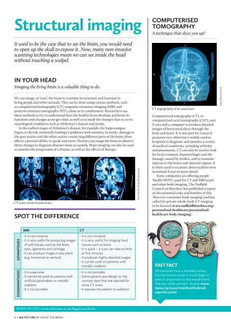
CT angiography of an aneurysm.<br />
Computerised tomography (CT), or<br />
computerised axial tomography (CAT), <strong>us</strong>es<br />
X-rays and a computer to produce detailed<br />
images of horizontal slices through <strong>the</strong><br />
body and <strong>brain</strong>. It is not <strong>us</strong>ed for <strong>research</strong><br />
purposes very often but is widely <strong>us</strong>ed in<br />
hospitals to diagnose and monitor a variety<br />
of medical conditions, including arthritis<br />
and pneumonia. CT can also be <strong>us</strong>ed to <strong>look</strong><br />
for <strong>brain</strong> tumours, haemorrhages and <strong>the</strong><br />
damage ca<strong>us</strong>ed by strokes, and to examine<br />
injuries to <strong>the</strong> bones and internal organs. It<br />
is often <strong>us</strong>ed to examine abnormalities seen<br />
in normal X-rays in more detail.<br />
Some companies are offering people<br />
‘health MOTs’: paid-for CT and MRI scans<br />
and o<strong>the</strong>r body imaging. The Nuffield<br />
Council on Bioethics has published a report<br />
on <strong>the</strong> potential risks and benefits of this<br />
‘direct-to-consumer body imaging’ and has<br />
called for private whole-body CT imaging<br />
to be banned (www.nuffieldbioethics.org/<br />
personalised-healthcare/personalisedhealthcare-body-imaging).<br />
FAST FACT<br />
All mammals have a wrinkled cortex,<br />
but <strong>the</strong> human cortex is much larger in<br />
area (in proportion to <strong>the</strong> overall <strong>brain</strong>)<br />
than any o<strong>the</strong>r animal’s. Source: www.<br />
dana.org/news/<strong>brain</strong>health/detail.<br />
aspx?id=10060<br />
hasa/Shutterstock