

MIND-BLOWING BRAINS How research helps us look inside the brain
MIND-BLOWING BRAINS How research helps us look inside the brain
MIND-BLOWING BRAINS How research helps us look inside the brain

You also want an ePaper? Increase the reach of your titles
YUMPU automatically turns print PDFs into web optimized ePapers that Google loves.
INSIDE THE BRAIN<br />
ISSUE 17 | SPRING 2013 bRINGING CUTTING-EDGE SCIENCE INTo THE CLASSRooM<br />
<strong>MIND</strong>-<strong>BLOWING</strong> <strong>BRAINS</strong><br />
<strong>How</strong> <strong>research</strong> <strong>helps</strong> <strong>us</strong><br />
<strong>look</strong> <strong>inside</strong> <strong>the</strong> <strong>brain</strong>
The <strong>brain</strong> is one of our<br />
most fascinating organs.<br />
Developments in technology<br />
and medicine mean that<br />
doctors and scientists can<br />
examine our <strong>brain</strong>s in more<br />
ways and more detail than<br />
ever before, all without<br />
having to open up <strong>the</strong> body.<br />
Join <strong>us</strong> as we find out more<br />
about how imaging <strong>research</strong><br />
has changed <strong>the</strong> way we can<br />
<strong>look</strong> <strong>inside</strong> <strong>the</strong> human <strong>brain</strong>.<br />
INSIDE<br />
NEURONS By NUMBERS<br />
A numerical tour of <strong>brain</strong>s<br />
and <strong>brain</strong> imaging.<br />
Why ThE BRAIN?<br />
Exploring <strong>the</strong> basics of <strong>brain</strong><br />
structure and function.<br />
STRUCTURAL IMAGING<br />
<strong>How</strong> <strong>research</strong>ers and clinicians<br />
can see <strong>inside</strong> our <strong>brain</strong>s.<br />
FUNCTIONAL IMAGING<br />
Ways to study what is happening<br />
in <strong>the</strong> <strong>brain</strong>.<br />
MyThBUSTING<br />
Putting to rest some common<br />
<strong>brain</strong>y misconceptions.<br />
KNOW yOUR <strong>MIND</strong><br />
Looking at some social, ethical<br />
and legal issues to do with <strong>the</strong> <strong>brain</strong>.<br />
REAL vOICES<br />
Three people talk about <strong>the</strong><br />
role of <strong>brain</strong>s in <strong>the</strong>ir lives.<br />
ONLINE<br />
2 | BIG PICTURE 17: INSIDE THE bRAIN<br />
2<br />
4<br />
6<br />
8<br />
10<br />
12<br />
14<br />
Go to www.wellcome.ac.uk/bigpicture/<br />
<strong>brain</strong> for more teaching resources, including<br />
extra articles, videos, image galleries,<br />
curriculum links, lesson ideas, a poster and<br />
an animation of <strong>the</strong> action potential. You can<br />
also download <strong>the</strong> PDF of this magazine or<br />
subscribe to <strong>the</strong> Big Picture series.<br />
Neurons by numbers<br />
The vital stats of <strong>brain</strong>s and <strong>brain</strong> imaging<br />
AvERAGE hUMAN BRAIN vOLUME<br />
1.5 l<br />
1.0 l<br />
0.5 l<br />
0.0 l<br />
Source: www.ncbi.nlm.nih.gov/pmc/articles/PMC2711771/<br />
BRAIN SIzE AND MASS<br />
Relative <strong>brain</strong> size and<br />
shape viewed from top<br />
1.13 l<br />
Elephant<br />
Human<br />
Chimp<br />
0.42 kg<br />
1.26 l<br />
1.4 kg 4.8 kg<br />
Sources: faculty.washington.edu/chudler/facts.html, www.frontiersin.org/neuroanatomy/10.3389/fnana.2011.00029/full<br />
TOTAL NUMBER OF SyNAPSES IN hUMAN NEOCORTEx<br />
150 000 00<br />
The neocortex is a six-layered part of <strong>the</strong> cerebral cortex found in mammals.<br />
Source: postcog.ucd.ie/files/Pakkenberg%202003.pdf
MRI units<br />
EqUIvALENT RADIATION DOSES<br />
SPEED OF NERvE TRANSMISSION<br />
From top: unmyelinated fibre, thin myelinated fibre, thick myelinated fibre, cruising commercial airliner.<br />
Sources: faculty.washington.edu/chudler/cv.html, forums.x-plane.org<br />
MRI UNITS (SCANNERS) PER MILLION PEOPLE<br />
50<br />
40<br />
30<br />
20<br />
10<br />
0<br />
10 mSv<br />
CT scan of <strong>the</strong><br />
whole body<br />
0.005 mSv<br />
135g bag of brazil nuts<br />
2.7 mSv<br />
Average annual radiation<br />
dose in <strong>the</strong> UK<br />
0.5–2 m/s<br />
43.1<br />
Japan<br />
5–35 m/s<br />
Source: www.oecd.org (2010 data)<br />
31.6<br />
USa<br />
0.02 mSv<br />
Chest X-ray<br />
7.8 mSv<br />
Average annual radon<br />
dose in Cornwall<br />
Area relates to dose in millisieverts.<br />
Source: www.hpa.org.uk/Topics/Radiation/UnderstandingRadiation/<br />
UnderstandingRadiationTopics/DoseComparisonsForIonisingRadiation<br />
22.6<br />
Greece<br />
1.4 mSv<br />
CT scan of <strong>the</strong> head<br />
0.07 mSv<br />
Transatlantic flight<br />
80–120 m/s<br />
5.9<br />
UK<br />
100 mSv<br />
Level at which<br />
changes in blood<br />
cells can be readily<br />
observed<br />
2.0<br />
Mexico<br />
(0.15 quadrillion)<br />
0 000 000<br />
NUMBER OF NEURONS IN ThE<br />
hUMAN BRAIN<br />
86bn<br />
There is roughly <strong>the</strong> same number of glia<br />
(supporting cells).<br />
Source: www.ncbi.nlm.nih.gov/pmc/articles/<br />
PMC3386878/?tool=pubmed<br />
80–120 m/s =<br />
179–268 mph<br />
FINDING DATA<br />
223.5 m/s<br />
Putting this diagram toge<strong>the</strong>r, we<br />
found that different sources gave<br />
different numbers for <strong>the</strong> same thing.<br />
Why don’t <strong>the</strong>y match?<br />
Well, data can be interpreted in<br />
different ways, and estimates can be<br />
made <strong>us</strong>ing different methods and/or<br />
baseline data. Definitions matter, too –<br />
different sources might define ‘volume’<br />
or ‘mass’ differently.<br />
Which should you choose? The source<br />
itself is important – is it reliable? Are<br />
<strong>the</strong> figures recent? <strong>How</strong> might an<br />
organisation’s ‘agenda’ affect how it<br />
calculates and presents data?<br />
SPRING 2013 | 3<br />
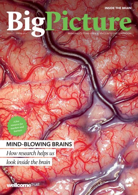
Cover image: The surface of <strong>the</strong> human <strong>brain</strong> during an operation. Robert Ludlow/Wellcome Images. Top and bottom row: Neuron ill<strong>us</strong>tration. VLADGRIN/iStockphoto.
Why <strong>the</strong> <strong>brain</strong>?<br />
The <strong>brain</strong> has mystified and inspired philosophers, doctors<br />
and scientists for centuries, and our fascination with it<br />
today only seems to be growing. So what exactly does this<br />
1.4 kg of tissue do for <strong>us</strong>?<br />
yOU AND yOUR BRAIN<br />
Your <strong>brain</strong> is your body’s control centre<br />
Your <strong>brain</strong> underpins who you are. It<br />
stores your knowledge and memories,<br />
gives you <strong>the</strong> capacity for thought<br />
and emotion, and enables you to<br />
control your body. The <strong>brain</strong> is j<strong>us</strong>t<br />
one part of <strong>the</strong> nervo<strong>us</strong> system.<br />
Toge<strong>the</strong>r with <strong>the</strong> spinal cord, it makes<br />
up <strong>the</strong> central nervo<strong>us</strong> system.<br />
The <strong>brain</strong> is <strong>the</strong> control centre,<br />
which sends and receives information to<br />
and from <strong>the</strong> body via <strong>the</strong> spinal cord.<br />
This involves <strong>the</strong> peripheral nervo<strong>us</strong><br />
system, which contains mixed nerves<br />
made up of sensory and motor nerve<br />
fibres. Sensory fibres carry information<br />
about what <strong>the</strong> body is sensing into <strong>the</strong><br />
spinal cord, and motor fibres transmit<br />
signals from <strong>the</strong> central nervo<strong>us</strong><br />
system to <strong>the</strong> m<strong>us</strong>cles and glands.<br />
WhITE AND GREy<br />
Inside <strong>the</strong> <strong>brain</strong>, only white and grey matter<br />
The <strong>brain</strong> is made of grey and white<br />
matter. Grey matter contains <strong>the</strong> cell<br />
bodies of neurons (nerve cells) and <strong>the</strong>ir<br />
local connections to each o<strong>the</strong>r. White<br />
matter contains bundles of nerve fibres<br />
that connect distant <strong>brain</strong> regions to<br />
one ano<strong>the</strong>r; it gets its name from <strong>the</strong><br />
fatty myelin that insulates <strong>the</strong> axons,<br />
which makes <strong>the</strong> tissue <strong>look</strong> white to<br />
<strong>the</strong> naked eye.<br />
The <strong>brain</strong> contains 86 billion<br />
neurons. They are specialised to<br />
produce electrical signals<br />
called action potentials. When<br />
resting, neurons produce a few<br />
action potentials (or ‘fire’) every<br />
FAST FACT<br />
MORE ONLINE: www.wellcome.ac.uk/bigpicture/<strong>brain</strong><br />
4 | BIG PICTURE 17: INSIDE THE bRAIN<br />
The autonomic<br />
nervo<strong>us</strong> system<br />
(divided into <strong>the</strong><br />
sympa<strong>the</strong>tic and<br />
parasympa<strong>the</strong>tic<br />
systems) is ano<strong>the</strong>r<br />
part of <strong>the</strong> peripheral<br />
nervo<strong>us</strong> system. It controls<br />
involuntary (autonomic) functions such<br />
as heart rate, breathing and digestion.<br />
The <strong>brain</strong> is incredibly complex and<br />
can be affected by many diseases, but we<br />
still know only a fraction of what <strong>the</strong>re<br />
is to know about how it works. A better<br />
comprehension of how <strong>the</strong> <strong>brain</strong> works<br />
will enable scientists to understand<br />
mental processes and behaviour better<br />
and, eventually, to prevent or treat any<br />
problems that occur.<br />
second, but this increases when <strong>the</strong>y<br />
become active. Neurons form intricate<br />
networks and communicate with each<br />
o<strong>the</strong>r across junctions called synapses.<br />
Action potentials ca<strong>us</strong>e <strong>the</strong> neuron<br />
on one side of <strong>the</strong> synapse to release a<br />
neurotransmitter that travels across<br />
<strong>the</strong> synapse to <strong>the</strong> neuron on <strong>the</strong> o<strong>the</strong>r<br />
side. Receptor proteins detect <strong>the</strong><br />
neurotransmitter and generate new<br />
action potentials in response.<br />
The world’s largest <strong>brain</strong> bank is at Harvard, where <strong>the</strong>re are over 7000 <strong>brain</strong>s.<br />
Source: www.youtube.com/watch?v=cvWb5OCgOr8<br />
Wellcome Images<br />
IN ThE SySTEM<br />
Exploring <strong>the</strong> nervo<strong>us</strong> system<br />
A cl<strong>us</strong>ter of neurons in culture.<br />
Like o<strong>the</strong>r systems in <strong>the</strong> body, <strong>the</strong> nervo<strong>us</strong><br />
system consists of tissue that is made of<br />
collections of cells, each of which contains<br />
tho<strong>us</strong>ands of different molecules. The cells<br />
of <strong>the</strong> nervo<strong>us</strong> system can be broadly divided<br />
into two types – neurons and glia.<br />
Neurons are specialised to produce<br />
electrical signals called action potentials.<br />
They form networks and communicate with<br />
each o<strong>the</strong>r by transmitting chemical signals<br />
across tiny gaps called synapses.<br />
Different projections from <strong>the</strong> cell body<br />
connect one neuron with o<strong>the</strong>rs. An axon<br />
carries <strong>the</strong> action potential away from <strong>the</strong> cell<br />
body, while many dendrites carry impulses<br />
from o<strong>the</strong>r neurons towards <strong>the</strong> cell body.<br />
Neurons have distinctive shapes that are<br />
closely related to <strong>the</strong>ir function. Primary<br />
sensory neurons, for example, carry<br />
information from <strong>the</strong> body into <strong>the</strong> spinal<br />
cord. They have a single fibre that splits<br />
into two. One branch goes out towards<br />
<strong>the</strong> body and <strong>the</strong> o<strong>the</strong>r goes into <strong>the</strong> spinal<br />
cord. Motor neurons in <strong>the</strong> spinal cord have<br />
dendrites that receive signals from o<strong>the</strong>r cells,<br />
and a long axon that extends to <strong>the</strong> m<strong>us</strong>cles<br />
through peripheral nerves. Interneurons<br />
connect sensory and motor neurons to<br />
each o<strong>the</strong>r and have short dendrites and a<br />
branched axon.<br />
The o<strong>the</strong>r cells of <strong>the</strong> nervo<strong>us</strong> system<br />
are called glia. They include Schwann cells,<br />
which form <strong>the</strong> myelin sheaths that wrap<br />
around <strong>the</strong> axons of sensory and motor<br />
neurons, principally in <strong>the</strong> peripheral<br />
nervo<strong>us</strong> system (<strong>the</strong> nerves and nerve cells<br />
outside of <strong>the</strong> <strong>brain</strong> and spinal cord). The<br />
myelin sheath is not continuo<strong>us</strong> but is<br />
interrupted by gaps called nodes of Ranvier.<br />
Although <strong>the</strong> myelin sheath is an electrical<br />
insulator, <strong>the</strong> gaps actually speed up nerve<br />
conduction beca<strong>us</strong>e <strong>the</strong>y permit saltatory<br />
conduction, where action potentials ‘jump’<br />
from one node to <strong>the</strong> next, and this increases<br />
<strong>the</strong>ir conduction speed.<br />
Ludovic Collin/Wellcome Images
FINDING yOUR WAy AROUND<br />
Below is an annotated guide to <strong>the</strong> <strong>brain</strong>. Remember, many simple and complex psychological<br />
functions are mediated by multiple <strong>brain</strong> regions and – at <strong>the</strong> same time – a single <strong>brain</strong> area<br />
may control many psychological functions.<br />
Cortex: The thin, folded structure on<br />
<strong>the</strong> outside surface of <strong>the</strong> <strong>brain</strong>.<br />
Pituitary gland: The ‘master gland’ of <strong>the</strong> body,<br />
which releases hormones that control growth,<br />
blood pressure, <strong>the</strong> stress response and <strong>the</strong><br />
function of <strong>the</strong> sex organs.<br />
Substantia nigra: The ‘black substance’<br />
contains cells that produce <strong>the</strong><br />
neurotransmitter dopamine and <strong>the</strong> pigment<br />
melatonin, giving it a black appearance.<br />
hypothalam<strong>us</strong>: The interface between <strong>the</strong><br />
<strong>brain</strong> and pituitary gland. It controls <strong>the</strong><br />
production and release of hormones.<br />
Frontal<br />
lobe<br />
Temporal<br />
lobe<br />
Parietal<br />
lobe<br />
Occipital<br />
lobe<br />
Spinal cord: A large bundle of millions of nerve<br />
fibres, which carries information back and forth<br />
between <strong>the</strong> <strong>brain</strong> and <strong>the</strong> body.<br />
Medulla oblongata: Controls vital involuntary<br />
functions such as breathing and heart rate.<br />
Cerebral hemispheres: The two halves of <strong>the</strong><br />
<strong>brain</strong>, each of which controls and receives<br />
information from <strong>the</strong> opposite side of <strong>the</strong> body.<br />
Cerebellum: The ‘little <strong>brain</strong>’ that controls<br />
balance and coordinates movements. It’s normally<br />
required for learning motor skills, such as riding a<br />
bike, and is involved in thought processes.<br />
Cranial nerve nuclei: Cl<strong>us</strong>ters of neurons in <strong>the</strong><br />
<strong>brain</strong> stem. Their axons form <strong>the</strong> cranial nerves.<br />
For even more on <strong>the</strong> <strong>brain</strong><br />
(including <strong>the</strong> limbic system)<br />
and neurons, synapses and<br />
neurotransmitters, order or<br />
download our poster at www.<br />
wellcome.ac.uk/bigpicture/<strong>brain</strong><br />
SPRING 2013 | 5
Structural imaging<br />
It <strong>us</strong>ed to be <strong>the</strong> case that to see <strong>the</strong> <strong>brain</strong>, you would need<br />
to open up <strong>the</strong> skull to expose it. Now, many non-invasive<br />
scanning technologies mean we can see <strong>inside</strong> <strong>the</strong> head<br />
without touching a scalpel.<br />
IN yOUR hEAD<br />
Imaging <strong>the</strong> living <strong>brain</strong> is a valuable thing to do<br />
We can image, or ‘scan’, <strong>the</strong> <strong>brain</strong> to examine its structure and function in<br />
living people and o<strong>the</strong>r animals. This can be done <strong>us</strong>ing vario<strong>us</strong> methods, such<br />
as computerised tomography (CT), magnetic resonance imaging (MRI) and<br />
positron emission tomography (PET), alone or in combination. Researchers <strong>us</strong>e<br />
<strong>the</strong>se methods to try to understand how <strong>the</strong> healthy <strong>brain</strong> develops, performs its<br />
functions and changes as we get older, as well as to study <strong>the</strong> changes that occur in<br />
neurological conditions such as Alzheimer’s disease and stroke.<br />
In <strong>the</strong> earliest stages of Alzheimer’s disease, for example, <strong>the</strong> hippocamp<strong>us</strong><br />
begins to shrink, eventually leading to problems with memory. In stroke, damage to<br />
<strong>the</strong> grey matter and <strong>the</strong> white matter connecting different parts of <strong>the</strong> <strong>brain</strong> often<br />
affects a person’s ability to speak and move. Doctors can image <strong>the</strong> <strong>brain</strong> to observe<br />
<strong>the</strong>se changes to diagnose diseases more accurately. Brain imaging can also be <strong>us</strong>ed<br />
to monitor <strong>the</strong> progression of a disease, as well as <strong>the</strong> effects of <strong>the</strong>rapy.<br />
CT scans of <strong>the</strong> human <strong>brain</strong>.<br />
SPOT ThE DIFFERENCE<br />
ADvANTAGES<br />
DISADvANTAGES<br />
MRI CT<br />
- It is non-invasive<br />
- It is very <strong>us</strong>eful for producing images<br />
of soft tissues, such as <strong>the</strong> <strong>brain</strong>,<br />
eyes, ligaments and cartilage<br />
- It can produce images in any plane<br />
(e.g. horizontal or vertical)<br />
- It is expensive<br />
- It cannot be <strong>us</strong>ed on patients with<br />
artificial pacemakers or metallic<br />
implants<br />
- It is not portable<br />
MORE MORE ONLINE: www.wellcome.ac.uk/bigpicture/<strong>brain</strong><br />
Fossilised bones show humans were butchering meat 3.5 million years ago. Read more at www.wellcome.ac.uk/bigpicture/food<br />
6 | BIG PICTURE 17: INSIDE THE bRAIN<br />
- It is non-invasive<br />
- It is very <strong>us</strong>eful for imaging hard<br />
tissues such as bone<br />
- It is quick – a scan can take as little<br />
as five minutes<br />
- It produces highly detailed images<br />
- It can be <strong>us</strong>ed on patients with<br />
metallic implants<br />
- It is not portable<br />
- Some patients are allergic to <strong>the</strong><br />
contrast dyes that are injected for<br />
some CT scans<br />
- It exposes <strong>the</strong> patient to radiation<br />
Sved Attila Oliver/Shutterstock<br />
COMPUTERISED<br />
TOMOGRAPhy<br />
A technique that slices you up?<br />
CT angiography of an aneurysm.<br />
Computerised tomography (CT), or<br />
computerised axial tomography (CAT), <strong>us</strong>es<br />
X-rays and a computer to produce detailed<br />
images of horizontal slices through <strong>the</strong><br />
body and <strong>brain</strong>. It is not <strong>us</strong>ed for <strong>research</strong><br />
purposes very often but is widely <strong>us</strong>ed in<br />
hospitals to diagnose and monitor a variety<br />
of medical conditions, including arthritis<br />
and pneumonia. CT can also be <strong>us</strong>ed to <strong>look</strong><br />
for <strong>brain</strong> tumours, haemorrhages and <strong>the</strong><br />
damage ca<strong>us</strong>ed by strokes, and to examine<br />
injuries to <strong>the</strong> bones and internal organs. It<br />
is often <strong>us</strong>ed to examine abnormalities seen<br />
in normal X-rays in more detail.<br />
Some companies are offering people<br />
‘health MOTs’: paid-for CT and MRI scans<br />
and o<strong>the</strong>r body imaging. The Nuffield<br />
Council on Bioethics has published a report<br />
on <strong>the</strong> potential risks and benefits of this<br />
‘direct-to-consumer body imaging’ and has<br />
called for private whole-body CT imaging<br />
to be banned (www.nuffieldbioethics.org/<br />
personalised-healthcare/personalisedhealthcare-body-imaging).<br />
FAST FACT<br />
All mammals have a wrinkled cortex,<br />
but <strong>the</strong> human cortex is much larger in<br />
area (in proportion to <strong>the</strong> overall <strong>brain</strong>)<br />
than any o<strong>the</strong>r animal’s. Source: www.<br />
dana.org/news/<strong>brain</strong>health/detail.<br />
aspx?id=10060<br />
hasa/Shutterstock
MAGNETIC RESONANCE IMAGING<br />
An imaging technique where protons get in a spin<br />
Magnetic resonance imaging (MRI)<br />
is well suited to visualising soft<br />
tissues such as <strong>the</strong> <strong>brain</strong>. It relies on<br />
<strong>the</strong> magnetic properties of atoms to<br />
produce images. An MRI scanner is a<br />
large cylinder containing an extremely<br />
powerful magnet. When a patient<br />
lies <strong>inside</strong> <strong>the</strong> scanner, <strong>the</strong> magnetic<br />
field it produces ca<strong>us</strong>es <strong>the</strong> protons<br />
in atomic nuclei to align <strong>the</strong>mselves<br />
with it. The scanner <strong>the</strong>n transmits<br />
radio waves through <strong>the</strong> body, making<br />
<strong>the</strong> protons alter <strong>the</strong>ir alignment.<br />
This ca<strong>us</strong>es <strong>the</strong> nuclei to produce tiny<br />
rotating magnetic fields that can be<br />
detected and recorded by <strong>the</strong> scanner to<br />
construct <strong>the</strong> image.<br />
Researchers <strong>us</strong>e MRI to <strong>look</strong> at <strong>the</strong><br />
structure of <strong>the</strong> <strong>brain</strong>s (both living<br />
and dead) of humans and animals. In<br />
2007, neuroscientists <strong>us</strong>ed it to scan<br />
<strong>the</strong> <strong>brain</strong>s of two stroke patients who<br />
died about 150 years ago. More recently,<br />
<strong>research</strong>ers transplanted human cells<br />
into <strong>the</strong> <strong>brain</strong>s of rats to help <strong>the</strong>m<br />
recover from stroke and <strong>us</strong>ed MRI to<br />
detect <strong>the</strong> structural changes ca<strong>us</strong>ed by<br />
<strong>the</strong> transplanted cells.<br />
Doctors <strong>us</strong>e MRI to visualise <strong>the</strong><br />
changes that occur in a wide variety<br />
of neurological diseases, including<br />
Alzheimer’s disease, epilepsy and<br />
multiple sclerosis. In Alzheimer’s<br />
disease, certain parts of <strong>the</strong> <strong>brain</strong> begin<br />
to shrink many years before symptoms<br />
appear. MRI scans can detect this<br />
shrinkage, which is important beca<strong>us</strong>e<br />
early detection can lead to earlier<br />
treatment. In addition, <strong>the</strong> method is<br />
<strong>us</strong>ed to diagnose <strong>brain</strong> tumours and<br />
to determine exactly where <strong>the</strong>y are so<br />
<strong>the</strong>y can be surgically removed.<br />
MRI scan of a human <strong>brain</strong> after a stroke (dead tissue in red) and 3D MRI of a dog’s head.<br />
ChECK ThE vOLUME<br />
Volume changes in <strong>the</strong> <strong>brain</strong> can tell <strong>us</strong> about disease and ageing<br />
Voxel-based morphometry (VBM) is a type of analysis applied to MRI images that<br />
is <strong>us</strong>ed to measure <strong>the</strong> volume of specific <strong>brain</strong> structures. By comparing healthy<br />
and diseased <strong>brain</strong>s, <strong>research</strong>ers can detect <strong>the</strong> subtle structural changes that<br />
occur in neurological and psychiatric conditions. It can detect <strong>the</strong> reduction of<br />
hippocamp<strong>us</strong> volume that occurs in Alzheimer’s disease and depression, as well<br />
as <strong>the</strong> thinning of <strong>the</strong> cerebral cortex that occurs as a normal part of ageing.<br />
Memory <strong>research</strong>ers in London have been investigating <strong>the</strong> changes that<br />
occur in taxi drivers’ <strong>brain</strong>s as a result of learning. To qualify for <strong>the</strong> job,<br />
London’s taxi drivers have to learn ‘<strong>the</strong> Knowledge’ (every street name, landmark<br />
and direction of traffic flow in a six-mile radi<strong>us</strong> of Charing Cross) so <strong>the</strong>y can<br />
navigate <strong>the</strong> city effectively. Using VBM, <strong>the</strong> <strong>research</strong>ers found that those who<br />
qualified had an increased volume of grey matter in <strong>the</strong> hippocamp<strong>us</strong>, a part<br />
of <strong>the</strong> <strong>brain</strong> known to be involved in generating maps and forming spatial<br />
memories. They have also found that <strong>the</strong> longer a person has been driving a taxi,<br />
<strong>the</strong> larger <strong>the</strong>ir hippocamp<strong>us</strong>.<br />
(L) Zephyr/Science Photo Library. (R) Thierry Berrod,<br />
Mona Lisa Production/Science Photo Library<br />
WELL CONNECTED<br />
Neuroscientists want to<br />
understand how our <strong>brain</strong><br />
pathways link up<br />
Neuroscientists see <strong>the</strong> <strong>brain</strong> as<br />
consisting of hundreds of specialised<br />
areas organised into multiple<br />
interconnected networks. They<br />
<strong>us</strong>e imaging to visualise <strong>the</strong> white<br />
matter tracts that form connections<br />
within and between networks and<br />
examine how <strong>the</strong> connections break in<br />
diseases such as stroke. One method<br />
that is increasingly being <strong>us</strong>ed for<br />
this is diff<strong>us</strong>ion tensor imaging<br />
(DTI) tractography, a form of MRI<br />
that detects <strong>the</strong> diff<strong>us</strong>ion of water<br />
molecules along axons.<br />
DTI tractography reveals <strong>the</strong><br />
<strong>brain</strong>’s largest pathways. Last year,<br />
Harvard <strong>research</strong>ers <strong>us</strong>ed it to image<br />
large fibres throughout <strong>the</strong> entire<br />
<strong>brain</strong> for <strong>the</strong> first time. Researchers<br />
at Cardiff University are developing<br />
a more advanced method, called DTI<br />
tractometry, to image <strong>the</strong> microscopic<br />
structure of <strong>the</strong> pathways and provide<br />
details such as <strong>the</strong> density and<br />
diameter of <strong>the</strong> axons in <strong>the</strong> tracts.<br />
Some of this work is part of a larger<br />
project called <strong>the</strong> Human Connectome<br />
Project, which some <strong>research</strong>ers say<br />
is too ambitio<strong>us</strong>. If it is eventually<br />
possible to produce a complete map of<br />
<strong>the</strong> connections in <strong>the</strong> <strong>brain</strong>, even that<br />
will not tell <strong>us</strong> everything about how<br />
<strong>the</strong> <strong>brain</strong> works.<br />
LOOKING AT<br />
ThE PAST<br />
<strong>How</strong> did people explore <strong>the</strong> <strong>brain</strong><br />
without imaging it?<br />
Before <strong>brain</strong> imaging was invented, doctors<br />
would observe <strong>the</strong> behaviour of people with<br />
<strong>brain</strong> damage and <strong>the</strong>n examine <strong>the</strong>ir <strong>brain</strong>s<br />
after <strong>the</strong>y died. This enabled <strong>the</strong>m to link<br />
<strong>the</strong> changes in behaviour to specific parts<br />
of <strong>the</strong> <strong>brain</strong>. One early example is Phineas<br />
Gage, who had a metre-long iron rod<br />
propelled by an explosion through his skull.<br />
His experience led doctors to link <strong>the</strong> frontal<br />
lobes to social behaviour. Read about Gage<br />
and o<strong>the</strong>r historical case studies at www.<br />
wellcome.ac.uk/bigpicture/<strong>brain</strong>.<br />
SPRING 2013 | 7
Functional imaging<br />
From reconstructing YouTube clips from <strong>brain</strong> scans to ‘watching’ a<br />
person’s <strong>brain</strong> as <strong>the</strong>y slip under an anaes<strong>the</strong>tic, functional <strong>brain</strong> imaging<br />
is producing fascinating insights into how our <strong>brain</strong>s work.<br />
EEG AND MEG<br />
Imaging techniques that detect electrical activity<br />
Electro-encephalography (EEG) and<br />
magneto-encephalography (MEG) are<br />
functional imaging methods <strong>us</strong>ed to<br />
measure <strong>brain</strong> activity directly and noninvasively<br />
(from outside <strong>the</strong> head). EEG<br />
detects synchronised electrical activity<br />
of large groups of neurons, whereas MEG<br />
detects <strong>the</strong> tiny changes in magnetic fields<br />
that this electrical activity is associated<br />
with. The images produced by EEG and<br />
MEG are not very localised, but <strong>the</strong>y can<br />
monitor how electrical activity changes<br />
with time very precisely.<br />
EEG requires electrodes to be attached<br />
to <strong>the</strong> scalp. It can be <strong>us</strong>ed to detect general<br />
patterns of electrical activity, such as<br />
BOLD ThINKING<br />
What does fMRI really measure?<br />
The most common form of fMRI is<br />
blood oxygenation level dependent<br />
(BOLD) fMRI. It is based on <strong>the</strong> idea<br />
that neurons require more energy when<br />
<strong>the</strong>y fire and that increased blood flow<br />
to active parts of <strong>the</strong> <strong>brain</strong> supplies <strong>the</strong><br />
oxygen required. This form of MRI<br />
measures changes in <strong>the</strong> concentration<br />
of oxygen in red blood cells.<br />
The main limitation of fMRI is that it<br />
measures <strong>brain</strong> activity indirectly, <strong>us</strong>ing<br />
blood flow as an indication that neurons<br />
are active. In 2009, <strong>research</strong>ers in <strong>the</strong><br />
USA published important work showing<br />
that parts of <strong>the</strong> <strong>brain</strong> that receive more<br />
oxygenated blood do not necessarily<br />
become more active. The <strong>research</strong>ers<br />
<strong>us</strong>ed fMRI to scan <strong>the</strong> <strong>brain</strong>s of monkeys<br />
while <strong>the</strong>y <strong>look</strong>ed at pictures, and <strong>the</strong>y<br />
found that <strong>the</strong> <strong>brain</strong> anticipates which<br />
of its parts will be activated over <strong>the</strong><br />
next few seconds and pre-empts itself by<br />
sending more blood to <strong>the</strong>m. Most areas<br />
that receive more blood are more active,<br />
but some areas that receive more blood<br />
do not become more active.<br />
MORE ONLINE: www.wellcome.ac.uk/bigpicture/<strong>brain</strong><br />
8 | BIG PICTURE 17: INSIDE THE bRAIN<br />
<strong>the</strong> <strong>brain</strong> waves that occur during sleep.<br />
Researchers <strong>us</strong>ed EEG to compare <strong>the</strong><br />
visual cortex activity of people who are<br />
born blind with those who are not blind,<br />
and <strong>the</strong>y found that <strong>the</strong> visual cortex<br />
of blind people was active.<br />
EEG can also be <strong>us</strong>ed to detect electrical<br />
signals associated with specific sensory<br />
stimuli, thought processes or movements.<br />
Detecting and measuring <strong>the</strong>se ‘eventrelated<br />
potentials’ can be done at a single<br />
electrode, but in practice, many are <strong>us</strong>ed.<br />
They are spread across <strong>the</strong> scalp, to help<br />
<strong>research</strong>ers pinpoint where in <strong>the</strong> <strong>brain</strong> <strong>the</strong><br />
neurons responsible for <strong>the</strong> potentials are.<br />
FUNCTIONAL MRI<br />
An imaging technique to show <strong>brain</strong> activation during tasks<br />
Functional magnetic resonance imaging<br />
(fMRI) is <strong>us</strong>ed to image <strong>the</strong> parts of <strong>the</strong><br />
<strong>brain</strong> that become active during different<br />
mental processes. In medicine, fMRI can<br />
be <strong>us</strong>ed to map <strong>brain</strong> functions in patients<br />
who are about to undergo neurosurgery<br />
to remove a <strong>brain</strong> tumour or abnormal<br />
tissue that ca<strong>us</strong>es epileptic seizures. This<br />
<strong>helps</strong> neurosurgeons to minimise <strong>the</strong> risk<br />
of accidentally removing or damaging <strong>the</strong><br />
parts of <strong>the</strong> <strong>brain</strong> involved in functions such<br />
as language and memory.<br />
fMRI studies show that different <strong>brain</strong><br />
areas are specialised for certain functions,<br />
often supporting data from previo<strong>us</strong><br />
neurological and psychological studies. For<br />
example, <strong>the</strong> frontal lobe contains areas<br />
that plan and control voluntary movements.<br />
The frontal lobe also contains areas<br />
specialised for complex functions such as<br />
making decisions and judgements, which<br />
are important for social interactions. fMRI<br />
studies show that activity in <strong>the</strong>se areas and<br />
<strong>the</strong>ir detailed structure is altered in people<br />
with autism, but fur<strong>the</strong>r <strong>research</strong> is needed<br />
to confirm this.<br />
fMRI can also be <strong>us</strong>ed to ‘decode’ <strong>brain</strong><br />
activity. In 2011, <strong>research</strong>ers in California<br />
scanned participants’ <strong>brain</strong>s while <strong>the</strong>y<br />
watched different YouTube clips; <strong>the</strong>y<br />
<strong>the</strong>n recorded activity in <strong>the</strong> primary<br />
visual cortex, which contains cells that<br />
detect contrast and <strong>the</strong> orientation of<br />
moving edges. Researchers <strong>the</strong>n showed<br />
<strong>the</strong> participants different film clips while<br />
scanning <strong>the</strong>ir <strong>brain</strong>s again and were able<br />
to construct low-quality copies of what <strong>the</strong><br />
participants saw by decoding primary visual<br />
cortex activity. Our thoughts are much<br />
more complex than this simple mapping<br />
of visual images, so we are a long way from<br />
decoding <strong>the</strong> content of minds.<br />
YouTube Pasieka/Science Photo Library
KNOW yOUR NEUROTRANSMITTERS<br />
Imaging can be <strong>us</strong>ed to study neurotransmitters too<br />
Imaging can be <strong>us</strong>ed to measure<br />
<strong>the</strong> levels of a neurotransmitter, its<br />
receptors and its transporters (which<br />
remove neurotransmitters from <strong>the</strong><br />
synapse after release). This is very<br />
<strong>us</strong>eful beca<strong>us</strong>e many neurological<br />
conditions involve <strong>the</strong> death of<br />
neurons that produce a specific<br />
neurotransmitter, or alterations in <strong>the</strong><br />
activity or distribution of receptors or<br />
transporters within certain parts of<br />
<strong>the</strong> <strong>brain</strong>. The relationship between<br />
diseases and neurotransmitters<br />
is complex. For example, o<strong>the</strong>r<br />
neurotransmitters than those disc<strong>us</strong>sed<br />
below could also be involved in <strong>the</strong><br />
conditions presented.<br />
In Parkinson’s disease, dopamineproducing<br />
neurons in <strong>the</strong> mid<strong>brain</strong> die.<br />
Alzheimer’s disease, depression and<br />
schizophrenia involve alterations in <strong>the</strong><br />
transporter for <strong>the</strong> neurotransmitter<br />
serotonin. Attention deficit<br />
hyperactivity disorder (ADHD) has<br />
been associated with malfunctions<br />
of dopamine receptors and dopa<br />
decarboxylase, one of <strong>the</strong> enzymes<br />
needed for making dopamine, in <strong>the</strong><br />
frontal cortex. ADHD, depression<br />
and schizophrenia can all involve<br />
disturbances in <strong>the</strong> function of<br />
receptors and transporters for <strong>the</strong><br />
neurotransmitter noradrenaline.<br />
The imaging methods <strong>us</strong>ed most<br />
often to measure neurotransmitter<br />
levels are positron emission tomography<br />
(PET) and single-photon emission<br />
computed tomography (SPECT). Both<br />
involve injecting a radioactive ‘tracer’<br />
substance that binds to <strong>the</strong> molecule<br />
being studied. The scan reveals where<br />
<strong>the</strong> tracer binds, and <strong>the</strong> intensity of<br />
<strong>the</strong> radioactive signal in each <strong>brain</strong> area<br />
is related to <strong>the</strong> level of <strong>the</strong> molecule<br />
being studied.<br />
Neurotransmitter receptors come in<br />
multiple forms, or subtypes, and each<br />
subtype has multiple variations. Some<br />
tracers bind to one particular subtype<br />
or more specifically to one variation of<br />
a subtype. To date, we know of at least<br />
five subtypes of receptor for dopamine<br />
– D1 to D5. Genetic mutations in <strong>the</strong><br />
D4 subtype of <strong>the</strong> dopamine receptor<br />
(DRD4), for example, are linked to<br />
ADHD, Parkinson’s disease and<br />
schizophrenia, as well as drug <strong>us</strong>e and<br />
personality traits such as aggression and<br />
impulsiveness. PET and SPECT can be<br />
<strong>us</strong>ed to examine exactly how alterations<br />
in <strong>the</strong> DRD4 receptor are linked to<br />
<strong>the</strong>se conditions and behaviours.<br />
OThER WAyS TO IMAGE<br />
Many different imaging methods are in <strong>us</strong>e and development<br />
One imaging technique that is now<br />
widely <strong>us</strong>ed by <strong>research</strong>ers is calcium<br />
imaging. When neurons fire, <strong>the</strong><br />
concentration of calcium ions <strong>inside</strong><br />
<strong>the</strong>m increases. This can be detected<br />
in slices of <strong>brain</strong> tissue or in live<br />
animals, <strong>us</strong>ing fluorescent dyes that<br />
are sensitive to <strong>the</strong> concentration<br />
increases combined with two-photon<br />
microscopy (a type of microscopy <strong>us</strong>ed<br />
particularly to image living cells).<br />
This technique requires an operation<br />
to expose <strong>the</strong> <strong>brain</strong> and is not <strong>us</strong>ed<br />
in humans.<br />
Near-infrared spectroscopy (NIRS)<br />
is a method that detects changes in<br />
blood flow around <strong>the</strong> <strong>brain</strong>. It works<br />
by transmitting light with a wavelength<br />
of 700–900 nanometres through<br />
<strong>the</strong> head; <strong>the</strong> light passes through<br />
skin, bones and <strong>brain</strong> tissue but is<br />
absorbed differently by oxygenated and<br />
deoxygenated blood. The person being<br />
scanned wears a cap containing lasers<br />
or light-emitting diodes; this allows<br />
people to do more things while <strong>the</strong>ir<br />
<strong>brain</strong> is examined beca<strong>us</strong>e <strong>the</strong>y are not<br />
enclosed in a scanner. NIRS is cheaper<br />
than fMRI and is particularly <strong>us</strong>eful in<br />
small babies, whose skulls are thinner<br />
than those of children or adults and<br />
transmit <strong>the</strong> light <strong>us</strong>ed by NIRS better.<br />
O<strong>the</strong>r techniques include<br />
functional electrical impedance<br />
tomography by evoke response<br />
(fEITER). This <strong>us</strong>es electrodes attached<br />
to <strong>the</strong> head, which transmit electrical<br />
currents that are interrupted by <strong>the</strong><br />
<strong>brain</strong>’s electrical activity. Researchers<br />
recently <strong>us</strong>ed fEITER to image how<br />
<strong>brain</strong> activity changes when people lose<br />
conscio<strong>us</strong>ness under anaes<strong>the</strong>sia.<br />
hANDS-ON RESEARCh<br />
Researchers can alter your <strong>brain</strong> function<br />
Some techniques enable <strong>research</strong>ers to alter<br />
activity in <strong>the</strong> living human <strong>brain</strong>. One of<br />
<strong>the</strong>se is transcranial magnetic stimulation (see<br />
above), which <strong>us</strong>es a figure-of-eight-shaped<br />
coil placed on <strong>the</strong> scalp to deliver magnetic<br />
pulses to parts of <strong>the</strong> <strong>brain</strong> directly under <strong>the</strong><br />
coil. This can be <strong>us</strong>ed in <strong>the</strong> lab to interrupt<br />
<strong>brain</strong> activity, to see how different <strong>brain</strong><br />
regions contribute to certain functions. In <strong>the</strong><br />
clinic, transcranial magnetic stimulation can<br />
also be <strong>us</strong>ed to assess <strong>brain</strong> damage in people<br />
who have suffered a stroke and to aid <strong>the</strong>ir<br />
rehabilitation.<br />
Deep <strong>brain</strong> stimulation is an invasive<br />
surgical procedure in which thin wire<br />
electrodes are implanted into <strong>the</strong> <strong>brain</strong>. By<br />
passing current through <strong>the</strong> wires from a<br />
control box outside <strong>the</strong> head, you can alter<br />
<strong>the</strong> activity of <strong>the</strong> region where <strong>the</strong> wires<br />
are implanted. Deep <strong>brain</strong> stimulation can<br />
help alleviate some symptoms of Parkinson’s<br />
disease, and so far it has been <strong>us</strong>ed to treat<br />
about 90 000 patients with <strong>the</strong> condition. It<br />
is also being <strong>us</strong>ed to treat o<strong>the</strong>r conditions,<br />
such as depression<br />
and obsessive–<br />
compulsive<br />
disorder, as well as<br />
drug addiction.<br />
FAST FACT<br />
A 2011 study suggests that <strong>the</strong> increase in<br />
general intelligence seen during adolescence<br />
can be attributed to increases in mental<br />
speed. Source: Coyle TR et al. Psychol Sci<br />
2011;22(10):1265–9.<br />
SPRING 2013 | 9<br />
University of Durham, Simon Fraser/Science Photo Library
Mythb<strong>us</strong>ting<br />
We’ve all heard <strong>the</strong> one about people <strong>us</strong>ing only 10 per cent<br />
of <strong>the</strong>ir <strong>brain</strong>, but is this j<strong>us</strong>t a myth or proven fact? Like any<br />
respectable science publication, we’ve gone directly to <strong>the</strong><br />
published evidence to help you separate scientific fact from<br />
<strong>brain</strong>y baloney.<br />
…WE ONLy USE 10 PER CENT<br />
OF OUR <strong>BRAINS</strong>?<br />
Many people think that we only <strong>us</strong>e 10<br />
per cent of our <strong>brain</strong>s and that we can<br />
harness <strong>the</strong> rest to boost our mental<br />
abilities. This is <strong>the</strong> most popular<br />
myth about <strong>the</strong> <strong>brain</strong>, and <strong>the</strong>re is no<br />
scientific evidence for it – we almost<br />
certainly <strong>us</strong>e all of it. The <strong>brain</strong> is a very<br />
hungry organ, consuming nearly<br />
10 | BIG PICTURE 17: INSIDE THE bRAIN<br />
IS IT TRUE ThAT…<br />
…LEARNING CAN PhySICALLy<br />
ALTER ThE BRAIN?<br />
Neuroscientists believe that learning<br />
and memory change <strong>the</strong> physical<br />
structure of <strong>the</strong> <strong>brain</strong>. Animal<br />
<strong>research</strong> shows that forming a new<br />
memory involves <strong>the</strong> streng<strong>the</strong>ning<br />
of synapses in a network of<br />
neurons. The same is probably true<br />
of humans, although we still do not<br />
have <strong>the</strong> techniques to observe <strong>the</strong>se<br />
changes directly in <strong>the</strong> human <strong>brain</strong>.<br />
Learning can ca<strong>us</strong>e o<strong>the</strong>r changes in <strong>the</strong><br />
<strong>brain</strong>, some of which can be seen in <strong>the</strong> human<br />
<strong>brain</strong> <strong>us</strong>ing vario<strong>us</strong> imaging techniques. In 2004, for example, <strong>research</strong>ers<br />
<strong>us</strong>ed MRI to show that learning to juggle for three months increases <strong>the</strong><br />
density of grey matter in parts of <strong>the</strong> visual cortex specialised for processing<br />
complex visual motion. More recently, ano<strong>the</strong>r group of <strong>research</strong>ers<br />
showed that learning to juggle also leads to changes in <strong>the</strong> <strong>brain</strong>’s white<br />
matter tracts. Using diff<strong>us</strong>ion tensor imaging, <strong>the</strong>y found that it increases<br />
<strong>the</strong> density of white matter underneath <strong>the</strong> intraparietal sulc<strong>us</strong>, which is<br />
involved in several mental functions, including memory for where things<br />
are in space and perceptual–motor skills (such as hand–eye coordination).<br />
Learning to read as an adult also ca<strong>us</strong>es significant changes in <strong>brain</strong><br />
structure. In a unique study, <strong>research</strong>ers in London and Spain examined <strong>the</strong><br />
<strong>brain</strong>s of former Colombian guerrillas who learned to read Spanish after<br />
returning to mainstream society. Using MRI, <strong>the</strong>y detected increases in <strong>the</strong><br />
volume of grey matter in <strong>the</strong> left frontal and temporal lobes, both of which<br />
contain areas specialised for processing language.<br />
MORE ONLINE: www.wellcome.ac.uk/bigpicture/<strong>brain</strong><br />
one-quarter of <strong>the</strong> body’s energy,<br />
despite accounting for j<strong>us</strong>t 2 per cent<br />
of body mass. From <strong>the</strong> point of view<br />
of evolution, it j<strong>us</strong>t doesn’t make sense<br />
to have a large organ that consumes so<br />
much energy if only 10 per cent of it is<br />
actually needed.<br />
Eric Boucher/Shutterstock<br />
…BRAIN GyM MAKES<br />
yOU SMARTER?<br />
The makers of Brain Gym claim that<br />
particular physical activities enable<br />
students to access parts of <strong>the</strong> <strong>brain</strong> that<br />
<strong>the</strong>y normally do not <strong>us</strong>e, and that p<strong>us</strong>hing<br />
“Brain Buttons” on parts of <strong>the</strong> body, or<br />
performing certain repetitive movements,<br />
can improve blood flow to certain parts<br />
of <strong>the</strong> <strong>brain</strong> or improve certain <strong>brain</strong><br />
functions.<br />
www.<strong>brain</strong>gym.org/faq states: “Our<br />
primary evidence comes from <strong>the</strong> countless<br />
anecdotal stories reported to <strong>us</strong> since 1986.”<br />
They also publish three publications of<br />
<strong>the</strong>ir own <strong>research</strong>. Several scientists have<br />
explained publicly why <strong>the</strong>y think some<br />
of <strong>the</strong> claims do not stand up scientifically<br />
(on senseaboutscience.org, for example).<br />
A 2007 paper by Dr Keith Hyatt from<br />
Western Washington University found that<br />
“a review of <strong>the</strong> <strong>the</strong>oretical foundations<br />
of Brain Gym and <strong>the</strong> associated peerreviewed<br />
<strong>research</strong> studies failed to<br />
support <strong>the</strong> contentions of <strong>the</strong> promoters<br />
of Brain Gym”.<br />
<strong>How</strong>ever, regular aerobic exercise is<br />
good beca<strong>us</strong>e it makes <strong>the</strong> heart work<br />
harder and increases blood flow to <strong>the</strong><br />
<strong>brain</strong>. There is evidence that it improves<br />
cognitive function and memory, and<br />
can be linked to academic achievement.<br />
Regular exercise throughout life may help<br />
to protect <strong>the</strong> <strong>brain</strong> against <strong>the</strong> changes<br />
that occur with age, and could slow <strong>the</strong>se<br />
changes down. In mice and rats, exercise<br />
also ca<strong>us</strong>es <strong>the</strong> growth of new <strong>brain</strong> cells,<br />
but we still don’t know whe<strong>the</strong>r this also<br />
happens in humans.<br />
…ThERE’S A JENNIFER<br />
ANISTON NEURON?<br />
While examining <strong>the</strong> <strong>brain</strong>s of people<br />
with epilepsy who were about to undergo<br />
neurosurgery, <strong>research</strong>ers discovered<br />
neurons that respond specifically when<br />
people <strong>look</strong> at pictures of celebrities like<br />
Jennifer Aniston and Halle Berry or famo<strong>us</strong><br />
landmarks like <strong>the</strong> Eiffel Tower. The cells<br />
also fired when <strong>the</strong> patients thought about<br />
<strong>the</strong> celebrities. They are located near <strong>the</strong><br />
hippocamp<strong>us</strong>, which is crucial for memory<br />
formation. Each cell is probably part of<br />
a network that encodes <strong>the</strong> memory, or<br />
concept, of <strong>the</strong> celebrity or landmark and<br />
probably contributes to hundreds of o<strong>the</strong>r<br />
networks, each of which encodes a different<br />
memory or concept. It is unlikely that<br />
single neurons map directly and uniquely<br />
onto single people or objects.
Thomas Deerinck, NCMIR/Science Photo Library<br />
…A BIGGER BRAIN IS A CLEvERER BRAIN?<br />
It was once thought that having a<br />
bigger <strong>brain</strong> makes you more clever,<br />
but that isn’t true. Women’s <strong>brain</strong>s are,<br />
on average, 9 per cent smaller than<br />
men’s (and so contain fewer neurons),<br />
but women are not less intelligent than<br />
men; <strong>the</strong> size difference j<strong>us</strong>t reflects <strong>the</strong><br />
fact that women’s bodies are generally<br />
smaller. Brain size and intelligence are<br />
definitely linked, however, but we still<br />
don’t know exactly how. What seems<br />
to be more important than overall size<br />
is how <strong>the</strong> neurons are connected to<br />
each o<strong>the</strong>r.<br />
Albert Einstein’s <strong>brain</strong> (right)<br />
ill<strong>us</strong>trates j<strong>us</strong>t how complicated <strong>the</strong><br />
relationship between <strong>brain</strong> size and<br />
intelligence is. Einstein is often said<br />
to be one of <strong>the</strong> most intelligent<br />
people that ever lived, and <strong>research</strong>ers<br />
naturally believed his <strong>brain</strong> might<br />
provide important clues about what<br />
made him so clever. When he died, <strong>the</strong>y<br />
were surprised to discover that his <strong>brain</strong><br />
was actually quite small. It weighed<br />
…ThE SIDES OF OUR BRAIN DO<br />
DIFFERENT ThINGS?<br />
1.23 kg, or about 200 g lighter than <strong>the</strong><br />
average of 1.4 kg, and <strong>the</strong> temporal<br />
lobes – which contain areas that are<br />
specialised for speech and language –<br />
were also smaller than average.<br />
Scientists who examined Einstein’s<br />
<strong>brain</strong> did find some un<strong>us</strong>ual differences<br />
that help to explain why he was so<br />
intelligent. For example, his parietal<br />
lobes, which are important for<br />
ma<strong>the</strong>matical abilities and visual and<br />
spatial functions, were about 15 per cent<br />
wider than average. They also had an<br />
un<strong>us</strong>ual pattern of grooves and ridges,<br />
and certain regions of <strong>the</strong> parietal<br />
lobes had more glial cells per neuron<br />
than normal.<br />
Ano<strong>the</strong>r popular myth about <strong>the</strong> <strong>brain</strong> is that <strong>the</strong> left hemisphere is ‘logical’<br />
and <strong>the</strong> right hemisphere is ‘artistic’. This probably comes from early studies of<br />
people with <strong>brain</strong> damage and from work on ‘split-<strong>brain</strong>’ patients, in whom <strong>the</strong><br />
connections between <strong>the</strong> left and right hemispheres were destroyed to prevent<br />
epileptic seizures from spreading throughout <strong>the</strong> <strong>brain</strong>. It’s true that <strong>the</strong> left and<br />
right hemispheres each contain areas that are specialised for different functions,<br />
but most things we do involve <strong>the</strong> coordinated activity of <strong>the</strong> two hemispheres.<br />
…ABUSE AND LOvE CAN<br />
ChANGE ThE BRAIN?<br />
There is plenty of evidence that childhood<br />
neglect and ab<strong>us</strong>e can ca<strong>us</strong>e changes in<br />
<strong>the</strong> <strong>brain</strong> that have long-lasting effects.<br />
Most of it comes from <strong>research</strong> on animal<br />
behaviour, but <strong>the</strong>re is evidence for this in<br />
humans as well.<br />
In recent years, it has been found<br />
that growing up in poverty has a direct<br />
effect on children’s mental abilities. One<br />
study showed that children raised in<br />
impoverished conditions have a reduced<br />
capacity for working memory – <strong>the</strong> ability<br />
to store small amounts of information<br />
for short periods of time – in comparison<br />
to better-off children. A follow-up study<br />
showed that this is probably beca<strong>us</strong>e stress<br />
affects <strong>the</strong> way in which children’s <strong>brain</strong>s<br />
develop. More recently, it has been found<br />
that childhood ab<strong>us</strong>e stunts growth of<br />
<strong>the</strong> hippocamp<strong>us</strong>,<br />
part of <strong>the</strong> medial<br />
temporal lobe<br />
that is crucial for<br />
memory formation.<br />
…WE’RE BORN WITh ALL ThE BRAIN CELLS WE’LL EvER hAvE?<br />
Nerve cells and glial cells, SEM<br />
Einstein’s <strong>brain</strong>. Wellcome Images<br />
Neuroscientists had always believed<br />
that <strong>the</strong> adult <strong>brain</strong> could not produce<br />
new cells and that we are born with<br />
all <strong>the</strong> neurons we will ever have. This<br />
viewpoint slowly began to change in <strong>the</strong><br />
1970s, when scientists discovered that <strong>the</strong><br />
<strong>brain</strong>s of rats and songbirds produce new<br />
cells throughout life. This is now widely<br />
accepted as being correct, and many<br />
scientists also take it for granted that <strong>the</strong><br />
same is true for <strong>the</strong> adult human <strong>brain</strong>.<br />
Actually, though, <strong>the</strong>re’s still very little<br />
evidence for it. According to <strong>the</strong> latest<br />
estimates, <strong>the</strong> <strong>brain</strong> contains about<br />
86 billion neurons and roughly <strong>the</strong> same<br />
FAST FACT<br />
The human <strong>brain</strong> accounts for 2 per cent<br />
of <strong>the</strong> total mass of <strong>the</strong> body, but 20 per<br />
cent of its oxygen consumption at rest.<br />
Source: www.ncbi.nlm.nih.gov/books/<br />
NBK28194/<br />
number of glial cells. As far as we know, <strong>the</strong><br />
vast majority of <strong>the</strong>se cells are produced in<br />
<strong>the</strong> womb – during early pregnancy, about<br />
250 000 <strong>brain</strong> cells are produced every<br />
minute. The rest are produced during a<br />
short period of time after birth – maybe as<br />
little as a few months, or as much as a year.<br />
As a result, <strong>the</strong> <strong>brain</strong> produces at least<br />
twice as many cells than it actually needs to<br />
work properly. Neurons begin to die before<br />
we have even been born and continue to<br />
die every day of our lives. Researchers have<br />
estimated that about 85 000 neurons die<br />
every day in <strong>the</strong> cerebral cortex. That’s<br />
equivalent to one every second.<br />
SPRING 2013 | 11
Know your mind<br />
New techniques and developments in <strong>brain</strong> imaging can bring big improvements for<br />
patients, including a quicker or more accurate diagnosis and better ways to monitor <strong>the</strong><br />
effectiveness of treatments. <strong>How</strong>ever, <strong>the</strong>se developments can also raise tricky ethical, legal<br />
and social questions. Read our examples below, based on situations that could arise today<br />
(or have already), and see what you would advise for <strong>the</strong> people involved.<br />
INCIDENTAL FINDINGS<br />
Imagine your friend is doing a PhD and you<br />
have volunteered to participate in a <strong>brain</strong><br />
scanning study as part of her <strong>research</strong>.<br />
You visit her in <strong>the</strong> lab and she gives you<br />
instructions about what you need to do while<br />
she scans your <strong>brain</strong>. When <strong>the</strong> results come<br />
in, your friend’s supervisor <strong>look</strong>s at your scans<br />
and spots something abnormal.<br />
UNLOCKING CONSCIOUSNESS<br />
Researchers have devised a clever way of<br />
<strong>us</strong>ing fMRI to try to communicate with<br />
patients diagnosed as being in a vegetative<br />
state. To do so, <strong>the</strong>y place a patient in <strong>the</strong><br />
scanner and ask <strong>the</strong>m simple questions.<br />
The patient is told to imagine playing a<br />
game of tennis if <strong>the</strong>y want to answer<br />
“yes” and to imagine walking around <strong>the</strong>ir<br />
ho<strong>us</strong>e if <strong>the</strong>y want to answer “no”. These<br />
thoughts produce different patterns of<br />
<strong>brain</strong> activity, which can be detected by<br />
<strong>the</strong> scanner: imagining playing tennis<br />
activates <strong>the</strong> premotor cortex, which is<br />
involved in planning movements, whereas<br />
imagining walking around a ho<strong>us</strong>e activates<br />
MORE ONLINE: www.wellcome.ac.uk/bigpicture/<strong>brain</strong><br />
12 | BIG PICTURE 17: INSIDE THE bRAIN<br />
What should she do?<br />
According to a recent survey, this happens<br />
to a small number of people. In 2009,<br />
neurologists reviewed <strong>the</strong> results of 31 MRI<br />
studies carried out between 1950 and 2008,<br />
involving about 35 000 people. According to<br />
<strong>the</strong>ir results, abnormalities were found in up<br />
to 1 in 50 people who had <strong>the</strong>ir <strong>brain</strong>s scanned<br />
for a non-diagnostic reason. These were more<br />
common in older people and included several<br />
different types of <strong>brain</strong> tumours, as well as<br />
abnormalities in blood vessels that might<br />
indicate an increased risk of stroke.<br />
A similar situation is found in genetics<br />
<strong>research</strong>. The cost of DNA sequencing has<br />
decreased significantly in recent years, and it<br />
is now common for <strong>research</strong>ers to sequence<br />
and compare <strong>the</strong> entire genomes of many<br />
tho<strong>us</strong>ands of people. As a result, <strong>the</strong>y are<br />
more likely to identify genetic mutations and<br />
variations that predispose people to certain<br />
diseases or behaviours.<br />
<strong>the</strong> hippocamp<strong>us</strong> and surrounding areas,<br />
which store maps of <strong>the</strong> environment and<br />
memories of how to navigate <strong>the</strong>m.<br />
Until recently, it was thought that<br />
patients in a vegetative state were<br />
completely unable to follow instructions<br />
like this. Using this method, however,<br />
<strong>research</strong>ers have found that at least one in<br />
ten of <strong>the</strong>m can willfully modify <strong>the</strong>ir <strong>brain</strong><br />
activity in this way and apparently answer<br />
simple questions correctly. Follow-up<br />
work has led some <strong>research</strong>ers to conclude<br />
that close to 20 per cent of patients<br />
who are thought to be vegetative are<br />
actually conscio<strong>us</strong>.<br />
Do <strong>research</strong>ers have a duty to share this kind<br />
of information with you? And would you<br />
want to know if your <strong>brain</strong> scan revealed<br />
an abnormality? Often, people participate<br />
in scientific <strong>research</strong> anonymo<strong>us</strong>ly, so <strong>the</strong><br />
<strong>research</strong>ers don’t know whose results <strong>the</strong>y<br />
are analysing. Is this right? What should<br />
guidelines suggest should be done when<br />
<strong>research</strong>ers think <strong>the</strong>y have found something<br />
un<strong>us</strong>ual about one of <strong>the</strong>ir participants?<br />
The apparent ability to communicate with<br />
<strong>the</strong>se patients raises some difficult ethical<br />
questions. Most of <strong>the</strong>se patients are<br />
unresponsive and cannot communicate in<br />
any o<strong>the</strong>r way, so who should be responsible<br />
for giving <strong>the</strong> <strong>research</strong>ers permission to do<br />
this? Patients who can answer questions in<br />
this way probably have a better chance of<br />
recovering, but <strong>brain</strong> scans are expensive.<br />
<strong>How</strong> should <strong>the</strong> <strong>research</strong>ers decide which<br />
patients to test? And what questions should<br />
<strong>the</strong>y ask <strong>the</strong> patients <strong>the</strong>y do scan?
FAST FACT<br />
From <strong>the</strong> 1930s to <strong>the</strong> 1960s psychiatrist Walter Freeman performed tho<strong>us</strong>ands of<br />
lobotomies (surgery on <strong>the</strong> frontal lobes of <strong>the</strong> <strong>brain</strong>), often <strong>us</strong>ing an ice pick. one<br />
patient was a 12-year-old, <strong>How</strong>ard Dully, who now works as a b<strong>us</strong> driver.<br />
Source: psychcentral.com/blog/archives/2011/03/21/<strong>the</strong>-surprising-history-of<strong>the</strong>-lobotomy/<br />
<strong>BRAINS</strong> TO BLAME<br />
<strong>brain</strong> injuries often affect a person’s<br />
behaviour and personality, ca<strong>us</strong>ing <strong>the</strong>m to<br />
do things that <strong>the</strong>y may not o<strong>the</strong>rwise do.<br />
one example is <strong>the</strong> US schoolteacher who<br />
began visiting prostitutes and collecting<br />
child pornography, <strong>the</strong>n molested his<br />
12-year-old stepdaughter. The man was<br />
eventually charged and remanded in<br />
SELF-IMPROvEMENT<br />
Adam is in <strong>the</strong> first year of a university<br />
degree in economics. His exams are<br />
approaching, but he hasn’t done much<br />
revision. He wants to enhance his <strong>brain</strong><br />
function <strong>us</strong>ing psychostimulants and <strong>brain</strong><br />
stimulation, so he can cram in as much<br />
as possible in <strong>the</strong> little time he has left<br />
before <strong>the</strong>y start. He knows someone<br />
who can get him some ‘smart drugs’ –<br />
modafinil, a stimulant given to people with<br />
narcolepsy, which enhances wakefulness<br />
and has memory-boosting effects, and<br />
methylphenidate (sold as Ritalin), a<br />
stimulant given to children with ADHD,<br />
which enhances attention.<br />
c<strong>us</strong>tody. He <strong>the</strong>n started complaining<br />
of severe headaches, which got so bad<br />
that he was taken to hospital. A <strong>brain</strong><br />
scan showed that he had a large tumour<br />
in his frontal lobe. Doctors removed<br />
<strong>the</strong> tumour and his inappropriate sexual<br />
behaviour stopped. but about a year later,<br />
<strong>the</strong> tumour grew back, and <strong>the</strong> behaviour<br />
returned. www.newscientist.com/<br />
article/dn2943-<strong>brain</strong>-tumour-ca<strong>us</strong>esuncontrollable-paedophilia.html<br />
Drug treatments can also ca<strong>us</strong>e<br />
profound changes in behaviour. The drug<br />
L-dopa, for instance, which is given to<br />
patients with Parkinson’s disease, can<br />
sometimes lead to compulsive gambling or<br />
impulsive sexual behaviour.<br />
Adam thinks that taking modafinil might<br />
help him to stay up all night, giving him<br />
precio<strong>us</strong> extra time for last-minute revision,<br />
and that methylphenidate might help<br />
him to stay foc<strong>us</strong>ed on his work for much<br />
longer than normal. He also knows that<br />
he can fur<strong>the</strong>r boost his capacity to learn<br />
and retain information <strong>us</strong>ing transcranial<br />
direct current stimulation (tDCS), a<br />
simple technique that involves applying<br />
small electrical currents to <strong>the</strong> <strong>brain</strong>, via<br />
electrodes on <strong>the</strong> scalp that are attached to<br />
a 9-volt battery. He can buy a cheap tDCS<br />
kit online and apply it himself.<br />
Are people responsible for <strong>the</strong>ir actions in<br />
cases like this? Should <strong>the</strong>y be punished<br />
in <strong>the</strong> same way as o<strong>the</strong>rs, or should<br />
<strong>the</strong>ir <strong>brain</strong> damage or drug treatment be<br />
taken into account? And is <strong>the</strong>re an ethical<br />
problem with prescribing a drug when it<br />
is known to have side-effects, like L-dopa?<br />
According to <strong>research</strong> published last year,<br />
judges are more lenient towards violent<br />
criminals who are known to be genetically<br />
predisposed towards violence. Do you<br />
think this is right?<br />
Do you think it’s OK for Adam to enhance<br />
his mental functions in this way? Would it<br />
give him an unfair advantage over o<strong>the</strong>r<br />
students who have worked hard throughout<br />
<strong>the</strong> year? What would you do if you were in<br />
his position? And how is this different from<br />
drinking coffee to keep yourself alert? If<br />
you were a potential employer, would <strong>the</strong>se<br />
actions influence <strong>the</strong> value you placed on<br />
his grades?<br />
SPRING 2013 | 13
Real voices<br />
Three people talk about <strong>the</strong> role of <strong>brain</strong>s in <strong>the</strong>ir lives. Meet Jessica Collis, who is<br />
being treated for OCD; Conor Mallucci, a surgeon who <strong>us</strong>es <strong>brain</strong> imaging; and<br />
Dr Mari<strong>us</strong> Kwint, a cultural historian.<br />
JESSICA COLLIS CONOR MALLUCCI<br />
Greengrocer’s assistant being treated<br />
for a neurological condition (OCD)<br />
Who are you?<br />
I’m 18 and I have worked at a<br />
greengrocer’s since I finished<br />
sixth form about a month ago.<br />
I have obsessive–compulsive<br />
disorder (OCD), which means<br />
I have to do certain things a<br />
lot, like washing my hands,<br />
checking things and sometimes<br />
self-harming.<br />
When did you find out you had<br />
OCD?<br />
I knew for a while before I<br />
was diagnosed a few months<br />
ago. I got really bad during<br />
my GCSEs; I <strong>us</strong>ed to check<br />
everything – fire alarms,<br />
burglar alarms, corners of<br />
rooms. I think I’ve had it since<br />
I was about five, but it’s only<br />
been confirmed recently.<br />
<strong>How</strong> does it affect your life?<br />
There are a lot of things in <strong>the</strong><br />
ho<strong>us</strong>e I don’t like touching:<br />
<strong>the</strong> side of <strong>the</strong> chair, <strong>the</strong> TV<br />
controls, a wall in my bedroom,<br />
light switches. The main things<br />
I do are check things and wash<br />
my hands, but when I’m really<br />
low I punch my leg over and<br />
over, and sometimes I scratch<br />
my arm until it breaks <strong>the</strong> skin.<br />
I have a thought in my head<br />
that my arm itches and I have<br />
to keep scratching it. I haven’t<br />
done those things for a while,<br />
though.<br />
<strong>How</strong> do o<strong>the</strong>r people react?<br />
I’ve only been able to tell<br />
about five people face-to-face<br />
beca<strong>us</strong>e I don’t know what to<br />
say. I recently made a friend<br />
on <strong>the</strong> OCD UK forum. That<br />
really <strong>helps</strong> beca<strong>us</strong>e I can learn<br />
from his experiences. I always<br />
14 | BIG PICTURE 17: INSIDE THE bRAIN<br />
thought I was <strong>the</strong> only person,<br />
so it’s good to talk to someone<br />
else who understands what I’m<br />
going through.<br />
It really annoys me when<br />
celebrities and o<strong>the</strong>r people<br />
talk about ‘having OCD’.<br />
Everybody thinks it’s j<strong>us</strong>t<br />
hand washing and making<br />
sure everything is straight.<br />
They don’t realise it affects<br />
you a lot and can make you<br />
feel really down.<br />
<strong>How</strong> do you think it will be in<br />
<strong>the</strong> future?<br />
I j<strong>us</strong>t started CBT (cognitive<br />
behaviour <strong>the</strong>rapy) yesterday.<br />
It was my first one, so we j<strong>us</strong>t<br />
talked about everything, and<br />
[<strong>the</strong> CBT specialist is] going to<br />
try to help me get better. I want<br />
to be more confident so I can<br />
go out more without worrying<br />
about my OCD. I think in <strong>the</strong><br />
future I would like to go out to<br />
schools and talk about OCD so<br />
more people understand.<br />
I know I’m not supposed to be<br />
like this, but I can’t help it. I’m<br />
hoping I will get a lot better<br />
soon, now I’m getting <strong>the</strong> help<br />
and support that I need.<br />
CBT is a highly effective<br />
treatment for OCD. For more<br />
info on this and <strong>the</strong> condition<br />
itself, see www.ocduk.org/,<br />
call 0845 120 3778 or email<br />
support@ocduk.org. Read an<br />
extended Q&A with Jessica and<br />
her mum at www.wellcome.<br />
ac.uk/bigpicture/<strong>brain</strong>.<br />
MORE ONLINE: www.wellcome.ac.uk/bigpicture/<strong>brain</strong><br />
Paediatric neurosurgeon at Alder<br />
Hey Children’s Hospital, Liverpool.<br />
What you do?<br />
I am a consultant paediatric<br />
[children’s] neurosurgeon<br />
at Alder Hey Hospital in<br />
Liverpool. I was appointed<br />
when I was 31 and I’m now 46,<br />
so I’ve been doing it for 14 years.<br />
My main interest is paediatric<br />
<strong>brain</strong> tumours, and that’s what<br />
I do 90 per cent of <strong>the</strong> time.<br />
<strong>How</strong> do you <strong>us</strong>e MRI?<br />
All kids with <strong>brain</strong> tumours<br />
need MRI [magnetic resonance<br />
imaging] scans at some point<br />
before <strong>the</strong>ir operation. It’s<br />
standard practice. When you<br />
take out a paediatric <strong>brain</strong><br />
tumour, you’ve only got <strong>the</strong><br />
surgeon’s word for it that<br />
it’s been removed. You need<br />
to confirm that with a postoperative<br />
[after <strong>the</strong> operation]<br />
MRI scan. This would be done<br />
<strong>the</strong> next day, and if <strong>the</strong>re is<br />
some residual tumour, you’d<br />
<strong>the</strong>n have to disc<strong>us</strong>s whe<strong>the</strong>r to<br />
take <strong>the</strong> child back to <strong>the</strong>atre<br />
within <strong>the</strong> next couple of weeks<br />
to remove that residual tumour.<br />
The vast majority of <strong>brain</strong><br />
tumours do better, and <strong>the</strong><br />
patient’s chances of survival are<br />
much improved, if you remove<br />
<strong>the</strong>m completely.<br />
<strong>How</strong> is intraoperative MRI<br />
different?<br />
Intraoperative [during <strong>the</strong><br />
operation] MRI brings <strong>the</strong><br />
scanner into <strong>the</strong>atre. When<br />
we think we’ve completed a<br />
resection [removal], we keep<br />
<strong>the</strong> head open and move <strong>the</strong><br />
patient into <strong>the</strong> scanner, which<br />
is next to <strong>the</strong>atre, through a<br />
special transport system. While<br />
<strong>the</strong> head is still open, we do an<br />
intraoperative MRI scan. Then<br />
we disc<strong>us</strong>s <strong>the</strong> results, and if<br />
we are happy with <strong>the</strong> scan we<br />
go back and close <strong>the</strong> patient.<br />
If we’re not happy, we do more<br />
surgery. In <strong>the</strong> past couple of<br />
years, we’ve done more surgery<br />
in 30 per cent of cases.<br />
What are <strong>the</strong> advantages?<br />
It means we can be sure that<br />
we’ve done exactly what we<br />
wanted to do – that we’ve<br />
resected <strong>the</strong> whole tumour – at<br />
<strong>the</strong> time of <strong>the</strong> first surgery<br />
without having to wait until <strong>the</strong><br />
next day for <strong>the</strong> post-operative<br />
scan.<br />
Using intraoperative MRI<br />
reduces <strong>the</strong> chance of having<br />
to bring <strong>the</strong> child back for<br />
repeated surgery over <strong>the</strong> next<br />
couple of weeks. We <strong>us</strong>ed to<br />
have a 12 per cent return-to<strong>the</strong>atre<br />
rate within six months<br />
for fur<strong>the</strong>r surgery. That rate is<br />
now zero per cent.<br />
What are <strong>the</strong> disadvantages?<br />
The problem with<br />
intraoperative MRI scanners<br />
is that <strong>the</strong>y’re extremely<br />
expensive, and paediatric<br />
<strong>brain</strong> tumours are rare. It’s<br />
an expensive luxury, which is<br />
paramount to <strong>the</strong> parent of a<br />
child with a <strong>brain</strong> tumour but<br />
not necessarily to <strong>the</strong> NHS.<br />
<strong>How</strong> would you sum up your<br />
job in a single sentence?<br />
It has to be <strong>the</strong> best job in <strong>the</strong><br />
world and <strong>the</strong> most privileged<br />
job in <strong>the</strong> world.
DR MARIUS KWINT<br />
Cultural historian and curator of<br />
Wellcome Collection’s Brains exhibition<br />
What do you do?<br />
I’m a cultural historian with<br />
particular interests in <strong>the</strong><br />
relationship between art,<br />
science and visual culture.<br />
I study changing attitudes<br />
to <strong>the</strong> world: how culture<br />
changes over time and in<br />
different places. I lecture at <strong>the</strong><br />
University of Portsmouth.<br />
<strong>How</strong> did you become a cultural<br />
historian?<br />
I did a degree in cultural history<br />
at Aberdeen University. It was<br />
an interdisciplinary degree,<br />
and Scottish universities allow<br />
you to do a mixture of subjects<br />
in <strong>the</strong> first couple of years,<br />
so I studied a combination of<br />
science, <strong>the</strong> arts and <strong>the</strong> social<br />
sciences. My A-levels weren’t all<br />
that promising, but I knuckled<br />
down for my degree – it really<br />
interested me and it was a new,<br />
experimental course – so I did<br />
pretty well and managed to get<br />
a grant to do a PhD at Oxford.<br />
<strong>How</strong> did you get involved with<br />
<strong>the</strong> Brains exhibition?<br />
I was invited by <strong>the</strong> senior<br />
curator of <strong>the</strong> Wellcome<br />
Collection, James Peto, and <strong>the</strong><br />
Head of Public Programmes<br />
at <strong>the</strong> Wellcome Tr<strong>us</strong>t, Ken<br />
Arnold, to guest curate it. My<br />
interest in <strong>the</strong> <strong>brain</strong> stems from<br />
an exhibition that I worked<br />
on in 2005 in Zurich. The<br />
exhibition was on branching<br />
forms – anything that has a<br />
tree shape that isn’t actually a<br />
tree (e.g. a <strong>brain</strong> cell). I co-wrote<br />
a paper with a neuroscientist,<br />
Richard Wingate, on that<br />
subject. That helped give me<br />
a bit of understanding of <strong>the</strong><br />
nature of <strong>brain</strong> anatomy.<br />
What was <strong>the</strong> exhibition<br />
about?<br />
The exhibition explored not<br />
so much what <strong>the</strong> <strong>brain</strong> is<br />
or what it does, but ra<strong>the</strong>r<br />
‘what we do to it’. I wanted to<br />
foc<strong>us</strong> on things that only an<br />
exhibition could show: objects<br />
and images. We concentrated<br />
on collecting <strong>brain</strong> matter,<br />
which has been central to <strong>brain</strong><br />
science since <strong>the</strong> 18th century,<br />
when preservation techniques<br />
(i.e. pickling body parts in<br />
alcohol or formaldehyde) were<br />
developed.<br />
What problems did you face?<br />
We had to consider <strong>the</strong> ethics<br />
of exhibiting human specimens<br />
and also <strong>the</strong> human tissue<br />
regulations, which govern<br />
what you can and can’t display<br />
to <strong>the</strong> public. But Wellcome<br />
Collection has a human tissue<br />
authority license and lots of<br />
experience in <strong>the</strong>se matters.<br />
The specimens were properly<br />
handled and complied with<br />
ethical requirements to protect<br />
<strong>the</strong> anonymity of <strong>the</strong> subjects.<br />
All <strong>the</strong> <strong>brain</strong>s we showed had<br />
been collected a long time ago,<br />
in some cases more than 100<br />
years before.<br />
Who would you swap <strong>brain</strong>s<br />
with?<br />
Mary Shelley. She had<br />
tremendo<strong>us</strong> experiences and<br />
an extraordinary imagination<br />
and was able to say so many<br />
interesting things about science<br />
in her novel Frankenstein.<br />
For more on Wellcome<br />
Collection’s Brains: The mind<br />
as matter exhibition, see www.<br />
wellcomecollection.org/<strong>brain</strong>s<br />
and <strong>the</strong> related article at www.<br />
wellcome.ac.uk/bigpicture/<br />
<strong>brain</strong>.<br />
ThE TEAM<br />
Education editor: Stephanie Sinclair<br />
Editor: Chrissie Giles<br />
Assistant editor: Kirsty Strawbridge<br />
Writers: Moheb Costandi, Chrissie Giles, Holly Story,<br />
Nancy Wilkinson<br />
Graphic designer: James Stride<br />
Ill<strong>us</strong>trators: Glen McBeth, Bret Syfert<br />
Teachers’ advisory board: Peter Anderson, Paul<br />
Connell, Alison Davies, Helen English, Ian Graham,<br />
Stephen Ham, Kim Hatfield, Jaswinder Kaur, Moss<br />
Newnham, Jonathan Schofield, Robert Rowney<br />
Advisory board: Tom Baldwin, James Batty, Gemma<br />
Calvert, Elena Dreosti, Paul <strong>How</strong>ard-Jones, Neil Levy,<br />
David Maxwell, Giles Newton, Emlyn Samuel, John<br />
Williams<br />
Wellcome Tr<strong>us</strong>t: We are a global charitable foundation<br />
dedicated to achieving extraordinary improvements in<br />
human and animal health. We support <strong>the</strong> brightest<br />
minds in biomedical <strong>research</strong> and <strong>the</strong> medical<br />
humanities. Our breadth of support includes public<br />
engagement, education and <strong>the</strong> application of <strong>research</strong><br />
to improve health. We are independent of both political<br />
and commercial interests.<br />
The future of science depends on <strong>the</strong> quality of science<br />
education today.<br />
All images, unless o<strong>the</strong>rwise indicated, are from<br />
Wellcome Images (images.wellcome.ac.uk).<br />
Big Picture is © <strong>the</strong> Wellcome Tr<strong>us</strong>t 2011 and is<br />
licensed under Creative Commons Attribution<br />
2.0 UK. ISSN 1745-7777. Cartoon ill<strong>us</strong>trations<br />
are © Glen McBeth.<br />
This is an open access publication and, with <strong>the</strong><br />
exception of images and ill<strong>us</strong>trations, <strong>the</strong> content<br />
may unless o<strong>the</strong>rwise stated be reproduced free<br />
of charge in any format or medium, subject to <strong>the</strong><br />
following conditions: content m<strong>us</strong>t be reproduced<br />
accurately; content m<strong>us</strong>t not be <strong>us</strong>ed in a misleading<br />
context; <strong>the</strong> Wellcome Tr<strong>us</strong>t m<strong>us</strong>t be attributed as<br />
<strong>the</strong> original author and <strong>the</strong> title of <strong>the</strong> document<br />
m<strong>us</strong>t be specified in <strong>the</strong> attribution.<br />
The Wellcome Tr<strong>us</strong>t is a charity registered in England<br />
and Wales, no. 210183. Its sole tr<strong>us</strong>tee is The Wellcome<br />
Tr<strong>us</strong>t Limited, a company registered in England and<br />
Wales, no. 2711000 (whose registered office is at 215<br />
E<strong>us</strong>ton Road, London NW1 2BE, UK).<br />
PU-5524/23K/11–2012/JS<br />
SPRING 2013 | 15
BigPicture free subscriptions<br />
Sign up to receive free regular<br />
copies of Big Picture at<br />
www.wellcome.ac.uk/<br />
bigpicture/order<br />
Here, you can also order more<br />
copies of this issue of<br />
Big Picture, or any of <strong>the</strong> past<br />
issues, which include<br />
Addiction and Food and Diet.<br />
Feedback<br />
Questions, comments, ideas? Share your thoughts on Big Picture<br />
by emailing <strong>us</strong>: bigpicture@wellcome.ac.uk<br />
Big Picture is a free post-16 resource that explores issues around biology and medicine.<br />
Or you can contact <strong>us</strong>:<br />
T +44 (0)20 7611 8651<br />
E publishing@wellcome.ac.uk<br />
Are you a teacher in <strong>the</strong> UK? If so, you can order a class set. Email publishing@wellcome.ac.uk<br />
Name:<br />
Job title:<br />
Organisation:<br />
Address:<br />
Email address:<br />
50%<br />
This document was printed on material<br />
made from 25 per cent post-consumer<br />
waste & 25 per cent pre-consumer waste.<br />
Big Picture<br />
Wellcome Tr<strong>us</strong>t<br />
Freepost RSYJ-HGEK-RGBX<br />
126 Fairlie Road, Slough, SL1 4PY