

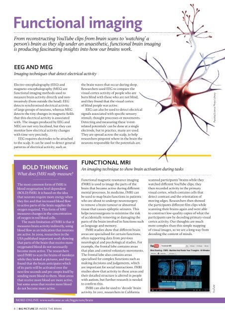
MIND-BLOWING BRAINS How research helps us look inside the brain
MIND-BLOWING BRAINS How research helps us look inside the brain
MIND-BLOWING BRAINS How research helps us look inside the brain

Create successful ePaper yourself
Turn your PDF publications into a flip-book with our unique Google optimized e-Paper software.
Functional imaging<br />
From reconstructing YouTube clips from <strong>brain</strong> scans to ‘watching’ a<br />
person’s <strong>brain</strong> as <strong>the</strong>y slip under an anaes<strong>the</strong>tic, functional <strong>brain</strong> imaging<br />
is producing fascinating insights into how our <strong>brain</strong>s work.<br />
EEG AND MEG<br />
Imaging techniques that detect electrical activity<br />
Electro-encephalography (EEG) and<br />
magneto-encephalography (MEG) are<br />
functional imaging methods <strong>us</strong>ed to<br />
measure <strong>brain</strong> activity directly and noninvasively<br />
(from outside <strong>the</strong> head). EEG<br />
detects synchronised electrical activity<br />
of large groups of neurons, whereas MEG<br />
detects <strong>the</strong> tiny changes in magnetic fields<br />
that this electrical activity is associated<br />
with. The images produced by EEG and<br />
MEG are not very localised, but <strong>the</strong>y can<br />
monitor how electrical activity changes<br />
with time very precisely.<br />
EEG requires electrodes to be attached<br />
to <strong>the</strong> scalp. It can be <strong>us</strong>ed to detect general<br />
patterns of electrical activity, such as<br />
BOLD ThINKING<br />
What does fMRI really measure?<br />
The most common form of fMRI is<br />
blood oxygenation level dependent<br />
(BOLD) fMRI. It is based on <strong>the</strong> idea<br />
that neurons require more energy when<br />
<strong>the</strong>y fire and that increased blood flow<br />
to active parts of <strong>the</strong> <strong>brain</strong> supplies <strong>the</strong><br />
oxygen required. This form of MRI<br />
measures changes in <strong>the</strong> concentration<br />
of oxygen in red blood cells.<br />
The main limitation of fMRI is that it<br />
measures <strong>brain</strong> activity indirectly, <strong>us</strong>ing<br />
blood flow as an indication that neurons<br />
are active. In 2009, <strong>research</strong>ers in <strong>the</strong><br />
USA published important work showing<br />
that parts of <strong>the</strong> <strong>brain</strong> that receive more<br />
oxygenated blood do not necessarily<br />
become more active. The <strong>research</strong>ers<br />
<strong>us</strong>ed fMRI to scan <strong>the</strong> <strong>brain</strong>s of monkeys<br />
while <strong>the</strong>y <strong>look</strong>ed at pictures, and <strong>the</strong>y<br />
found that <strong>the</strong> <strong>brain</strong> anticipates which<br />
of its parts will be activated over <strong>the</strong><br />
next few seconds and pre-empts itself by<br />
sending more blood to <strong>the</strong>m. Most areas<br />
that receive more blood are more active,<br />
but some areas that receive more blood<br />
do not become more active.<br />
MORE ONLINE: www.wellcome.ac.uk/bigpicture/<strong>brain</strong><br />
8 | BIG PICTURE 17: INSIDE THE bRAIN<br />
<strong>the</strong> <strong>brain</strong> waves that occur during sleep.<br />
Researchers <strong>us</strong>ed EEG to compare <strong>the</strong><br />
visual cortex activity of people who are<br />
born blind with those who are not blind,<br />
and <strong>the</strong>y found that <strong>the</strong> visual cortex<br />
of blind people was active.<br />
EEG can also be <strong>us</strong>ed to detect electrical<br />
signals associated with specific sensory<br />
stimuli, thought processes or movements.<br />
Detecting and measuring <strong>the</strong>se ‘eventrelated<br />
potentials’ can be done at a single<br />
electrode, but in practice, many are <strong>us</strong>ed.<br />
They are spread across <strong>the</strong> scalp, to help<br />
<strong>research</strong>ers pinpoint where in <strong>the</strong> <strong>brain</strong> <strong>the</strong><br />
neurons responsible for <strong>the</strong> potentials are.<br />
FUNCTIONAL MRI<br />
An imaging technique to show <strong>brain</strong> activation during tasks<br />
Functional magnetic resonance imaging<br />
(fMRI) is <strong>us</strong>ed to image <strong>the</strong> parts of <strong>the</strong><br />
<strong>brain</strong> that become active during different<br />
mental processes. In medicine, fMRI can<br />
be <strong>us</strong>ed to map <strong>brain</strong> functions in patients<br />
who are about to undergo neurosurgery<br />
to remove a <strong>brain</strong> tumour or abnormal<br />
tissue that ca<strong>us</strong>es epileptic seizures. This<br />
<strong>helps</strong> neurosurgeons to minimise <strong>the</strong> risk<br />
of accidentally removing or damaging <strong>the</strong><br />
parts of <strong>the</strong> <strong>brain</strong> involved in functions such<br />
as language and memory.<br />
fMRI studies show that different <strong>brain</strong><br />
areas are specialised for certain functions,<br />
often supporting data from previo<strong>us</strong><br />
neurological and psychological studies. For<br />
example, <strong>the</strong> frontal lobe contains areas<br />
that plan and control voluntary movements.<br />
The frontal lobe also contains areas<br />
specialised for complex functions such as<br />
making decisions and judgements, which<br />
are important for social interactions. fMRI<br />
studies show that activity in <strong>the</strong>se areas and<br />
<strong>the</strong>ir detailed structure is altered in people<br />
with autism, but fur<strong>the</strong>r <strong>research</strong> is needed<br />
to confirm this.<br />
fMRI can also be <strong>us</strong>ed to ‘decode’ <strong>brain</strong><br />
activity. In 2011, <strong>research</strong>ers in California<br />
scanned participants’ <strong>brain</strong>s while <strong>the</strong>y<br />
watched different YouTube clips; <strong>the</strong>y<br />
<strong>the</strong>n recorded activity in <strong>the</strong> primary<br />
visual cortex, which contains cells that<br />
detect contrast and <strong>the</strong> orientation of<br />
moving edges. Researchers <strong>the</strong>n showed<br />
<strong>the</strong> participants different film clips while<br />
scanning <strong>the</strong>ir <strong>brain</strong>s again and were able<br />
to construct low-quality copies of what <strong>the</strong><br />
participants saw by decoding primary visual<br />
cortex activity. Our thoughts are much<br />
more complex than this simple mapping<br />
of visual images, so we are a long way from<br />
decoding <strong>the</strong> content of minds.<br />
YouTube Pasieka/Science Photo Library