Comparison of UVA protection afforded by high sun protection factor ...
Comparison of UVA protection afforded by high sun protection factor ...
Comparison of UVA protection afforded by high sun protection factor ...

Create successful ePaper yourself
Turn your PDF publications into a flip-book with our unique Google optimized e-Paper software.
Ultraviolet A (<strong>UVA</strong>, 320-400 nm) makes up the<br />
major portion <strong>of</strong> ultraviolet radiation reaching<br />
the surface <strong>of</strong> the earth. <strong>UVA</strong> has been<br />
shown to play a role in skin carcinogenesis, photodermatosis<br />
induction, and other <strong>sun</strong>-induced skin<br />
diseases. It can induce mutations in cultured cells 1 as<br />
well as squamous cell carcinoma in hairless mice. 2<br />
<strong>UVA</strong> has been recognized to be involved in the genesis<br />
<strong>of</strong> solar elastosis 3 and is the major waveband<br />
responsible for polymorphous light eruption, the<br />
most common <strong>of</strong> the idiopathic photodermatoses. 4<br />
It is therefore relevant to be able to evaluate <strong>UVA</strong><br />
<strong>protection</strong> <strong>afforded</strong> <strong>by</strong> <strong>sun</strong>screens. The <strong>sun</strong> <strong>protection</strong><br />
<strong>factor</strong> (SPF) is a simple and internationally used<br />
method to compare <strong>sun</strong>screen <strong>protection</strong> against UV<br />
erythema, which is predominantly induced <strong>by</strong> UVB.<br />
The SPF can be used as a guide to select a <strong>sun</strong>screen<br />
to avoid <strong>sun</strong>burn. However, there is no universally<br />
accepted method to evaluate <strong>UVA</strong> <strong>protection</strong> <strong>afforded</strong><br />
<strong>by</strong> <strong>sun</strong>screens. UVB/<strong>UVA</strong>-labeled <strong>sun</strong>screens are<br />
thus sold with no indication <strong>of</strong> the level <strong>of</strong> <strong>UVA</strong> <strong>protection</strong><br />
they provide. In this study we used the pigmentation<br />
darkening method to compare <strong>UVA</strong> <strong>protection</strong><br />
<strong>afforded</strong> <strong>by</strong> 6 commercially available <strong>sun</strong>screens<br />
with an SPF <strong>of</strong> 20 or more that claimed on<br />
their label to <strong>of</strong>fer <strong>UVA</strong> and UVB <strong>protection</strong>.<br />
From the Division <strong>of</strong> Dermatology, University <strong>of</strong> Montreal Hospital<br />
Centre, Montreal, a and L’Oréal Research, Clichy. b<br />
Supported <strong>by</strong> a grant from La Roche-Posay Pharmaceutical<br />
Laboratories.<br />
Accepted for publication May 24, 2000.<br />
Reprint requests: Robert Bissonnette, MD, MSc, FRCPC, Division <strong>of</strong><br />
Dermatology, University <strong>of</strong> Montreal Hospital Centre, Notre-<br />
Dame Hospital, 1560 Sherbrooke St East, Montreal, Quebec,<br />
Canada H2L 4M1. E-mail: rbissonn@globetrotter.qc.ca.<br />
Copyright © 2000 <strong>by</strong> the American Academy <strong>of</strong> Dermatology, Inc.<br />
0190-9622/2000/$12.00 + 0 16/1/109299<br />
doi:10.1067/mjd.2000.109299<br />
1036<br />
<strong>Comparison</strong> <strong>of</strong> <strong>UVA</strong> <strong>protection</strong> <strong>afforded</strong> <strong>by</strong> <strong>high</strong><br />
<strong>sun</strong> <strong>protection</strong> <strong>factor</strong> <strong>sun</strong>screens<br />
Robert Bissonnette, MD, MSc, FRCPC, a Soraya Allas, MD, PhD, a Dominique Moyal, PhD, b<br />
and Nathalie Provost, MD, FRCPC a Montreal, Québec, Canada, and Clichy, France<br />
<strong>UVA</strong> <strong>protection</strong> <strong>afforded</strong> <strong>by</strong> 6 different <strong>sun</strong>screens with a <strong>sun</strong> <strong>protection</strong> <strong>factor</strong> <strong>of</strong> 21 or more was compared<br />
<strong>by</strong> means <strong>of</strong> the persistent pigmentation darkening method. Colorimetric and visual assessment showed<br />
significant differences in UV radiation–induced pigmentation at 2 hours. The labeled <strong>sun</strong> <strong>protection</strong> <strong>factor</strong><br />
<strong>of</strong> the tested <strong>sun</strong>screens was not predictive <strong>of</strong> <strong>UVA</strong> <strong>protection</strong> level. (J Am Acad Dermatol 2000;43:1036-8.)<br />
METHODS<br />
After ethics board approval, informed consent<br />
was obtained from 12 volunteers with skin phototype<br />
III or IV and with no history <strong>of</strong> <strong>sun</strong> or artificial<br />
light exposure on their back for at least 3 months.<br />
Volunteers who were taking photosensitizing medication<br />
or who had a history <strong>of</strong> lupus, porphyria, or<br />
another light-sensitive dermatosis were excluded.<br />
Six commercially available <strong>sun</strong>screens with an SPF <strong>of</strong><br />
21 or <strong>high</strong>er were studied (Table I). Sunscreens were<br />
selected among frequently used brands to include 2<br />
<strong>sun</strong>screens with physical agents only, 2 <strong>sun</strong>screens<br />
with at least Parsol 1789 as a chemical agent for <strong>UVA</strong><br />
<strong>protection</strong>, and 2 <strong>sun</strong>screens with chemical agents<br />
but without Parsol 1789 and Mexoryl SX. Three <strong>of</strong><br />
the 6 <strong>sun</strong>screens were applied at a rate <strong>of</strong> 2 mg/cm 2<br />
on the back <strong>of</strong> each volunteer in a double-blind fashion<br />
so that each <strong>sun</strong>screen was applied on exactly 6<br />
volunteers. After 15 minutes the back <strong>of</strong> each volunteer<br />
was exposed to UV light generated <strong>by</strong> a metal<br />
halide lamp (<strong>UVA</strong> spot 400/T, Dr K Hönle UV-<br />
Technologie, Munich, Germany). For each <strong>sun</strong>screen,<br />
five 1.5 × 1.5 cm squares <strong>of</strong> normal skin on<br />
the back were exposed to one <strong>of</strong> the following <strong>UVA</strong><br />
doses: 20, 29, 44, 67, and 100 J/cm 2 . The ultraviolet<br />
spectral output <strong>of</strong> this source includes both <strong>UVA</strong> and<br />
UVB (290-400 nm) and is similar to the spectral output<br />
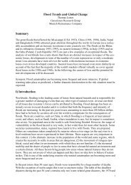
<strong>of</strong> the <strong>sun</strong> (Fig 1). Irradiance was measured with<br />
a radiometer (Centra Osram, Berlin, Germany)<br />
equipped with <strong>UVA</strong> and UVB detectors and was 20<br />
mW/cm 2 for <strong>UVA</strong> and 0.55 mW/cm 2 for UVB. Two<br />
hours after UV exposure, the pigmentation <strong>of</strong> the<br />
exposed sites was compared visually with adjacent<br />
normal skin and graded on a scale <strong>of</strong> 0 to 4, where 0<br />
meant no difference from adjacent skin and 4 meant<br />
marked pigmentation. Pigmentation intensity was<br />
also measured with a colorimeter (Minolta CR200) in<br />
the L*a*b mode. Measurements were taken in tripli-
J AM ACAD DERMATOL<br />
VOLUME 43, NUMBER 6<br />
Fig 1. Spectral irradiance <strong>of</strong> the metal halide lamp used in this study (dotted line) as measured<br />
with a spectroradiometer (model SR3010-PC, Macam Photometrics, Livingston, UK) and compared<br />
with a zenithal standard <strong>sun</strong> spectrum (DIN 67501) (solid line).<br />
cate from each exposed site as well as from adjacent<br />
normal skin. The pigmentation intensity <strong>of</strong> each<br />
square was calculated <strong>by</strong> subtracting the mean L<br />
(luminance) value <strong>of</strong> the square <strong>by</strong> the mean L value<br />
<strong>of</strong> adjacent skin. Mean pigmentation for both the<br />
visual and colorimetric method was calculated for<br />
each <strong>sun</strong>screen at each <strong>of</strong> the 5 <strong>UVA</strong> exposure intensity<br />
and compared using an analysis <strong>of</strong> variance for<br />
repeated measures method.<br />
RESULTS<br />
Two hours after exposure, intense pigmentation<br />
could be seen on some <strong>of</strong> the sites exposed to UV<br />
radiation. For visual assessment differences were<br />
observed between the various <strong>sun</strong>screens at 20<br />
J/cm 2 , but they were not statistically significant (Fig<br />
2, A). At a dose <strong>of</strong> 100 J/cm 2 , pigmentation intensity<br />
was lowest for <strong>sun</strong>screen A, followed in order <strong>of</strong><br />
increasing pigmentation <strong>by</strong> <strong>sun</strong>screens D, F, E, C,<br />
and B (Fig 2, A). The difference in visual pigmentation<br />
between <strong>sun</strong>screen A and all other <strong>sun</strong>screens<br />
at 100 J/cm 2 was statistically significant (P < .001).<br />
For colorimetric analysis the pigmentation was lowest<br />
for <strong>sun</strong>screen A followed in order <strong>of</strong> increasing<br />
pigmentation <strong>by</strong> <strong>sun</strong>screens D, F, E, C, and B (Fig 2,<br />
B). The difference in pigmentation intensity at 100<br />
J/cm 2 between <strong>sun</strong>screen A and all other <strong>sun</strong>screens,<br />
as measured with the colorimeter, was statistically<br />
significant (P < .017) except for <strong>sun</strong>screen D.<br />
DISCUSSION<br />
Several techniques have been used to compare <strong>UVA</strong><br />
<strong>protection</strong> <strong>afforded</strong> <strong>by</strong> <strong>sun</strong>screens including assess-<br />
A<br />
B<br />
Bissonnette et al 1037<br />
Fig 2. Pigmentation intensity according to <strong>UVA</strong> doses<br />
assessed with the visual (A) and colorimetric (B) method<br />
2 hours after exposure to UV radiation. For colorimetric<br />
measurements the luminance (L) <strong>of</strong> the UV-exposed skin<br />
was subtracted from the luminance <strong>of</strong> the adjacent covered<br />
skin.
1038 Bissonnette et al J AM ACAD DERMATOL<br />
DECEMBER 2000<br />
Table I. Active <strong>sun</strong>screening agents <strong>of</strong> <strong>sun</strong>screens<br />
studied<br />
Sunscreen Chemical agent Physical agent<br />
A Mexoryl SX 3.3% Titanium<br />
4-Methylbenzylidene dioxide 4.1%<br />
camphor 5.0%<br />
Parsol 1789 (butylmethoxydibenzoylmethane)<br />
3.5%<br />
B Octyl methoxycinnamate Titanium<br />
7.5% dioxide<br />
Octyl salicylate 5.0%<br />
C Homosalate 8.0%<br />
Ethythexyl p-methoxycinnamate<br />
7.5%<br />
Oxybenzone 6.0%<br />
Octyl salicylate 5.0%<br />
D Parsol 1789 (butyl methoxydibenzoylmethane)<br />
3.0%<br />
Octyl methoxycinnamate<br />
7.5%<br />
Octyl salicylate 5.0%<br />
Oxybenzone 3.0%<br />
E — Titanium<br />
dioxide 9.6%<br />
Zinc oxide 1.5%<br />
F — Titanium<br />
dioxide<br />
12.0%<br />
ment <strong>of</strong> immediate pigmentation darkening (IPD), 5<br />
persistent pigmentation darkening (PPD) at 2 to 24<br />
hours, 6 <strong>UVA</strong>-induced erythema, 7 erythema induced<br />
after topical psoralens application and <strong>UVA</strong> exposure, 8<br />
and in vitro spectroscopic methods. 9 The action spectrum<br />
<strong>of</strong> PPD is maximum at short <strong>UVA</strong> wavelengths<br />
and gradually decreases from 320 to 400 nm. 10,11 PPD<br />
response has been shown not to be influenced <strong>by</strong> <strong>UVA</strong><br />
irradiance, as opposed to IPD response, suggesting<br />
that PPD may be a better method to compare <strong>UVA</strong> <strong>protection</strong><br />
<strong>afforded</strong> <strong>by</strong> <strong>sun</strong>screens. 10 At 100 J/cm 2 , the<br />
maximal <strong>UVA</strong> dose applied in this study, photostability<br />
was indirectly studied with the PPD method, as photodegradation<br />
<strong>of</strong> <strong>sun</strong>screens occurs after only 18 J/cm 2<br />
<strong>of</strong> UV exposure. 9<br />
Sunscreen A, which induced the lowest pigmentation<br />
intensity, contained Parsol 1789, Mexoryl SX,<br />
and titanium dioxide for <strong>UVA</strong> <strong>protection</strong>. The second<br />
best, <strong>sun</strong>screen D, was the only other <strong>sun</strong>screen<br />
with Parsol 1789 and was followed <strong>by</strong> the 2 physical<br />
<strong>sun</strong>screens and the 2 <strong>sun</strong>screens with only benzophenones<br />
for <strong>UVA</strong> <strong>protection</strong>. The <strong>sun</strong>screens<br />
that protected the least against <strong>UVA</strong>-induced pigmentation<br />
were the <strong>sun</strong>screens with the second and<br />
third <strong>high</strong>est SPF (45 and 50), showing that selecting<br />
a <strong>high</strong> SPF <strong>sun</strong>screen cannot be used as the only<br />
guide to compare <strong>UVA</strong> <strong>protection</strong> <strong>afforded</strong> <strong>by</strong> <strong>sun</strong>screens.<br />
Although the visual and colorimetric assessments<br />
rated <strong>UVA</strong> pigmentation <strong>of</strong> the 6 tested <strong>sun</strong>screens<br />
in the same order, there were differences in<br />
the shape <strong>of</strong> the pigmentation curves for a given<br />
<strong>sun</strong>screen. These differences and the negative values<br />
observed at 20 J/cm 2 with colorimetry could be related<br />
to the precision limit <strong>of</strong> the technique as well as<br />
the difficulty <strong>of</strong> performing colorimetric measurements<br />
on a small skin area with our colorimeter.<br />
In conclusion, labeled SPF is not predictive <strong>of</strong> <strong>UVA</strong><br />
<strong>protection</strong> as assessed <strong>by</strong> pigmentation induced at 2<br />
hours after exposure to UV radiation. In addition to<br />
SPF, another labeling method to specifically compare<br />
<strong>UVA</strong> <strong>protection</strong> could help in <strong>sun</strong>screen selection.<br />
REFERENCES<br />
1. Drobetsky EA, Turcotte J, Chateauneuf A. A role for ultraviolet A<br />
in solar mutagenesis. Proc Natl Acad Sci 1995;92:2350-4.<br />
2. de Gruijl FR, Sterenborg HJ, Forbes PD, Davies RE, Cole C,<br />
Kelfkens G, et al.Wavelength dependence <strong>of</strong> skin cancer induction<br />
<strong>by</strong> ultraviolet irradiation <strong>of</strong> albino hairless mice. Cancer Res<br />
1993;53:53-60.<br />
3. Fourtanier A, Labat-Robert J, Kern P, Berrebi C, Gracia AM, Boyer<br />
B. In vivo evaluation <strong>of</strong> photo<strong>protection</strong> against chronic ultraviolet-A<br />
irradiation <strong>by</strong> a new <strong>sun</strong>screen Mexoryl SX. Photochem<br />
Photobiol 1992;55:549-60.<br />
4. Neumann RA, Pohl-Markl H, Knobler RM. Polymorphous light<br />
eruption: experimental reproduction <strong>of</strong> skin lesions <strong>by</strong> wholebody<br />
<strong>UVA</strong> irradiation. Photodermatology 1987;4:252-6.<br />
5. Kaidbey KH, Barnes A. Determination <strong>of</strong> <strong>UVA</strong> <strong>protection</strong> <strong>factor</strong>s<br />
<strong>by</strong> means <strong>of</strong> immediate pigment darkening in normal skin. J<br />
Am Acad Dermatol 1991;25:262-6.<br />
6. Roelandts R, Sohrabvand N, Garmyn M. Evaluating the <strong>UVA</strong> <strong>protection</strong><br />
<strong>of</strong> <strong>sun</strong>screens. J Am Acad Dermatol 1989;21:56-62.<br />
7. Cole C, VanFossen R. Measurement <strong>of</strong> <strong>sun</strong>screen <strong>UVA</strong> <strong>protection</strong>:<br />
an unsensitized human model. J Am Acad Dermatol 1992;<br />
26:178-84.<br />
8. Lowe NJ, Dromgoole SH, Sefton J, Bourget T, Weingarten D.<br />
Indoor and outdoor efficacy testing <strong>of</strong> a broad-spectrum <strong>sun</strong>screen<br />
against ultraviolet A radiation in psoralen-sensitized<br />
subjects. J Am Acad Dermatol 1987;17:224-30.<br />
9. Diffey BL SR, Forestier S, Mazilier C, Rougier A. Suncare product<br />
photostability: a key parameter for a more realistic in vitro efficacy<br />
evaluation. Part I: in vitro efficacy assessment. Eur J<br />
Dermatol 1997;7:226-8.<br />
10. Chardon A, Moyal D, Hourseau C. Persistent pigment darkening<br />
response as a method for evaluation <strong>of</strong> <strong>UVA</strong> <strong>protection</strong> assays.<br />
In: Lowe NJ, Shaath N, Pathak M, editors. Sunscreens: development,<br />
evaluation and regulatory aspects. New York: Marcel<br />
Dekker; 1994. p. 559-82.<br />
11. Irwin C, Barnes A, Veres D, Kaidbey K. An ultraviolet radiation<br />
action spectrum for immediate pigment darkening. Photochem<br />
Photobiol 1993;57:504-7.