Anisolpidium saprobium and Rhizidiomyces hirsutus, new records of ...
Anisolpidium saprobium and Rhizidiomyces hirsutus, new records of ...
Anisolpidium saprobium and Rhizidiomyces hirsutus, new records of ...

You also want an ePaper? Increase the reach of your titles
YUMPU automatically turns print PDFs into web optimized ePapers that Google loves.
Fung. Sci. 22(3, 4): 79–83, 2007<br />
<strong>Anisolpidium</strong> <strong>saprobium</strong> <strong>and</strong> <strong>Rhizidiomyces</strong> <strong>hirsutus</strong>,<br />
<strong>new</strong> <strong>records</strong> <strong>of</strong> Hyphochytriomycetes<br />
(Hyphochytriales) in Taiwan<br />
Shu-Fen Chen<br />
Department <strong>of</strong> Health <strong>and</strong> Nutrition, Chia Nan University <strong>of</strong> Pharmacy <strong>and</strong> Science, Tainan 71710, Taiwan<br />
(Accepted: December 17, 2007)<br />
ABSTRACT<br />
<strong>Anisolpidium</strong> <strong>saprobium</strong> Karling <strong>and</strong> <strong>Rhizidiomyces</strong> <strong>hirsutus</strong> Karling are species <strong>of</strong> Hyphochytriales. These are<br />
first time reported from Taiwan.<br />
Keywords: <strong>Anisolpidium</strong>, Hyphochytriomycetes (Hyphochytriales), <strong>Rhizidiomyces</strong>.<br />
Introduction<br />
Hyphochytriomycetes (Hyphochytriales) is a<br />
group chytrid-like organisms with anteriorly<br />
uniflagellate zoospores <strong>and</strong> contains only about<br />
23 known species. Fuller (1990) included them<br />
in the class Hyphochytriomycetes, phylum Hyphochytriomycota.<br />
According to Berbee <strong>and</strong><br />
Taylor (1999) the Hyphochytriomycota, Labyrinthulomycota<br />
<strong>and</strong> Oomycota belong to the<br />
Kingdom Stramenopila. Hyphochytriomycota,<br />
consisting <strong>of</strong> a single order Hyphochytriales<br />
which have been classified into three families,<br />
namely Anisolpidiaceae, Rhizidiomycetaceae<br />
<strong>and</strong> Hyphochytriaceae on the basis <strong>of</strong> thallus<br />
structure <strong>and</strong> development (Karling, 1943,<br />
1967; Fuller, 1990; Alexopoulos et al, 1996).<br />
The purpose <strong>of</strong> this paper is to describe <strong>and</strong> illustrate<br />
<strong>Anisolpidium</strong> <strong>saprobium</strong> <strong>and</strong> <strong>Rhizidiomyces</strong><br />
<strong>hirsutus</strong> as two species <strong>new</strong>ly recorded<br />
in Taiwan.<br />
Materiales <strong>and</strong> Methods<br />
Samples <strong>of</strong> water <strong>and</strong> soil were baited with<br />
pine pollen. Emerson’s 1/4YpSs agar (containing<br />
antibiotics) was used to isolate <strong>and</strong> culture<br />
the organisms (Chen <strong>and</strong> Chien, 1998). Pure<br />
cultures were maintained in 1/4YpSs slush in<br />
screw-cap tube <strong>and</strong> transfered each three<br />
months. All pure cultures were deposited at the<br />
mycological laboratory in Chia Nan University<br />
<strong>of</strong> Pharmacy <strong>and</strong> Science, Tainan, Taiwan.<br />
Developmental stages <strong>and</strong> morphological<br />
characters were examined using the light microscope<br />
<strong>and</strong> scanning electron microcope<br />
(ABT DS-130S).<br />
Taxonomy<br />
<strong>Anisolpidium</strong> <strong>saprobium</strong> Karling, J. <strong>of</strong><br />
Mitchell Society, 84: 166–178, 1968. (Fig.1)<br />
On pine pollen: Sporangium holocarpic, en-
80 Fung. Sci. 22(3, 4), 2007<br />
dobiotic, spherical, 25–32 µm in diam, with a<br />
long discharge tube extending out pollen.<br />
In 1/4YpSs slush: Sporangium spherical,<br />
17.5–30 µm in diam, sometimes with absorbing<br />
tubes. Protoplasm oozing from an exit pore, 5–<br />
10 µm in diam, or exit tube, 25–60 µm in<br />
length, the tube begins to enlarge at its tip as the<br />
protoplasm move into it. Zoospores delimited<br />
within or outside <strong>of</strong> sporangium. Zoospores<br />
fusiform or elongate, 2.5–4 × 5–7.5 µm, with<br />
anteriorly uniflugellum.<br />
On 1/4YpSs agar: Color <strong>of</strong> colony white.<br />
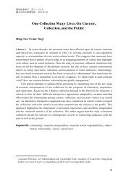
Fig. 1. <strong>Anisolpidium</strong> <strong>saprobium</strong>. A–E: in 1/4YpSs slush. F & G: on 1/4YpSs agar, scanning electron microscopy. A. Incipient<br />
holocarpic thalli. B. Some sporangium with absorbing tube. C. Three exit tubes enlarge at tip. D. Sporangium with one<br />
exit canal <strong>and</strong> some absorbing tubes. E. An empty sporangium (left), zoospores delimiting within <strong>and</strong> outside the other sporangium.<br />
F. Three holocarpic thalli. G. One sporangium with exit tube; <strong>and</strong> an escaping zoospore (arrow) on nearly sporangium.<br />
Bars = 10 µm for A–E, 2.0 µm for F, <strong>and</strong> 3.33 µm for G.
Specimen examined. Yulin: farm soil, 22<br />
Jul. 1992, CSF S02a; Tainan: Paiho, pond water,<br />
11 Jul. 1998, CHNA 3801a. Isolated on pine<br />
pollen from soil <strong>and</strong> water.<br />
Notes. Our isolates include soil-inhabiting<br />
<strong>and</strong> aquatic strains. <strong>Anisolpidium</strong> <strong>saprobium</strong> is<br />
the first known saprophytic species <strong>of</strong> the ge-<br />
New <strong>records</strong> <strong>of</strong> Hyphochytriomycetes in Taiwan 81<br />
nus. However, all other known species are parasited<br />
<strong>of</strong> algae (Karling, 1943). The place <strong>of</strong><br />
zoospores cleavage in this species is varied<br />
markedly (Fig. 1E).<br />
<strong>Rhizidiomyces</strong> <strong>hirsutus</strong> Karling, Bull. Torrey<br />
Bot. Club, 72: 47, 1945. (Fig. 2)<br />
Fig. 2. <strong>Rhizidiomyces</strong> <strong>hirsutus</strong>. In 1/4YpSs slush. A. Young thalli with several rhizoids around the sporangium. B. Sporangium<br />
with several rhizoids <strong>and</strong> hair-like setae. C. Sporangium with a long exit canal. D. Sporangium with one simple rhizoid<br />
<strong>and</strong> two long hairs. E. Tubular, branched thalli. Bars = 10 µm.
82 Fung. Sci. 22(3, 4), 2007<br />
In 1/4YpSs slush: Sporangium spherical, 40–<br />
128 µm in diam, one to several short, tubular<br />
rhizoidal main axis arising from the peripheral<br />
<strong>of</strong> sporangium. On surface <strong>of</strong> mature sporangium<br />
wall with 2–18 or more hair-like setae, up<br />
to 450 µm in length. Zoospore spherical or<br />
elongate, 3–4 × 6–8 µm, with an anteriorly flagellum.<br />
On 1/4YpSs agar: Color <strong>of</strong> colony white.<br />
Specimens examined. Tainan: Kwantien,<br />
water-caltrop pond, 14 Jun. 1998, CHNA 3701f.<br />
Yilan: Tung-Shan River, water, 28 Sep. 2000,<br />
CHNA 5001a; Fushan Botanical Garden, stream<br />
water, 29 Sep. 2000, CHNA 5201a. Isolated on<br />
pine pollen from all water samples.<br />
Notes. Monocentric, eucarpic, sporangium is<br />
the characterics <strong>of</strong> Rhizidiomycetaceae (Karling,<br />
1977). Some thalli develope into elongate,<br />
simple or branched tubular structures. Many<br />
variations <strong>of</strong> thallus structure occurs in axenic<br />
culture, it is the same as Karling’s described<br />
(1968). Mature sporangial wall with several<br />
elongate hairs-like setae is the main character <strong>of</strong><br />
this species (Fig. 2B).<br />
Acknowledgements<br />
This study was partly supported by the Council<br />
<strong>of</strong> Agriculture, Taipei, Taiwan, R.O.C., grant<br />
number 89-AST-1.5-FOD-05(5-9-2).<br />
References<br />
Alexopoulos, C.J., C.W. Mims, <strong>and</strong> M. Blackwell.<br />
1996. Introductory Mycology, 4th ed.,<br />
John Wiley <strong>and</strong> Sons, Inc.<br />
Berbee M.L. <strong>and</strong> J.W. Taylor. 1999. Fungal<br />
pgylogeny. Pp. 21–77. In: Molecular Fungal<br />
Biology. Eds., R.P. Oliver <strong>and</strong> M. Schweizer.<br />
Cambridge University Press.<br />
Chen, S.F. <strong>and</strong> C.Y. Chien. 1998. Some chytrids<br />
<strong>of</strong> Taiwan (II). Bot. Bull. Acad. Sin. 39: 47–<br />
56.<br />
Fuller, M.S. 1990. Phylum Hyphochytriomycota.<br />
Pp. 380–287. In: H<strong>and</strong>book <strong>of</strong> Protoctista.<br />
Eds., L. Margulis, J.O. Corliss, M.<br />
Melkonian, <strong>and</strong> D.J. Chapman. John <strong>and</strong><br />
Bartlett, Boston, MA.<br />
Karling, J.S. 1943. The life history <strong>of</strong> <strong>Anisolpidium</strong><br />
ectocarpii gen. nov. et sp. nov., <strong>and</strong> a<br />
synopsis <strong>and</strong> classification <strong>of</strong> other fungi<br />
with anteriorly uniflagellate zoospores.<br />
Amer. J. Bot. 30: 637–648.<br />
Karling, J.S. 1967. Some zoosporic fungi <strong>of</strong><br />
New Zeal<strong>and</strong>. IX. Hyphochytriales or Anisochytridiales.<br />
Sydowia 20: 137–143.<br />
Karling, J.S. 1968. Zoosporic fungi <strong>of</strong> Oceania<br />
I. J. <strong>of</strong> Mitchell Soc. 84: 166–178.<br />
Karling, J.S. 1977. Chytridiomycetarum<br />
Iconorgraphia. Luberecht I Cramer. Monticello,<br />
New York, 414 pp.
New <strong>records</strong> <strong>of</strong> Hyphochytriomycetes in Taiwan 83<br />
臺灣產腐生異壺菌及粗毛根前毛菌二種新紀錄<br />
陳 淑 芬<br />
嘉南藥理科技大學保健營養系,臺南縣仁德鄉二仁路一段 60 號,臺灣<br />
摘 要<br />
本文描述二種具有前生單鞭毛游孢子的絲壺菌,分別是:腐生異壺菌 (<strong>Anisolpidium</strong> <strong>saprobium</strong> Karling)<br />
及粗毛根前毛菌 (<strong>Rhizidiomyces</strong> <strong>hirsutus</strong> Karling),均為臺灣新記錄。<br />
關鍵詞:根前毛菌屬、異壺菌屬、絲壺菌綱 (絲壺菌目)。
84 Fung. Sci. 22(3, 4), 2007