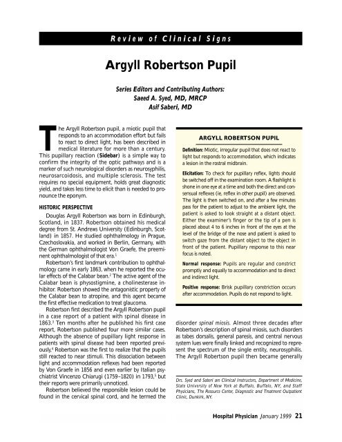
Argyll Robertson Pupil - Turner White Communications
Argyll Robertson Pupil - Turner White Communications
Argyll Robertson Pupil - Turner White Communications

Create successful ePaper yourself
Turn your PDF publications into a flip-book with our unique Google optimized e-Paper software.
The <strong>Argyll</strong> <strong>Robertson</strong> pupil, a miotic pupil that<br />
responds to an accommodation effort but fails<br />
to react to direct light, has been described in<br />
medical literature for more than a century.<br />
This pupillary reaction (Sidebar) is a simple way to<br />
confirm the integrity of the optic pathways and is a<br />
marker of such neurological disorders as neurosyphilis,<br />
neurosarcoidosis, and multiple sclerosis. The test<br />
requires no special equipment, holds great diagnostic<br />
yield, and takes less time to elicit than is needed to pronounce<br />
the eponym.<br />
HISTORIC PERSPECTIVE<br />
Douglas <strong>Argyll</strong> <strong>Robertson</strong> was born in Edinburgh,<br />
Scotland, in 1837. <strong>Robertson</strong> obtained his medical<br />
degree from St. Andrews University (Edinburgh, Scotland)<br />
in 1857. He studied ophthalmology in Prague,<br />
Czechoslovakia, and worked in Berlin, Germany, with<br />
the German ophthalmologist Von Graefe, the preeminent<br />
ophthalmologist of that era. 1<br />
<strong>Robertson</strong>’s first landmark contribution to ophthalmology<br />
came in early 1863, when he reported the ocular<br />
effects of the Calabar bean. 2 The active agent of the<br />
Calabar bean is physostigmine, a cholinesterase inhibitor.<br />
<strong>Robertson</strong> showed the antagonistic property of<br />
the Calabar bean to atropine, and this agent became<br />
the first effective medication to treat glaucoma.<br />
<strong>Robertson</strong> first described the <strong>Argyll</strong> <strong>Robertson</strong> pupil<br />
in a case report of a patient with spinal disease in<br />
1863. 3 Ten months after he published his first case<br />
report, <strong>Robertson</strong> published four more similar cases.<br />
Although the absence of pupillary light response in<br />
patients with spinal disease had been reported previously,<br />
4 <strong>Robertson</strong> was the first to realize that the pupils<br />
still reacted to near stimuli. This dissociation between<br />
light and accommodation reflexes had been reported<br />
by Von Graefe in 1856 and even earlier by Italian psychiatrist<br />
Vincenzo Chiarugi (1759–1820) in 1793, 5 but<br />
their reports were primarily unnoticed.<br />
<strong>Robertson</strong> believed the responsible lesion could be<br />
found in the cervical spinal cord, and he termed the<br />
Review of Clinical Signs<br />
<strong>Argyll</strong> <strong>Robertson</strong> <strong>Pupil</strong><br />
Series Editors and Contributing Authors:<br />
Saeed A. Syed, MD, MRCP<br />
Asif Saberi, MD<br />
ARGYLL ROBERTSON PUPIL<br />
Definition: Miotic, irregular pupil that does not react to<br />
light but responds to accommodation, which indicates<br />
a lesion in the rostral midbrain.<br />
Elicitation: To check for pupillary reflex, lights should<br />
be switched off in the examination room. A flashlight is<br />
shone in one eye at a time and both the direct and consensual<br />
reflexes (ie, reflex in other pupil) are observed.<br />
The light is then switched on, and after a few minutes<br />
pass for the patient to adjust to the ambient light, the<br />
patient is asked to look straight at a distant object.<br />
Either the examiner’s finger or the tip of a pen is<br />
placed about 4 to 6 inches in front of the eyes at the<br />
level of the bridge of the nose and patient is asked to<br />
switch gaze from the distant object to the object in<br />
front of the patient. <strong>Pupil</strong>lary response to this near<br />
focus is noted.<br />
Normal response: <strong>Pupil</strong>s are regular and constrict<br />
promptly and equally to accommodation and to direct<br />
and indirect light.<br />
Positive response: Brisk pupillary constriction occurs<br />
after accommodation. <strong>Pupil</strong>s do not respond to light.<br />
disorder spinal miosis. Almost three decades after<br />
<strong>Robertson</strong>’s description of spinal miosis, such disorders<br />
as tabes dorsalis, general paresis, and central nervous<br />
system lues were finally linked and recognized to represent<br />
the spectrum of the single entity, neurosyphilis.<br />
The <strong>Argyll</strong> <strong>Robertson</strong> pupil then became generally<br />
Drs. Syed and Saberi are Clinical Instructors, Department of Medicine,<br />
State University of New York at Buffalo, Buffalo, NY, and Staff<br />
Physicians, The Resource Center, Diagnostic and Treatment Outpatient<br />
Clinic, Dunkirk, NY.<br />
Hospital Physician January 1999 21
Syed & Saberi : <strong>Argyll</strong> <strong>Robertson</strong> <strong>Pupil</strong> : pp. 21–22<br />
accepted as a pathognomonic sign of neurosyphilis,<br />
and this sign has since been identified with other central<br />
nervous system diseases as well.<br />
PHYSIOLOGY<br />
Light Reflex<br />
From an inverted image on the light-sensitive cells<br />
of the retina, impulses pass through the optic nerve to<br />
the optic tract via the optic chiasma. The fibers partially<br />
cross at the chiasma. The optic tracts then pass to the<br />
lateral geniculate body; some fibers pass to the midbrain<br />
as the afferent limb of the pupillary light reflex.<br />
From the midbrain, information is relayed to the parasympathetic<br />
preganglionic neurons located in the<br />
Edinger-Westphal nucleus. The efferent fibers from<br />
the Edinger-Westphal nucleus pass through the ciliary<br />
ganglion and supply the sphincter pupillae muscles.<br />
The partial crossing of the optic nerve fibers at the<br />
optic chiasma explains the consensual reflex. Also,<br />
optic tract fibers pass through the pretectal nuclei and<br />
are then redistributed to both sides.<br />
Accommodation Reflex<br />
A blurred image of a near object is formed on the<br />
visual cortex. Cortical connections are then relayed<br />
through the frontal cortex to the oculomotor nerves<br />
that control the medial rectus muscles, causing the eye<br />
to converge. Fibers from the visual cortex are also<br />
relayed through the temporal lobe to efferent parasympathetic<br />
fibers originating in the ciliary ganglion. This<br />
causes ciliary muscle contraction, which increases lens<br />
convexity and brings the near object into focus on the<br />
central retina. Concomitantly, pupillary constriction<br />
occurs, mediated by oculomotor parasympathetic outflow,<br />
which enhances optic resolution.<br />
Pathophysiology<br />
In an <strong>Argyll</strong> <strong>Robertson</strong> pupil, the pupil’s better<br />
response to accommodation than to light stimuli<br />
occurs because the lesion involves the more dorsally<br />
located fibers that subserve the pupil’s response to<br />
light. The lesion spares the more ventrally located<br />
fibers that subserve the pupil reaction to near stimuli.<br />
CLINICAL PRESENTATION<br />
Typical <strong>Argyll</strong> <strong>Robertson</strong> pupils are small and irregular<br />
and react to accommodation but not to light.<br />
Initially, the pupil’s response to light may only be sluggish,<br />
but the accommodation reflex is always more pro-<br />
22 Hospital Physician January 1999<br />
nounced and the light reflex eventually disappears.<br />
The exact site of the lesion is debated. The lesion is<br />
generally believed to be in the rostral midbrain proximal<br />
to the oculomotor nuclei. In some patients, magnetic<br />
resonance imaging studies have localized the<br />
lesion to the Edinger-Westphal nuclei.<br />
The <strong>Argyll</strong> <strong>Robertson</strong> pupil has become a rare diagnostic<br />
sign of neurosyphilis. If neurosyphilis is suspected,<br />
examination may reveal clues such as ptosis; ataxia<br />
(positive Romberg’s test); tremors of the mouth,<br />
tongue, outstretched hands, and whole body; diminished<br />
or absent tendon reflexes; or impaired vibratory<br />
and joint position sense. Severe cases of neurosyphilis<br />
may even demonstrate aortic incompetence, gastric crisis,<br />
and Charcot’s arthropathy. Finally, <strong>Argyll</strong> <strong>Robertson</strong><br />
pupils in the patient with neurosyphilis are invariably<br />
bilateral. However, with the declining incidence of neurosyphilis,<br />
the <strong>Argyll</strong> <strong>Robertson</strong> pupil is increasingly<br />
likely to indicate another disorder, such as sarcoidosis,<br />
multiple sclerosis, and, occasionally, diabetes mellitus. 6<br />
DIFFERENTIAL DIAGNOSIS<br />
<strong>Argyll</strong> <strong>Robertson</strong> pupil should not be confused with<br />
Adie’s pupil, which may yield a similar result on cursory<br />
examination. In Adie’s pupil, which is caused by ciliary<br />
ganglion destruction, the reaction to light is absent<br />
or greatly diminished when tested in the routine examination;<br />
however, Adie’s pupil does react slowly with<br />
prolonged maximal stimulation. Furthermore, once<br />
the Adie’s pupil reacts to accommodation, the pupil<br />
tends to remain tonically constricted and dilates very<br />
slowly. HP<br />
REFERENCES<br />
1. Ravin JG: <strong>Argyll</strong> <strong>Robertson</strong>: ‘twas better to be his pupil<br />
than to have his pupil. Ophthalmology 1998;105:867–870.<br />
2. <strong>Robertson</strong> DA: The Calabar bean as a new agent in ophthalmic<br />
medicine. Edinburgh Medical Journal 1863;8:<br />
815–820.<br />
3. <strong>Robertson</strong> DA: On an interesting series of eye symptoms<br />
in a case of spinal disease, with remarks on the action of<br />
belladonna on the iris. Edinburgh Medical Journal 1863;<br />
14:696–708.<br />
4. Rucker CW: Knowledge of the pupillary reactions in <strong>Argyll</strong><br />
<strong>Robertson</strong>’s time. Surv Ophthalmol 1969;14:162–171.<br />
5. Loewenfeld JE: The <strong>Argyll</strong> <strong>Robertson</strong> pupil, 1869–1969.<br />
A critical survey of the literature. Surv Ophthalmol 1969;<br />
14:199–299.<br />
6. Dacso CC, Bortz DL: Significance of the <strong>Argyll</strong> <strong>Robertson</strong><br />
pupil in clinical medicine. Am J Med 1989;86:199–202.<br />
Copyright 1999 by <strong>Turner</strong> <strong>White</strong> <strong>Communications</strong> Inc., Wayne, PA. All rights reserved.