Clinical Signs of Low Back Pain - Turner White
Clinical Signs of Low Back Pain - Turner White
Clinical Signs of Low Back Pain - Turner White

You also want an ePaper? Increase the reach of your titles
YUMPU automatically turns print PDFs into web optimized ePapers that Google loves.
ack pain has been cited as the fifth most common<br />
reason that patients visit a physician. 1<br />
BCommon causes <strong>of</strong> back pain include musculoskeletal<br />
injuries, degenerative disease, herniated<br />
nucleus pulposus, and spinal stenosis. Less common<br />
causes include metastatic cancer, spinal infections,<br />
ankylosing spondylitis, and referred pain from visceral<br />
organs. Although the majority <strong>of</strong> causes <strong>of</strong> back pain<br />
are benign, clinicians must be alert to potential red<br />
flags indicating serious pathology.<br />
HISTORY<br />
A patient’s medical history can provide major clues<br />
to a potential diagnosis. The essential components <strong>of</strong> a<br />
thorough history include onset, location, quality, and<br />
radiation <strong>of</strong> the pain, factors that relieve or aggravate<br />
the pain, and associated symptoms. Mechanical low<br />
back pain is characterized by increased pain with<br />
motion and decreased pain with rest, whereas the pain<br />
<strong>of</strong> nonmechanical low back pain generally occurs at<br />
rest and is less affected by motion. 2<br />
There are 2 important questions to consider when<br />
evaluating a patient with low back pain: (1) Is there a<br />
serious disease, such as metastatic cancer, causing the<br />
pain? (2) Is there any neurologic compromise? Assessment<br />
<strong>of</strong> historical clues, such as accompanying fever,<br />
weight loss, history <strong>of</strong> cancer, nocturnal pain, morning<br />
stiffness, and radicular pain, is also necessary. 2<br />
PHYSICAL EXAMINATION<br />
The patient’s spine should be inspected first for abnormal<br />
curvatures, and the gait should be observed.<br />
The gait is best assessed while the patient walks across<br />
the room, turns around, and then comes back. The<br />
physician should carefully observe the patient’s posture<br />
and movement. The patient should then be observed<br />
in the seated position; patients with localized<br />
pain and muscle spasm may exhibit abnormal posture.<br />
Palpation and percussion <strong>of</strong> the vertebrae should be<br />
performed to elicit localized tenderness. Range <strong>of</strong> motion,<br />
including flexion, extension, and rotation, should<br />
also be assessed. 3<br />
Review <strong>of</strong> <strong>Clinical</strong> <strong>Signs</strong><br />
Series Editor: Bernard Karnath, MD<br />
<strong>Clinical</strong> <strong>Signs</strong> <strong>of</strong> <strong>Low</strong> <strong>Back</strong> <strong>Pain</strong><br />
Bernard Karnath, MD<br />
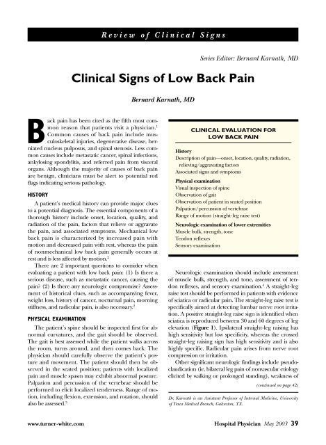
CLINICAL EVALUATION FOR<br />
LOW BACK PAIN<br />
History<br />
Description <strong>of</strong> pain—onset, location, quality, radiation,<br />
relieving/aggravating factors<br />
Associated signs and symptoms<br />
Physical examination<br />
Visual inspection <strong>of</strong> spine<br />
Observation <strong>of</strong> gait<br />
Observation <strong>of</strong> patient in seated position<br />
Palpation/percussion <strong>of</strong> vertebrae<br />
Range <strong>of</strong> motion (straight-leg raise test)<br />
Neurologic examination <strong>of</strong> lower extremities<br />
Muscle bulk, strength, tone<br />
Tendon reflexes<br />
Sensory examination<br />
Neurologic examination should include assessment<br />
<strong>of</strong> muscle bulk, strength, and tone, assessment <strong>of</strong> tendon<br />
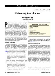
reflexes, and sensory examination. 4 A straight-leg<br />
raise test should be performed in patients with evidence<br />
<strong>of</strong> sciatica or radicular pain. The straight-leg raise test is<br />
specifically aimed at detecting lumbar nerve root irritation.<br />
A positive straight-leg raise sign is identified when<br />
sciatica is reproduced between 30 and 60 degrees <strong>of</strong> leg<br />
elevation (Figure 1). Ipsilateral straight-leg raising has<br />
high sensitivity but low specificity, whereas the crossed<br />
straight-leg raising sign has high sensitivity and is also<br />
highly specific. Radicular pain arises from nerve root<br />
compression or irritation.<br />
Other significant neurologic findings include pseudoclaudication<br />
(ie, bilateral leg pain <strong>of</strong> nonvascular etiology<br />
elicited by walking or prolonged standing), weakness <strong>of</strong><br />
(continued on page 42)<br />
Dr. Karnath is an Assistant Pr<strong>of</strong>essor <strong>of</strong> Internal Medicine, University<br />
<strong>of</strong> Texas Medical Branch, Galveston, TX.<br />
www.turner-white.com Hospital Physician May 2003 39
Karnath : <strong>Clinical</strong> <strong>Signs</strong> <strong>of</strong> <strong>Low</strong> <strong>Back</strong> <strong>Pain</strong> : pp. 39–44, 56<br />
(from page 39)<br />
Table 1. Neurologic Testing for Lumbosacral Nerve Root Compression<br />
Figure 1. Illustration <strong>of</strong> the straight-leg<br />
raise test. (Reprinted with permission<br />
from Judge RD, Woolliscr<strong>of</strong>t JO, Zelenock<br />
GB, Zuidema GD, editors. The<br />
Michigan manual <strong>of</strong> clinical diagnosis:<br />
the basis <strong>of</strong> cost-effective medical practice.<br />
Philadelphia: Lippincott-Raven;<br />
1998:298.)<br />
Nerve Root Intervertebral Space Motor Function Reflex<br />
L4 L3-4 Dorsiflexion <strong>of</strong> foot Knee jerk<br />
L5 L4-5 Dorsiflexion <strong>of</strong> great toe None<br />
S1 L5-S1 Eversion <strong>of</strong> foot and plantar flexion Ankle jerk<br />
NOTE: Protrusion <strong>of</strong> an intervertebral disc is usually posterolateral. A posterolaterally protruded disc may compress the roots<br />
<strong>of</strong> a spinal nerve and usually compresses the next lower nerve root (eg, an L3-4 disc herniation will compress the L4 nerve<br />
root, an L4-L5 disc herniation will compress the L5 nerve root).<br />
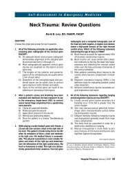
Figure 2. Illustration <strong>of</strong> the dermatomes.<br />
(Available at http://www.chiropage.com/<br />
dermatomes.htm. Reprinted with permission<br />
from Chiropage. Copyright ©1999.)<br />
42 Hospital Physician May 2003 www.turner-white.com
Karnath : <strong>Clinical</strong> <strong>Signs</strong> <strong>of</strong> <strong>Low</strong> <strong>Back</strong> <strong>Pain</strong> : pp. 39–44, 56<br />
Table 2. Differential Diagnosis and Accompanying <strong>Signs</strong> <strong>of</strong> <strong>Low</strong> <strong>Back</strong> <strong>Pain</strong><br />
Type <strong>of</strong> <strong>Low</strong> <strong>Back</strong> <strong>Pain</strong> Quality <strong>of</strong> <strong>Pain</strong> <strong>Signs</strong><br />
Mechanical<br />
<strong>Back</strong> strain<br />
Disk herniation<br />
Osteoarthritis<br />
Spinal stenosis<br />
Spondylolisthesis<br />
Nonmechanical<br />
Ankylosing spondylitis<br />
Infection (abscess,<br />
osteomyelitis, discitis)<br />
Malignancy<br />
Visceral<br />
Nephrolithiasis<br />
Pyelonephritis<br />
Aortic aneurysm<br />
Diseases <strong>of</strong> the pelvic organs<br />
and gastrointestinal tract<br />
Spasms<br />
Sharp<br />
Ache<br />
Ache<br />
Ache<br />
Ache<br />
Sharp<br />
Dull<br />
Colicky<br />
Dull<br />
Sharp<br />
Varies<br />
the lower extremities, and diminished reflexes. 4 The<br />
leg pain <strong>of</strong> pseudoclaudication is usually bilateral, although<br />
the pain on one side may be worse than on the<br />
other. Motor and reflex deficits for specific lumbar<br />
nerve roots are outlined in Table 1. Nerve root compression<br />
or irritation may also lead to a dermatomal<br />
distribution <strong>of</strong> pain (Figure 2), paresthesia, and numbness.<br />
The differential diagnosis and accompanying<br />
signs <strong>of</strong> low back pain are presented in Table 2.<br />
MECHANICAL BACK PAIN<br />
Causes <strong>of</strong> mechanical back pain include lumbar<br />
strain, herniated disks, spondylosis, spondylolisthesis,<br />
spinal stenosis, and fractures. <strong>Pain</strong> from mechanical<br />
causes is typically aggravated with motion and relieved<br />
with rest.<br />
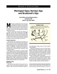
Disk Herniation<br />
Disk herniation (Figure 3) occurs most commonly<br />
between the fourth and fifth lumbar vertebrae and<br />
between the fifth lumbar and first sacral vertebrae. 5<br />
Patients with L5 radiculopathy (at the L4-5 disk)<br />
report low back, hip, and lateral thigh and leg pain<br />
Increased pain with activity; tenderness to palpation; limited range<br />
<strong>of</strong> motion; abnormal posture<br />
Increased pain with sitting; radicular pain on straight-leg raising<br />
Increased pain with activity; decreased range <strong>of</strong> motion<br />
Pseudoclaudication; decreased pain with back flexion; increased<br />
pain with extension<br />
Exaggeration <strong>of</strong> lumbar curve; pain with spine extension<br />
Decreased range <strong>of</strong> motion; diagnosed by Schober’s test<br />
Localized tenderness to percussion; fever<br />
Constant pain; nocturnal pain; localized tenderness; weight loss<br />
Flank pain radiating into the inguinal region and testicle; hematuria;<br />
writhing<br />
Fever; flank pain; chills; dysuria; costovertebral angle tenderness<br />
Pulsating abdominal mass; severe back, abdominal, or flank pain if<br />
ruptured<br />
Referred pain; associated abdominal pain; pain associated with menstrual<br />
cycle; pain unrelated to movement; cramping or colicky pain<br />
Figure 3. Herniated nucleus pulposus.<br />
and have weakness <strong>of</strong> dorsiflexion <strong>of</strong> the foot and toes.<br />
Patients with S1 nerve root compression (at the L5-S1<br />
disk) report pain in the posterior thigh, lateral calf,<br />
and foot, and have weakness <strong>of</strong> eversion and plantar<br />
www.turner-white.com Hospital Physician May 2003 43
Karnath : <strong>Clinical</strong> <strong>Signs</strong> <strong>of</strong> <strong>Low</strong> <strong>Back</strong> <strong>Pain</strong> : pp. 39–44, 56<br />
Fibrous tissue<br />
Figure 4. Mechanism <strong>of</strong> spondylolisthesis. (Reprinted with permission<br />
from Medtronic [http://www.medtronics<strong>of</strong>amordanek.<br />
com]. Available at http://www.back.com/causes-mechanicalspondylolisthesis.html.<br />
Copyright ©2003.)<br />
flexion <strong>of</strong> the foot with diminished ankle reflexes. Patients<br />
with disk herniation have pain with forward flexion,<br />
whereas patients with spinal stenosis have pain<br />
with extension. Cauda equina syndrome can be caused<br />
by a massive midline disk herniation. Patients may report<br />
bilateral sciatica with sensory deficits over the perineal<br />
regions (so-called “saddle anesthesia”).<br />
Spinal Stenosis<br />
Spinal stenosis occurs when the spinal cord is compressed.<br />
This condition should be suspected in patients<br />
with low back pain that is aggravated by walking<br />
and with hyperextension <strong>of</strong> the back and that is relieved<br />
by rest or flexion <strong>of</strong> the back. These patients<br />
<strong>of</strong>ten have fewer symptoms walking uphill than downhill,<br />
because the volume <strong>of</strong> the spinal canal increases<br />
with back flexion and decreases with extension. Patients<br />
may also report pseudoclaudication and sciatica.<br />
Pseudoclaudication or bilateral leg pain can occur with<br />
walking or prolonged standing.<br />
Spondylolisthesis<br />
Spondylolisthesis occurs when there is forward displacement<br />
<strong>of</strong> 1 or more lumbar vertebrae (Figure 4).<br />
Spondylitic spondylolisthesis is the most common type<br />
and occurs because <strong>of</strong> a defect in the pars interarticularis<br />
(ie, a defect in the thin isthmus <strong>of</strong> bone connecting the<br />
Figure 5. Illustration <strong>of</strong> Schober’s test. (Reprinted from McRae<br />
R. <strong>Clinical</strong> orthopaedic examination. 4th ed. New York:<br />
Churchill Livingstone; 1997:133, with permission from Elsevier.)<br />
superior and inferior facets); anterior displacement <strong>of</strong> L5<br />
over S1 occurs most commonly. 6 This type <strong>of</strong> spondylolisthesis<br />
is more common in patients who repeatedly lift<br />
heavy objects, thereby placing strain on this connection.<br />
Patients typically report low back pain that is worse with<br />
activity and spine extension but is relieved by flexion.<br />
Spondylolisthesis can occur at any age.<br />
Fracture<br />
Spinal compression fractures <strong>of</strong>ten occur in patients<br />
older than 70 years who have a history <strong>of</strong> osteoporosis.<br />
Patients with a history <strong>of</strong> long-term corticosteroid use are<br />
also at risk. Most patients who sustain spinal compression<br />
fractures do not have a history <strong>of</strong> trauma. Percussion tenderness<br />
over the involved vertebra can be elicited.<br />
NONMECHANICAL BACK PAIN<br />
Causes <strong>of</strong> nonmechanical low back pain include<br />
neoplasia, infection, and inflammatory arthritis. Typically,<br />
patients with a nonmechanical cause <strong>of</strong> low back<br />
pain report pain that occurs at rest and is less affected<br />
by motion.<br />
Ankylosing Spondylitis<br />
Ankylosing spondylitis is an inflammatory arthropathy<br />
affecting the spine and pelvis. Patients typically have<br />
morning stiffness that improves with activity. The condition<br />
has an insidious onset, typically starting in patients<br />
younger than 40 years. Affected patients have decreased<br />
range <strong>of</strong> motion <strong>of</strong> the lumbar and sacroiliac joints.<br />
Schober’s test can be used to measure the degree <strong>of</strong> impairment<br />
<strong>of</strong> range <strong>of</strong> motion. This test is performed by<br />
(continued on page 56)<br />
44 Hospital Physician May 2003 www.turner-white.com
Karnath : <strong>Clinical</strong> <strong>Signs</strong> <strong>of</strong> <strong>Low</strong> <strong>Back</strong> <strong>Pain</strong> : pp. 39–44, 56<br />
(from page 44)<br />
marking the lumbar spine at 0 cm and at 15 cm; more<br />
specifically, a mark is made 10 cm above S1 and another<br />
5 cm below. The patient is then asked to flex as far forward<br />
as possible, and the degree to which the marks<br />
separate is noted. In patients without impaired range <strong>of</strong><br />
motion, the points normally separate at least 5 cm<br />
(Figure 5).<br />
Neoplastic Disease<br />
Malignant neoplasm accounts for fewer than 1% <strong>of</strong><br />
episodes <strong>of</strong> low back pain. However, metastatic cancer<br />
should be considered as a potential etiology in any<br />
patient with a previous history <strong>of</strong> cancer, until proved<br />
otherwise. The most common primary sites are the<br />
breasts, lungs, or prostate; primary neoplasms such as<br />
multiple myeloma are less commonly the cause. A key<br />
historical finding is that back pain due to cancer is<br />
unrelieved by bedrest and typically worsens at night.<br />
Onset is usually slow and progressive. 7<br />
Infection<br />
Infectious etiologies <strong>of</strong> acute low back pain include<br />
osteomyelitis, septic discitis, and paraspinal or epidural<br />
abscess, whereas infectious etiologies <strong>of</strong> chronic low<br />
back pain include fungal or tuberculous infections. Patients<br />
typically first report fever and sharp focal pain in<br />
the lumbar spine. Physical examination reveals tenderness<br />
to percussion. A history <strong>of</strong> intravenous drug abuse<br />
is commonly elicited.<br />
VISCERAL DISEASE<br />
Common diseases causing referred back pain include<br />
renal diseases (eg, nephrolithiasis, pyelonephritis),<br />
vascular diseases (eg, abdominal aortic aneurysms),<br />
diseases <strong>of</strong> the pelvis (eg, endometriosis), and<br />
gastrointestinal diseases (eg, pancreatitis, cholecystis).<br />
Patients with back pain caused by visceral diseases<br />
<strong>of</strong>ten have pain unrelated to activity and pain that is<br />
worse when they are lying down. 8,9<br />
The back pain associated with nephrolithiasis can<br />
be severe and can cause the affected patient to writhe;<br />
pain is typically colicky and radiates to the groin.<br />
Pyelonephritis is another urologic cause <strong>of</strong> back pain;<br />
costovertebral angle tenderness can be elicited by percussing<br />
the paraspinal areas <strong>of</strong> affected patients.<br />
CONCLUSION<br />
<strong>Low</strong> back pain is one <strong>of</strong> the most common reasons<br />
that patients seek care from a primary care physician. In<br />
most cases, it has a benign etiology. However, a thorough<br />
history should be obtained and physical examination<br />
performed in patients with low back pain, because<br />
they can elicit warning signs that indicate the need for<br />
further work-up. Serious causes <strong>of</strong> low back pain, such<br />
as malignancy and infection, should not be missed. HP<br />
REFERENCES<br />
1. Borenstein D. Epidemiology, etiology, diagnostic evaluation,<br />
and treatment <strong>of</strong> low back pain. Curr Opin Rheumatol<br />
1996;8:124–9.<br />
2. Deyo RA, Rainville J, Kent DL. What can the history and<br />
physical examination tell us about low back pain? JAMA<br />
1992;268:760–5.<br />
3. Liang MH. Acute low back pain: diagnosis and management<br />
<strong>of</strong> mechanical back pain. Prim Care 1988;15:<br />
827–47.<br />
4. Deyo RA, Weinstein JN. <strong>Low</strong> back pain. N Engl J Med<br />
2001;344:363–70.<br />
5. Atlas SJ, Deyo RA. Evaluating and managing acute low<br />
back pain in the primary care setting. J Gen Intern Med<br />
2001;16:120–31.<br />
6. Rose-Innes AP, Engstrom JW. <strong>Low</strong> back pain: an algorithmic<br />
approach to diagnosis and management. Geriatrics<br />
1998;53:26–8, 33–6, 39–40.<br />
7. Borenstein DG. A clinician’s approach to acute low back<br />
pain. Am J Med 1997;102(1A):16S–22S.<br />
8. Patel AT, Ogle AA. Diagnosis and management <strong>of</strong> acute low<br />
back pain. Am Fam Physician 2000;61:1779–86,1789–90.<br />
9. Bratton RL. Assessment and management <strong>of</strong> acute low<br />
back pain. Am Fam Physician 1999;60:2299–308.<br />
Copyright 2003 by <strong>Turner</strong> <strong>White</strong> Communications Inc., Wayne, PA. All rights reserved.<br />
56 Hospital Physician May 2003 www.turner-white.com