Stabilis™ Surgical Technique (PDF) - Tornier DX
Stabilis™ Surgical Technique (PDF) - Tornier DX
Stabilis™ Surgical Technique (PDF) - Tornier DX
You also want an ePaper? Increase the reach of your titles
YUMPU automatically turns print PDFs into web optimized ePapers that Google loves.
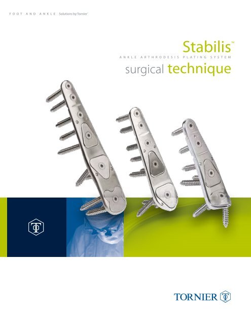
F O O T A N D A N K L E Solutions by <strong>Tornier</strong> ®<br />
Stabilis<br />
A N K L E A r T h r O D E S i S p L A T i N g S Y S T E M<br />
surgical technique
T O r N i E r ® S t a b i l i S a n k l e a r t h r o d e S i S P l a t i n g S y S t e m<br />
Indications for Use:<br />
The <strong>Tornier</strong> Stabilis Ankle Arthrodesis Plating System is intended for arthrodesis of the ankle<br />
joint and distal tibia, fractures, osteotomies, and fusions of small bones including the foot<br />
and ankle.<br />
Contraindications:<br />
The Stabilis Ankle Arthrodesis Plating System is contraindicated for the following conditions:<br />
Anterior Tibio-Talar Plate (AFPATTL and AFPATTH) . . . . . . . . . . . . . . . . . . . . . . . . . . . . . . . . . . . . . . . . . . . . . . . . . . . . . . . . . . . . . . . . . . . . . . . . . . 3-9<br />
Lateral Joint Preparation for Lateral TT and TTC Plates . . . . . . . . . . . . . . . . . . . . . . . . . . . . . . . . . . . . . . . . . . . . . . . . . . . . . . . . . . . . . . . . . 10-13<br />
Lateral Tibio-Talar Plate (AFPTTL and AFPTTH) . . . . . . . . . . . . . . . . . . . . . . . . . . . . . . . . . . . . . . . . . . . . . . . . . . . . . . . . . . . . . . . . . . . . . . . . . . . . 14-18<br />
Lateral Tibio-Talar-Calcaneal Plate (AFPTTC). . . . . . . . . . . . . . . . . . . . . . . . . . . . . . . . . . . . . . . . . . . . . . . . . . . . . . . . . . . . . . . . . . . . . . . . . . . . . . . . 19-25<br />
Instrument Trays<br />
• Sepsis, systemic infection, elevated White Blood Cell count, fever and/or local inflammation.<br />
• Complete talar necrosis or inadequate bone stock to permit stabilization of the arthrodesis.<br />
• Persisting skin lesion or poor skin coverage around the ankle joint that would make the<br />
procedure unjustifiable.<br />
• Neuromuscular or psychiatric disorders which might jeopardize fixation and post-operative care.<br />
• Suspected or documented metal allergy or intolerance.<br />
• Inadequate neurovascular status.<br />
• Severe longitudinal deformity.<br />
Preoperative Planning:<br />
It is important for the physician to evaluate the patient regarding leg length discrepancy and axial alignment<br />
of the entire involved lower extremity. Sometimes a prior proximal fracture in the femur or the tibia may<br />
leave elements of malalignment that may need to be recognized and planned for in the later performance<br />
of an ankle or hindfoot arthrodesis. Evaluation of the patient in a standing and sitting position, with special<br />
attention to translation at the ankle joint, varus/valgus of the hindfoot, rotational abnormalities and axial<br />
malalignment must all be recognized with both clinical and radiographic evaluation. Uncommonly, a more<br />
proximal osteotomy may be necessary to realign the extremity prior to the definitive ankle or hindfoot<br />
surgery. On occasion, a compensatory arthrodesis may realign the extremity at the ankle joint or hindfoot.<br />
Note: all system components (implants and instruments), are intended to be sterilized in the instrumentation tray.<br />
Top Tray . . . . . . . . . . . . . . . . . . . . . . . . . . . . . . . . . . . . . . . . . . . . . . . . . . . . . . . . . . . . . . . . . . . . . . . . . . . . . . . . . . . . . . . . . . . . . . . . . . . . . . . . . . . . . . . . . . . . . . . . . . . . . . . . . . . . . . . . 26<br />
Bottom Tray . . . . . . . . . . . . . . . . . . . . . . . . . . . . . . . . . . . . . . . . . . . . . . . . . . . . . . . . . . . . . . . . . . . . . . . . . . . . . . . . . . . . . . . . . . . . . . . . . . . . . . . . . . . . . . . . . . . . . . . . . . . . . . . . . . 27
Anterior Tibio-Talar Plate (AFPATTL and AFPATTH)<br />
Step 1<br />
Position the patient in the supine position on the table, with<br />
the heel at end of the table. Place a bump proximal to the<br />
ankle. The leg is exsanguinated and a thigh tourniquet<br />
is inflated.<br />
An extensile approach to the ankle is made between the<br />
anterior tibial and extensor hallucis longus tendons. The<br />
incision is followed by careful subcutaneous dissection.<br />
The superficial peroneal nerve is identified and retracted.<br />
The extensor retinaculum is divided between the anterior<br />
tibial and extensor hallucis longus tendons. Retraction of<br />
the tendons exposes the anterior aspect of the distal tibia,<br />
ankle joint, and talonavicular joint. The deep neurovascular<br />
structures are identified, mobilized, and protected. This allows<br />
for an anterior release and broad arthrolysis with resection of<br />
all the osteophytes. The top of the dome as well as the angles<br />
between the pilon and each of the malleoli can be identified<br />
precisely using this incision.<br />
Step 2<br />
Put the foot in plantar flexion to facilitate distraction. Distract the<br />
joint with the Joint Distractor and accompanying 2.5 x 100 mm<br />
Pins. Place the two Distractors on either side of the ankle at an<br />
approximate 70° angle from each other. Insert one pin in the talus<br />
and one pin in the tibia to hold each Distractor in place. Push the<br />
Distractor against the bone. Tighten the knobs on the handles to<br />
secure the Distractors to the pins. Distract the joint by squeezing<br />
the handles. Tighten the thumb screws on the proximal ends to<br />
lock the ratchets in place.<br />
2.5 x 100 mm Pins<br />
Step 3<br />
Joint Distractor<br />
Prepare the joint by removing osteophytes and cartilage on<br />
both the tibial and talar articular surfaces. Prepare the entire<br />
tibial side of the joint, and as much of the talar side as necessary.<br />
Prepare the gutter of the medial malleolus if necessary. The<br />
plate will not compromise the tibio-fibular joint.<br />
3
4<br />
T O r N i E r ® S t a b i l i S a n k l e a r t h r o d e S i S P l a t i n g S y S t e m<br />
Step 4<br />
If appropriate, micro-fracture the joint surfaces with a pick,<br />
drill or burr to the depth of the soft, cancellous bone. Pack the<br />
joint with bone graft as necessary. Remove the Distractors and<br />
associated pins.<br />
Step 5<br />
Step 6<br />
Drill Guide 2.7 mm Drill Bit<br />
Align the foot in the appropriate position for fusion. Hold the<br />
foot with the Foot Holding Device if desired. Verify alignment<br />
with fluoroscopy.<br />
Foot Holding Device<br />
If desired, a cross-joint compression screw can be inserted, such<br />
as the NexFix 6.5 Compression Screw with patented threaded<br />
washers. Insert the screw posteriorly from the distal tibia to the<br />
talus to help with the compression of the joint. Follow specific<br />
surgical steps pertaining to inserting the compression screw.<br />
Take care that the additional hardware does not hinder plate<br />
and screw placement.<br />
Temporarily fix the joint in place with 2.0 x 150 mm Pins<br />
medial and lateral of where the plate will sit. Avoid the<br />
subtalar joint.<br />
2.0 x 150 mm Pins NexFix 6.5 mm Compression Screw Tray
Step 7<br />
Use the X-ray template to select the appropriate Anterior Plate<br />
and position the plate for implantation.<br />
Two Anterior plates are available:<br />
- 107.5° distally angled portion against the talus (AFPATTL)<br />
- 95° distally angled portion against the talus (AFPATTH)<br />
Anterior Tibio-Talar Plate<br />
Step 8<br />
Remove all ostheophytes and prepare the bone of the talar<br />
neck for a proper fit of the plate.<br />
Generally, the plate should be positioned immediately<br />
posterior to the dorsal margin of the talonavicular joint<br />
avoiding impingement when dorsiflexing the navicular.<br />
Evaluate plate fit to the tibia. Bend the plate with the<br />
Plate Benders if necessary. Prepare bone surfaces additionally<br />
if necessary.<br />
Plate Benders<br />
Step 9<br />
Temporarily fix the plate to the tibia with a 2.0 x 150 mm Pin<br />
through the proximal pin hole. The pin hole is oriented from<br />
anterior-proximal to posterior-distal to help hold the plate<br />
against the bone. If desired, use additional pins through the<br />
other pin holes in the plate to further fix the plate to the tibia.<br />
2.0 x 150 mm Pins<br />
Anterior TT Plate<br />
High Talar Neck Low Talar Neck<br />
Part # AFPATTH Part # AFPATTL<br />
5
6<br />
T O r N i E r ® S t a b i l i S a n k l e a r t h r o d e S i S P l a t i n g S y S t e m<br />
Step 10<br />
Attach the Talus Hole Guide to the distal portion of the plate.<br />
Pivot the post to the side to avoid interference with the<br />
other instrumentation.<br />
Anterior Talus Hole Guide<br />
Step 11<br />
Insert the Drill Guide into the most distal hole in the Talus Hole<br />
Guide. Make sure that the laser etched line on the Drill Guide<br />
sits flush with the Hole Guide. Drill the screw hole in the talus,<br />
avoiding penetration into the subtalar joint.<br />
Use the Depth Gauge to measure the hole’s depth.<br />
Step 12<br />
Drill Guide 2.7 mm Drill Bit Depth Gauge<br />
Select the appropriate screw length. Use the Star Screw Driver<br />
and Ratcheting AO Driver to insert the Screw.<br />
Bone Screw<br />
Star Screw Driver<br />
Ratcheting AO Driver Handle
Step 13<br />
Drill and insert the two proximal screws through the Talus<br />
Hole Guide in the same fashion. Verify positions in the bone<br />
with fluoroscopy.<br />
Step 14<br />
Remove the Talus Hole Guide. Use the screw driver for a final<br />
tightening of the talar bone screws. Attach the Talar CoverLoc®<br />
to the plate. Use the driver to tightly secure the CoverLoc and<br />
stabilize each bone screw underneath it.<br />
Step 15<br />
Drill Guide 2.7 mm Drill Bit<br />
Depth Gauge<br />
Talar CoverLoc<br />
for Anterior Plate<br />
Star Screw Driver Bone Screw<br />
Star Screw Driver<br />
Check the plate fit against the tibia. If necessary, place the foot<br />
in plantar flexion and use the Plate Benders to bend the plate<br />
at the bending points.<br />
Attach the Tibia Hole Guide to the plate. Pivot the<br />
post to the side to avoid interference with the<br />
other instrumentation.<br />
Plate Benders Tibia Hole Guide<br />
Anterior TT Plate<br />
7
8<br />
T O r N i E r ® S t a b i l i S a n k l e a r t h r o d e S i S P l a t i n g S y S t e m<br />
Step 16<br />
Remove all cross-joint pins. Insert one end of the Compressor<br />
in the proximal hole of the Tibia Hole Guide as shown. Push it<br />
down fully to the bottom of the hole. Position the other distal<br />
end of the Compressor on the tibia, above the proximal end<br />
of the plate, leaving sufficient room so not to interfere with<br />
the plate when compression is applied. Typically a 10 mm gap<br />
or more is adequate between the proximal end of the plate<br />
and the Compressor. Position the proximal Compressor collet<br />
longitudinally in line with the plate. Insert a 2.5 x 100 mm Pin<br />
through the collet on this compressor end and drive it into the<br />
tibia bicortically. Turn the knob to tighten the collet against<br />
the pin. Keep the Compressor fully seated in the hole of the<br />
Tibial Hole Guide during this step, and avoid rotating the<br />
Compressor handles towards the proximal pin. If necessary,<br />
loosen the knob and realign the Compressor and tighten the<br />
knob again. Squeeze the Compressor handles. Tighten the<br />
thumb screw on the back of the Compressor to lock the<br />
ratchet in place.<br />
2.5 x 100 mm Pins Joint Compressor<br />
<strong>Surgical</strong> Note:<br />
Keeping the Compressor straight will typically result in a small<br />
gap between the tibial cortex and the proximal Compressor<br />
end on the pin. This is acceptable.<br />
Take care to compress the joint evenly. Avoid rotating the tibia<br />
during compression and creating a gap in the posterior joint<br />
space. Verify reduction under AP and lateral fluoroscopy. If<br />
necessary, release the compression, remove the Compressor,<br />
plantarflex the ankle and use the Plate Benders to bend the<br />
plate at the two bending zones for a better fit or even compression.<br />
Step 17<br />
Insert the Drill Guide into each of the proximal screw holes.<br />
Seat the guide squarely on the shoulder in the hole. Drill the<br />
proximal plate screw hole. Use the Depth Gauge to measure<br />
the hole’s depth. Select the appropriate screw length and<br />
place the screw. Repeat the process for the other screw hole.<br />
Drill Guide 2.7 mm Drill Bit<br />
Depth Gauge Bone Screw<br />
Ratcheting AO<br />
Driver Handle<br />
Star Screw Driver<br />
Alternatively, two Compressors may be used to compress the<br />
joint. Position one on either side of the plate, across the joint, at<br />
approximately 70° from each other – like the Distractors were<br />
positioned in Step 2. Use 2.5 x 100 mm Pins to secure one end<br />
of each Compressor into the tibia and other end in the talus.<br />
Tighten the collets against the pins. Squeeze both handles to<br />
compress the joint.
Step 18<br />
Insert Drill Guide into one of the distal holes in the Tibia Hole<br />
Guide. Make sure that the laser etched line on the Drill Guide<br />
sits flush with the Hole Guide. Drill the screw hole in the tibia.<br />
Use the Depth Gauge to measure the hole’s depth. Select<br />
the appropriate screw length. Use the Driver Bit and Driver<br />
Handle to insert the screw. Drill and insert the other distal<br />
screw through the Tibia Hole Guide in the same fashion.<br />
Verify positions in the bone with fluoroscopy.<br />
Drill Guide 2.7 mm Drill Bit<br />
Depth Gauge Bone Screw<br />
Step 19<br />
Remove the Compressor and associated pin. Insert the Drill<br />
Guide into the proximal hole of the Tibia Hole Guide and drill<br />
the proximal screw hole. Use the Depth Gauge to measure the<br />
hole’s depth. Select the appropriate screw length and insert<br />
the screw.<br />
Drill Guide 2.7 mm Drill Bit<br />
Depth Gauge Bone Screw<br />
Step 20<br />
Remove the Tibia Hole Guide. Use the Driver for final tightening<br />
of each tibial bone screw. Attach the Tibial CoverLoc® to the<br />
plate. Use the driver to tightly secure the CoverLoc and stabilize<br />
each bone screw underneath it. Verify positions in the<br />
bone with fluoroscopy.<br />
Close the incision with appropriate surgical technique. A closed<br />
suction drainage system may be used at the surgeon’s discretion.<br />
Tibial CoverLoc<br />
Star Screw Driver<br />
Anterior TT Plate<br />
9
10<br />
T O r N i E r ® T O r N i E r S t a b i l i S a n k l e a r t h r o d e S i S P l a t e S y S t e m<br />
® S t a b i l i S a n k l e a r t h r o d e S i S P l a t i n g S y S t e m<br />
Lateral Joint Preparation for Lateral TT and TTC Plates<br />
Step 1<br />
Position the patient in a semisupine position with a bump under<br />
the ipsilateral hip. This allows exposure of both the medial and<br />
the lateral ankle and assists in assessing correction of rotational<br />
deformities. It also allows for placement of medial and lateral<br />
compression and distraction clamps. A thigh tourniquet is<br />
placed, the leg is exsanguinated and the tourniquet is inflated.<br />
Palpate the lateral ankle and foot to identify the fibula.<br />
Make a lateral longitudinal incision to expose the fibula and<br />
tibio talar (TT) joint. Have the incision extend approximately<br />
10 to 11 cm up proximally from the distal tip of the fibula. Avoid<br />
the peroneal tendons and sural and superficial peroneal nerves.<br />
Step 2<br />
Dissect and remove the distal fibula (approximately 9 to<br />
10 cm). The fibula and bone shavings may be morselized<br />
if an autogenous graft is desired.<br />
Step 3<br />
Prepare a flat surface on the lateral surface of the exposed<br />
distal tibia and adjacent part of the talus with rongeurs<br />
and/or other appropriate surgical instruments.<br />
Align the foot in the appropriate position for fusion. Support the<br />
foot with the Foot Holding Device to help determine appropriate<br />
plantar/dorsiflexion of the fusion if desired. A wrap can be used to<br />
hold the foot to the Foot Holding Device.<br />
Foot Holding Device
Step 4<br />
Evaluate the arc of the tibio-talar joint and select the appropriate<br />
Lateral Cutting Guide set (radius of 25 mm or 40 mm) that<br />
best approximates the joint curvature. Position the Cutting Guide<br />
on the exposed lateral walls of the tibia and talus. The longer<br />
piece of the Cutting Guide with the concave surface aligns with<br />
the tibial wall and the shorter piece aligns with the talar wall. The<br />
curved intersection between the two guide pieces should align<br />
with the tibio-talar joint. Pull each half slightly back from the ends<br />
of the bones to ensure that the drill holes align with subchondral<br />
bone, so that all cartilage will be removed after drilling. Either end<br />
of the Cutting Guide may be pulled further back from the joint<br />
area if the removal of additional bone is needed.<br />
Lateral Cutting Guide<br />
Step 5<br />
Make sure the Cutting Guide sits flush on the lateral wall of the<br />
tibia and talus. If necessary, further prepare the tibial and talar<br />
surfaces to fit the guide. Fix the tibial component of the cutting<br />
guide to the bone by inserting two 2.5 x 60 mm Pins in the<br />
X-marked holes in the tibial half.<br />
2.5 x 60 mm Pins<br />
Step 6<br />
Put the 2.7 mm Drill Bit in the tibial portion of the Cutting<br />
Guide. Verify alignment with AP fluoroscopy. Change the position<br />
of the Cutting Guide by lifting it proximally or distally and<br />
reinserting the pin if varus or valgus misalignment is noticed.<br />
Insert the Drill to the appropriate depth to remove the distal<br />
tibial cartilage and make note of the depth.<br />
2.7 mm Drill Bit<br />
Lateral Joint Preparation<br />
11
12<br />
T O r N i E r ® S t a b i l i S a n k l e a r t h r o d e S i S P l a t i n g S y S t e m<br />
Step 7<br />
Hold the guide to the bone. Insert 2.5 x 100 mm Pins on the<br />
lateral sides of the Cutting Guide to secure the guide.<br />
Drill through each distal cutting hole and across the underlying<br />
bone. Hold the drill straight, advance slowly. Be careful not to<br />
drill too far medially, beyond the talus or across the medial gutter<br />
and into the medial malleolus. Fluoroscopy or direct visualization<br />
can be used to determine the accuracy of this osteotomy.<br />
2.5 x 100 mm Pins<br />
Step 8<br />
Attach the talar component of the Cutting Guide with the<br />
U-rail to the tibial component. Open the space between the<br />
tibial and talar component to the desired width so that all<br />
the cartilage will be removed after drilling. Place two 2.5 x<br />
60 mm Pins in the marked holes in the talar half. Insert pins<br />
deep enough so a drill can be fully driven into the cutting<br />
holes in the guide.<br />
2.5 x 60 mm Pins<br />
Step 9<br />
2.7 mm Drill Bit<br />
Hold the guide against the bone. Insert 2.5 x 100 mm Pins<br />
on the lateral sides of the Cutting Guide to secure the guide.<br />
Remove the U-rail.<br />
Drill through each distal cutting hole and across the underlying<br />
bone. Hold the drill straight, advance slowly, and use an “in and<br />
out” motion to prevent the drill from skiving. Drill through the<br />
entire width of the talus.<br />
Remove Cutting Guide and pins from each side except for<br />
two that can be used to anchor the Distractor.<br />
2.5 x 100 mm Pins<br />
2.7 mm Drill Bit
Step 13<br />
Use a Distractor and two accompanying 2.5 x 100 mm Pins to<br />
distract the joint. Place the Distractor across the tibio-talar joint.<br />
Open the Distractor enough so there is access to the joint for<br />
instrumentation. Insert one pin in the talus and one in the tibia<br />
to hold the Distractor in place. Push the Distractor’s ends down<br />
against the bone. Tighten the knobs to secure the Distractor to<br />
the pins. Squeeze the handles to distract the joint. Tighten the<br />
thumb screw on the proximal end of the Distractor to lock the<br />
ratchet in place.<br />
Joint Distractor<br />
Step 14<br />
Use osteotomes and mallet to remove the arc segments of<br />
bone from both sides of the joint.<br />
Step 15<br />
2.5 x 100 mm Pins<br />
Test the reduction of the joint. If there is either excessive<br />
valgus or varus, the resection of the tibial or talar surface may<br />
vary. Further bone may need to be resected to correct coronal<br />
plane deformities of the ankle joint. Intraoperative fluoroscopy<br />
is helpful from a lateral aspect to inspect the curvature of the<br />
resection and from an anterior position to assess the varus/<br />
valgus position of the alignment. Remove the Distractor. Verify<br />
the alignment with fluoroscopy.<br />
Align the foot in the appropriate position for arthrodesis.<br />
Temporarily fix the ankle joint in position with pins on either<br />
side of where the plate will sit (anteriorly and posteriorly to<br />
the plate). Avoid inserting pins into the talonavicular joint<br />
and subtalar space. The blue posterior foot holding device<br />
is helpful in ensuring appropriate dorsiflexion and plantar<br />
flexions have been obtained. At this point assess rotation of<br />
the joint, the presence of anterior posterior translation as well<br />
as the varus/valgus position of the proposed arthrodesis.<br />
Separate fixation screws or pins may be applied prior to<br />
attaching the plate. Ensure that additional hardware does<br />
not hinder plate and screw placement.<br />
Lateral Joint Preparation<br />
13
14<br />
T O r N i E r ® S t a b i l i S a n k l e a r t h r o d e S i S P l a t i n g S y S t e m<br />
Lateral Tibio-Talar Plate (AFPTTL and AFPTTH)<br />
For Lateral Joint preparation, see pages 10-13.<br />
Step 16<br />
Use the X-ray template to select the Lateral TT Plate that best<br />
fits the Talar anatomy.<br />
Two TT plates are available:<br />
- 15˚ distally angled portion against the talus (AFPTTL)<br />
- 30˚ distally angled portion against the talus (AFPTTH)<br />
Step 18<br />
Lateral Tibio-Talar Plate<br />
Step 17<br />
Use 2.0 x 150 mm Pins to temporarily fix the joint. Position the<br />
plate on the bone. Ensure that the distal end of the plate sits<br />
fully on the talus and does not extend down into the subtalar<br />
space or onto the calcaneus. Evaluate plate fit to the tibia. Bend<br />
the plate with the Plate Benders if necessary. Pin the plate to<br />
the talus (it is not recommended to use two pins in the talar<br />
part of the plate to prevent interference with the bone screw<br />
insertions later in the procedure). Attach the Talus Hole Guide<br />
to the Plate. Attach Driver Handle to the Guide Post if desired.<br />
Remove any protuberances that interfere with plate fit.<br />
Lateral Talus Hole Guide 2.5 x 150 mm Pins<br />
Insert Drill Guide into one of the proximal holes in the Talus<br />
Hole Guide. Make sure that the laser etched line on the Drill<br />
Guide sits flush with the Hole Guide. Drill the screw hole in<br />
the talus, avoiding penetration into the subtalar joint.<br />
Pivot the post to the side to avoid interference with the<br />
other instrumentation.<br />
Use the Depth Gauge to measure the hole. Select the<br />
appropriate screw length. Use the Driver Bit and Ratcheting<br />
AO Driver Handle to insert the screw.<br />
Drill Guide 2.7 mm Drill Bit<br />
Depth Gauge<br />
Star Screw Driver<br />
Plate Benders<br />
Bone Screw<br />
Ratcheting<br />
AO Driver Handle<br />
High Talar Flare<br />
Part # AFPTTH<br />
Low Talar Flare<br />
Part # AFPTTL
Step 19<br />
Drill and insert the other two talar screws in the same<br />
fashion. Verify position of these screws in the bone with<br />
fluoroscopy. Multiple views are necessary to ensure that the<br />
talar screws do not intrude or penetrate the subtalar joint.<br />
Step 20<br />
Remove the Talus Hole Guide. Use the screw driver for a final<br />
tightening of the talar bone screws. Attach the Talus CoverLoc®<br />
to the plate. Use the driver to tightly secure the CoverLoc<br />
and stabilize each bone screw underneath it. Remove all<br />
temporary fixation pins from the plate and the joint.<br />
Lateral Talus<br />
CoverLoc<br />
Star Screw Driver<br />
Lateral TT Plate<br />
15
T O r N i E r ® S t a b i l i S a n k l e a r t h r o d e S i S P l a t i n g S y S t e m<br />
Step 21<br />
2.5 x 225 mm Pins are now placed from anterior lateral to<br />
anterior medial across the tibia and the talus in order to<br />
compress the ankle arthrodesis site. These pins should be<br />
placed just anterior to the placement of the tibial portion of the<br />
plate and just anterior to the talar portion of the plate. These<br />
pins will penetrate the medial aspect of the distal tibia and<br />
talus and puncture wounds are made as these pins penetrate<br />
the skin. Care is taken to avoid injury to the posterior tibial<br />
neurovascular bundle. The compression device is used to<br />
determine the distance separating these two cross pins. (The<br />
Achilles complex tends to give counter compression forces to<br />
oppose the anterior compression devices.) Joint Compressors<br />
are placed both medially and laterally.<br />
Step 22<br />
Joint Compressors<br />
2.5 x 225 mm Pins<br />
Squeeze the Compressor handles to compress the joint. Take<br />
care to compress the joint evenly. Tighten the thumb screws<br />
on the back of the Compressors to lock the ratchets in place.<br />
Verify reduction with fluoroscopy.<br />
More compression may be placed either medially or laterally<br />
depending upon the joint preparation, and alignment desired.<br />
Bone graft may be inserted to assist in the alignment process<br />
prior to adding compression. Tighten the knobs on the<br />
Compressor to hold the pins in place.<br />
At this point a Guide Pin may be placed from the anterior<br />
cortex of the distal tibia in a distal posterior direction into<br />
the talus. A 6.5 mm cannulated screw such as the NexFix <br />
Compression Screw with patented threaded washers can be<br />
placed to strengthen compression of the construct. Care must<br />
be taken to avoid the cross-screws in the talus. Attention must<br />
be directed to the proposed screw route in the tibia as well.<br />
This screw may also be placed following the placement of the<br />
cross-screws fixating the plate.<br />
NexFix 6.5 mm Compression Screw Tray<br />
16
Step 23<br />
Attach the Tibia Hole Guide to the Plate. Insert the Drill<br />
Guide into one of the distal holes in the Tibia Hole Guide.<br />
Make sure that the laser etched line on the Drill Guide sits<br />
flush with the Hole Guide. Drill the screw hole in the tibia.<br />
Pivot the post to the side to avoid interference with the<br />
other instrumentation.<br />
Step 24<br />
Use the Depth Gauge to measure the hole. Select the<br />
appropriate screw length. Use the Driver Bit and Driver<br />
Handle to insert the screw. Insert the other screws in the<br />
same fashion. Verify positions in the bone with fluoroscopy.<br />
Depth Gauge<br />
Step 25<br />
Drill Guide 2.7 mm Drill Bit Tibia Hole Guide<br />
Star Screw Driver Bone Screw<br />
Remove Tibia Hole Guide. Use the Driver for final tightening of<br />
all tibial bone screws. Attach the Tibial CoverLoc® to the plate.<br />
Use the driver to tightly secure the CoverLoc and stabilize each<br />
bone screw underneath it.<br />
Tibial CoverLoc<br />
Star Screw Driver<br />
Lateral TT Plate<br />
17
18<br />
T O r N i E r ® S t a b i l i S a n k l e a r t h r o d e S i S P l a t i n g S y S t e m<br />
Step 26<br />
Insert the Drill Guide into each of the remaining proximal screw<br />
holes. Seat the guide squarely on the shoulder in the hole. Drill<br />
the screw hole. Measure, select and insert each screw using the<br />
technique described above. Repeat the process for the other<br />
screw hole.<br />
Step 27<br />
Remove the Compressors and pins. Verify implant<br />
position both clinically and with fluoroscopic control or<br />
with biplanar fluoroscopy.<br />
If desired, use the Augmentation Plate on the anterior lateral<br />
aspect of the tibio-talar fusion site. Slide the plate through<br />
the lateral incision onto the appropriate anterior surface.<br />
If necessary, bend the plate to fit to the bone surface. Drill<br />
the screw holes and insert screws using the same steps<br />
described above.<br />
Close the incision with appropriate surgical technique.<br />
A closed suction drainage system may be used at the<br />
surgeon’s discretion.<br />
Augmentation Plate
Lateral Tibio-Talar-Calcaneal Plate (AFPTTC)<br />
prepare the TT joint prior to preparing the subtalar (TC)<br />
joint (see pages 10-13 for Steps 1-15.)<br />
Step 16<br />
The incision for a TTC plate must be longer than for a lateral<br />
TT plate to expose the TC joint. Prepare the talocalcaneal (TC)<br />
joint. Use a Distractor and accompanying 2.5 x 100 mm Pins<br />
to distract the joint. Follow the same procedure for Distractor<br />
operation detailed in Lateral Joint Preparation, pages 11-13.<br />
Care is taken to avoid injuring the medial neurovascular<br />
structures with the placement of the pins. Alternatively, one or<br />
two lamina spreaders may be used to distract the joint.<br />
Joint Distractor<br />
Step 17<br />
2.5 x 100 mm Pins<br />
Use osteotomes and a mallet to remove the cartilage from<br />
the subtalar joint.<br />
If appropriate, micro-fracture the joint surfaces with a pick,<br />
drill or burr to the depth of the soft, cancellous bone. Pack the<br />
joint with bone graft as necessary. Remove the Distractors and<br />
associated pins.<br />
Step 18<br />
Align the foot in the appropriate position for fusion. Support<br />
the foot with the Foot Holding Device to help determine<br />
appropriate plantar/dorsiflexion of the fusion if desired. A<br />
wrap can be used to hold the foot to the Foot Holding Device.<br />
Foot Holding Device<br />
Lateral TTC Plate<br />
For Lateral Joint preparation, see pages 10-13.<br />
19
20<br />
T O r N i E r ® S t a b i l i S a n k l e a r t h r o d e S i S P l a t i n g S y S t e m<br />
Step 19<br />
Position the TTC plate on the bone. Remove any protuberances<br />
that interfere with plate fit. Evaluate plate fit to the tibia. Bend<br />
the plate with the Plate Benders if necessary. It is important<br />
to avoid excessive valgus or varus in the placement of the<br />
TTC plate.<br />
Lateral Tibio-Talo<br />
Calcaneal Plate<br />
<strong>Surgical</strong> Note:<br />
With an arthrodesis of both the ankle and subtalar joint,<br />
excessive varus or valgus is generally poorly tolerated with<br />
ambulation since there is no motion at these joints.<br />
Step 20<br />
Temporarily fix the joint with 2.0 x 150 mm Pins and fit the<br />
Plate to the bone with 2.0 x 150 mm Pins. (Note: it is not<br />
recommended to use two pins in the talar part of the<br />
plate due to the chance of interference with the bone<br />
screw insertions later in the procedure.)<br />
2.0 x 150 mm Pins<br />
Step 21<br />
Attach the Talus Hole Guide loosely to the TTC plate using<br />
the guidepost. The talus is fixed initially followed by the<br />
adjacent calcaneus and tibial bones.<br />
Lateral Talus Hole Guide<br />
Plate Benders
Step 22<br />
Insert the Drill Guide into one of the proximal holes in the Talus<br />
Hole Guide. Make sure that the laser etched line on the Drill<br />
Guide sits flush with the Hole Guide. Drill the screw hole in the<br />
talus, avoiding penetration into the subtalar joint. Pivot the post<br />
to the side to avoid interference with the other instrumentation.<br />
If the temporary cross joint pin interferes with the Drill or screw<br />
insertion, remove the pin, insert an additional pin in the tibial<br />
part of the plate, and take care to keep the plate in its intended<br />
position throughout the process.<br />
Use the Depth Gauge to measure the hole. Select the<br />
appropriate length screw. Use the Driver Bit and Ratcheting<br />
AO Driver Handle to insert the screw.<br />
Step 23<br />
Drill and insert the other two talar screws in the same fashion.<br />
Verify position of these screws in the bone with fluoroscopy.<br />
Multiple views are necessary to ensure that the talar screws<br />
do not intrude or penetrate the subtalar joint.<br />
Step 24<br />
Drill Guide 2.7 mm Drill Bit<br />
Star Screw Driver<br />
Remove the Talus Hole Guide. Use the screw driver for a final<br />
tightening of the talar bone screws. Attach the Talus CoverLoc®<br />
to the plate. Use the driver to tightly secure the CoverLoc and<br />
stabilize each bone screw underneath it. Remove the temporary<br />
fixation pins only from the subtalar joint.<br />
Lateral Talus<br />
CoverLoc<br />
Bone Screw<br />
Star Screw Driver<br />
Depth Gauge<br />
Ratcheting<br />
AO Driver Handle<br />
Lateral TTC Plate<br />
21
22<br />
T O r N i E r ® S t a b i l i S a n k l e a r t h r o d e S i S P l a t i n g S y S t e m<br />
Step 25<br />
2.5 x 225 mm Pins are now placed from anterior lateral to<br />
anterior medial across the talus and the calcaneus in order to<br />
compress the subtalar arthrodesis site. These pins should be placed<br />
just anterior to the placement of the talar portion of the plate and<br />
just anterior to the calcaneal portion of the plate. These pins will<br />
penetrate the medial aspect of the distal talus and calcaneus and<br />
puncture wounds are made as these pins penetrate the skin. Care<br />
is taken to avoid injury to the posterior tibial neurovascular<br />
bundle. The compression device is used to determine the<br />
distance separating these two cross pins. Joint Compressors are<br />
placed both medially and laterally.<br />
Step 26<br />
Squeeze the Compressor handles to compress the joint. Take<br />
care to compress the joint evenly. Tighten the thumb screws<br />
on the back of the Compressors to lock the ratchets in place.<br />
Verify reduction with fluoroscopy.<br />
More compression may be placed either medially or laterally<br />
depending upon the joint preparation, and alignment desired.<br />
Bone graft may be inserted to assist in the alignment process<br />
prior to adding compression. Tighten the knobs on the<br />
Compressor to hold the pins in place.<br />
Step 27<br />
Joint Compressors<br />
2.5 x 225 mm Pins<br />
Attach the Calcaneal Hole Guide to the Plate. Insert Drill Guide<br />
into one of the distal holes in the Hole Guide. Make sure that<br />
the laser etched line on the Drill Guide sits flush with the Hole<br />
Guide. Drill the screw hole in the calcaneus. Pivot the post to<br />
the side to avoid interference with the other instrumentation.<br />
Use the Depth Gauge to measure the hole. Select the<br />
appropriate screw length. Use the Driver Bit and Driver<br />
Handle to insert the screw.<br />
Drill and insert the other three calcaneal screws in the same<br />
fashion. Verify position of these screws in the bone with<br />
fluoroscopy. Verify position with fluoroscopy.<br />
Calcaneal Hole Guide<br />
Drill Guide<br />
2.7 mm Drill Bit Depth Gauge<br />
Bone Screw
Step 28<br />
Remove the Calcaneal Hole Guide. Use the screw driver for a final<br />
tightening of the calcaneal bone screws. Attach the Calcaneal<br />
CoverLoc® to the plate. Use the driver to tightly secure the CoverLoc<br />
and stabilize each bone screw underneath it. Remove Compressors<br />
and Pins. Remove the temporary fixation pins from the TT joint.<br />
Step 29<br />
A similar compression technique is now used to compress the<br />
TT joint. 2.5 x 225 mm Pins are now placed from anterior lateral<br />
to anterior medial across the tibia and the talus in order to<br />
compress the ankle arthrodesis site. These pins should be placed<br />
just anterior to the placement of the tibial portion of the plate<br />
and just anterior to the talar portion of the plate. These pins will<br />
penetrate the medial aspect of the distal tibia and talus and<br />
puncture wounds are made as these pins penetrate the skin.<br />
Care is taken to avoid injury to the posterior tibial neurovascular<br />
bundle. The compression device is used to determine the distance<br />
separating these two cross pins. (The Achilles complex tends to give<br />
counter compression forces to oppose the anterior compression<br />
devices.) Compressors are placed both medially and laterally.<br />
Step 30<br />
Calcaneal CoverLoc<br />
Joint Compressors<br />
2.5 x 225 mm Pins<br />
Squeeze the Compressor handles to compress the joint. Take<br />
care to compress the joint evenly. Tighten the thumb screws<br />
on the back of the Compressors to lock the ratchets in place.<br />
Verify reduction with fluoroscopy.<br />
More compression may be placed either medially or laterally<br />
depending upon the joint preparation, and alignment desired.<br />
Bone graft may be inserted to assist in the alignment process<br />
prior to adding compression. Tighten the knobs on the<br />
Compressor to hold the pins in place.<br />
At this point a Guide Pin may be placed from the anterior<br />
cortex of the distal tibia in a distal posterior direction into<br />
the talus. A 6.5 mm cannulated screw such as the NexFix <br />
Compression Screw with patented threaded washers can be<br />
placed to strengthen compression of the construct. Care must<br />
be taken to avoid the cross-screws in the talus. Attention<br />
must be directed to the proposed screw route in the tibia as<br />
well. This screw may also be placed following the placement<br />
of the cross-screws fixating the plate.<br />
NexFix 6.5 mm Compression Screw Tray<br />
Lateral TTC Plate<br />
23
24<br />
T O r N i E r ® S t a b i l i S a n k l e a r t h r o d e S i S P l a t i n g S y S t e m<br />
Step 31<br />
Attach the Tibia Hole Guide to the Plate. Insert the Drill Guide<br />
into one of the distal holes in the Tibia Hole Guide. Make sure<br />
that the laser etched line on the Drill Guide sits flush with the<br />
Hole Guide. Drill the screw hole in the tibia. Pivot the post to<br />
the side to avoid interference with the other instrumentation.<br />
Tibia Hole Guide Drill Guide 2.7 mm Drill Bit<br />
Step 32<br />
Use the Depth Gauge to measure the hole. Select the<br />
appropriate screw length. Use the Driver Bit and Driver Handle<br />
to insert the screw. Insert the other screws in the same fashion.<br />
Verify positions in the bone with fluoroscopy.<br />
Depth Gauge<br />
Step 33<br />
Remove Tibia Hole Guide. Use the Driver for final tightening<br />
of all tibial bone screws. Attach the Tibial CoverLoc® to<br />
the plate. Use the driver to tightly secure the CoverLoc and<br />
stabilize each bone screw underneath it.<br />
Tibial CoverLoc<br />
Star Screw Driver Bone Screw<br />
Star Screw Driver
Step 34<br />
Insert the Drill Guide into each of the remaining proximal<br />
screw holes. Seat the guide squarely on the shoulder in the<br />
hole. Drill the screw hole. Measure, select and insert each<br />
screw using the technique described above. Repeat the<br />
process for the other screw hole.<br />
Step 35<br />
Drill Guide 2.7 mm Drill Bit<br />
Depth Gauge<br />
Star Screw Driver<br />
Bone Screw<br />
Remove the Compressors and Pins. Verify implant position<br />
both clinically and with fluoroscopic control or with<br />
biplanar fluoroscopy.<br />
If desired, use the Augmentation Plate on the anterior lateral<br />
aspect of the tibio-talar fusion site. Slide the plate through<br />
the lateral incision onto the appropriate anterior surface.<br />
If necessary, bend the plate to fit to the bone surface. Drill<br />
the screw holes and insert screws using the same steps<br />
described above.<br />
Close the incision with appropriate surgical technique.<br />
A closed suction drainage system may be used at the<br />
surgeon’s discretion.<br />
Augmentation Plate<br />
Lateral TTC Plate<br />
25
26<br />
T O r N i E r ® S t a b i l i S a n k l e a r t h r o d e S i S P l a t i n g S y S t e m<br />
Top Tray:<br />
2<br />
Item Description Part # Quantity<br />
iNSTruMENTS<br />
1 Joint Distractor AFPDISTR 2<br />
2 Joint Compressor AFPCOMPR 2<br />
3 Lateral Cutting Guide 40 mm AFPCG40 1<br />
4 Lateral Cutting Guide 25 mm AFPCG25 1<br />
5 Pins Ø2.5 X 225 mm AFPK25225 5<br />
6 Pins Ø2.0 X 150 mm AFPK20150 5<br />
7 Pins Ø2.5 X 100 mm AFPK25100 5<br />
8 Pins Ø2.5 X 60 mm AFPK2560 5<br />
9 Hole Guide, Anterior Talus AFPHGDAT 1<br />
10 Hole Guide, Lateral Talus AFPHGDLT 1<br />
11 Hole Guide, Tibia AFPHGDT 1<br />
12 Hole Guide, Calcaneus AFPHGDC 1<br />
13 Screw Pick-Up Forceps NCS-45SF 1<br />
14 AO-Trinkle Power Driver Adaptor NCS-PA45 1<br />
15 2.7 mm Drill Bit Ø2.7 mm AFPDR 2<br />
16 Star Driver AFPSDRIV 2<br />
17 Drill Guide AFPDRG 1<br />
18 Ratcheting AO Driver Handle AFPDRAO 2<br />
19 Plate Bender AFPBEND 2<br />
20 Depth Gauge AFPDGA 1<br />
1<br />
4<br />
3<br />
5<br />
6<br />
7<br />
8<br />
15 16 17<br />
9<br />
13<br />
10<br />
18<br />
11<br />
12<br />
19<br />
14<br />
20
Bottom Tray:<br />
Item Description Part # Quantity<br />
pLATES AND COvErS<br />
1 Plate, Anterior TT, High Talar Neck (95°) AFPATTH 2<br />
Plate, Anterior TT, Low Talar Neck (107.5°) AFPATTL 2<br />
Plate, Lateral TT, Low Talar Flare (15°) AFPTTL 2<br />
Plate, Lateral TT, High Talar Flare (30°) AFPTTH 2<br />
Plate, TTC AFPTTC 2<br />
Plate, Augmentation AFPAUG 2<br />
CoverLoc® , Tibia AFPCPT 4<br />
CoverLoc, Lateral Talus AFPCPLT 2<br />
CoverLoc, Calcaneus AFPCPC 2<br />
CoverLoc, Anterior Talus AFPCPAT 3<br />
1<br />
Item Description Part # Quantity<br />
SCrEwS<br />
2 Bone Screw, Ø4.5x14 mm AFPSC4514 2<br />
Bone Screw, Ø4.5x16 mm AFPSC4516 2<br />
Bone Screw, Ø4.5x18 mm AFPSC4518 3<br />
Bone Screw, Ø4.5x20 mm AFPSC4520 4<br />
Bone Screw, Ø4.5x22 mm AFPSC4522 4<br />
Bone Screw, Ø4.5x24 mm AFPSC4524 4<br />
Bone Screw, Ø4.5x26 mm AFPSC4526 4<br />
Bone Screw, Ø4.5x28 mm AFPSC4528 4<br />
Bone Screw, Ø4.5x30 mm AFPSC4530 6<br />
Bone Screw, Ø4.5x32 mm AFPSC4532 6<br />
Bone Screw, Ø4.5x34 mm AFPSC4534 6<br />
Bone Screw, Ø4.5x36 mm AFPSC4536 6<br />
Bone Screw, Ø4.5x38 mm AFPSC4538 6<br />
Bone Screw, Ø4.5x40 mm AFPSC4540 6<br />
Bone Screw, Ø4.5x45 mm AFPSC4545 4<br />
Bone Screw, Ø4.5x50 mm AFPSC4550 4<br />
Bone Screw, Ø4.5x55 mm AFPSC4555 4<br />
Bone Screw, Ø4.5x60 mm AFPSC4560 2<br />
Bone Screw, Ø4.5x65 mm AFPSC4565 2<br />
Off-Plate Screw Washer AFPW 2<br />
MiSC<br />
4<br />
3 Foot Holding Device AFPALGN 1<br />
4 Space for Additional Instruments<br />
2 3<br />
27
<strong>Tornier</strong>, Inc.<br />
Edina, MN 55435<br />
USA<br />
+ 1 888 867 6437<br />
+ 1 281 494 7900<br />
www.tornier.com/us<br />
© 2010 <strong>Tornier</strong>, Inc. All rights reserved. Stabilis and NexFix are trademarks of <strong>Tornier</strong>, Inc. CoverLoc and <strong>Tornier</strong> are registered trademarks of <strong>Tornier</strong>, Inc.<br />
19-5126 rev. B 12/10


