Lab 7: Molluscs & Echinoderms - Brazosport
Lab 7: Molluscs & Echinoderms - Brazosport
Lab 7: Molluscs & Echinoderms - Brazosport

Create successful ePaper yourself
Turn your PDF publications into a flip-book with our unique Google optimized e-Paper software.
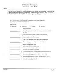
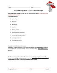
Name: _____________________________ Date: _________________<br />
1<br />
Gen Bio 2 <strong>Lab</strong> #7: <strong>Echinoderms</strong> and Mollusks<br />
Pre-lab Reading: Read pages 652-656 and 676-680 from your textbook. Read the entire lab ahead of time<br />
and complete all vocabulary and Pre-<strong>Lab</strong> activity before attending.<br />
Pre-<strong>Lab</strong> Vocabulary:<br />
1. Echinoderm –<br />
2. Pentaradial symmetry –<br />
3. Water vascular system –<br />
4. Tube feet –<br />
5. Ampulla –<br />
6. Endoskeleton –<br />
7. Pedicellariae –<br />
Pre-<strong>Lab</strong> activity: Look in your text and online and write the predominant traits for each of the<br />
following classification levels:<br />
A. Class Crinoidea –<br />
B. Subclass Asteroidea –<br />
C. Subclass Ophiuroidea –<br />
D. Class Echinoidea –<br />
E. Class Holothuroidea –<br />
Introduction:
<strong>Echinoderms</strong> are radially symmetrical animals that are only found in the sea (there are none on land or<br />
in fresh water). <strong>Echinoderms</strong> mean "spiny skin" in Greek. Many, but not all, echinoderms have spiny skin.<br />
There are over 6,000 species. <strong>Echinoderms</strong> usually have five appendages (arms or rays), but there are some<br />
exceptions.<br />
Radial symmetry means that the body is a hub, like a bicycle wheel, and tentacles are spokes coming<br />
out of it (think of a starfish). As larvae, echinoderms are bilaterally symmetrical. As they mature, they become<br />
radially symmetrical. Most adult echinoderms live on the bottom of the ocean floor. Many echinoderms have<br />
suckers on the ends of their feet that are used to capture and hold prey, and to hold onto rocks in a swift current.<br />
Sea Stars<br />
Sea stars (group name Stelleroidea) are sometimes called starfish, however<br />
they are not really fish (they lack both vertebrae and fins). There are two subtypes<br />
of sea stars:<br />
2<br />
Asteroids include: true sea stars and sun stars (A)<br />
Ophiuroids include: brittle stars (B) and basket stars (C)<br />
The differences between the two sub-types lie in how the arms connect to the<br />
central disk. Ophiuroids have arms that do not connect with each other. There<br />
is a distinct boundary between arm and central disk. Asteroids have arms that<br />
are connected to each other. Also, it is harder to tell with asteroids where the<br />
central disk ends and the arms begin. The sea star's top surface (aboral) is very<br />
spiny, to resist predation. If you look very closely, however, you will notice<br />
that there are different types of growths on the surface. Some bumps are used<br />
to absorb oxygen, they are called dermal branchiae. Pedicellaria are<br />
pincher-like organs used to clean the surface of the skin. Barnacle larvae could<br />
land on a sea star and start growing if it were not for these organs.<br />
How Do Sea Stars Move?<br />
Each sea star had hundreds of tiny feet on the bottom of each ray. These are tube feet, or podia. These tiny feet<br />
can be filled with sea water. The vascular system of the sea star is also filled with sea water. By moving water<br />
from the vascular system into the tiny feet, the sea star can make a foot move by expanding it. This is how sea<br />
stars move around. Muscles within the feet are used to retract them. Each ray of a sea star has a light sensitive<br />
organ called an eyespot. Though it cannot see nearly as well as we do, sea stars can detect light and its general<br />
direction. In other words, they have some idea of where they are going.<br />
B<br />
C<br />
A
Procedure 1: Echinoderm Gross Anatomy<br />
Procedure 1A: Fossilized echinoderms. Write down your observations about these fossilized organisms.<br />
Note any you can identify as relatives to ones in your Biology Atlas.<br />
Question: Why are these animals easy to fossilize?<br />
Procedure 1B: Examine the sand dollars and sea urchins. Write down your observations about the sand<br />
dollar and sea urchins, include descriptions of the morphology (external appearance), differences between the<br />
2, and then use the chart on page 4 of this handout and write down their classification system.<br />
Question: Do they have radial symmetry with 5, or multiples of 5, arms?<br />
Procedure 1C: Examine the sea cucumber. Write down your observations down about the sea cucumber,<br />
including descriptions of the morphology and the sea cucumbers’ classification system.<br />
Questions: a) Why would someone eat a sea cucumber? (It’s an Asian delicacy.)<br />
3<br />
b) What is the unique thing that sea cucumbers will do when harassed by a predator?
Echinoderm classification “key”:<br />
Kingdom Animalia - They're animals like us<br />
4<br />
o Phylum Echinodermata – or <strong>Echinoderms</strong><br />
Subphylum Crinozoa - Radially symmetric as ADULTS, with a dorsal (upward) mouth<br />
Class Crinoidea - Feather stars<br />
Subphylum Asterozoa - Radially symmetric as ADULTS, with a star shaped ventral<br />
(downward) mouth<br />
Class Stelleroidea<br />
Subclass Asteroidea - Sea stars<br />
Subclass Ophiuroidea - Brittle stars<br />
Subphylum Echinozoa - Globular or disc-shaped, ventral (downward) mouth<br />
Class Holothuroidea - Sea cucumbers<br />
Class Echinoidea - Sea urchins, sand dollars<br />
Procedure 2: Self-Guided Starfish Dissection<br />
Materials: Preserved starfish, dissecting pan, scissors, scalpel, forceps, T-pins, pencil, lab apron, safety<br />
glasses<br />
Procedure (Aboral Surface):<br />
1. Obtain a preserved starfish and rinse off preservative with tap water.<br />
2. Place the starfish in the dissecting pan with its dorsal or aboral (top) surface upward.<br />
3. Observe the starfish and determine its symmetry. ______<br />
4. Locate the central disc. Count and record the number of arms or rays the starfish has. ______<br />
5. Locate the small, round hard plate called the madreporite on top of the central disc. Water enters<br />
through this into the water vascular system. <strong>Lab</strong>el the central disc, arms, and madreporite on Figure<br />
1A.<br />
6. The anus, for excretion following digestion, is a smaller opening right next to the madreporite. <strong>Lab</strong>el<br />
this on Figure 1A.<br />
7. Feel the upper surface of the starfish for spines. These spines protect the starfish and are part of their<br />
internal skeleton. <strong>Lab</strong>el this on Figure 1A.
Figure 1: A = Aboral Surface, B = Oral Surface<br />
Procedure (Oral Surface):<br />
5<br />
7. Turn the starfish over to its ventral or oral surface (underside).<br />
8. Look at the tip of each arm and find the eyespot. <strong>Lab</strong>el this on Figure 1B.<br />
9. Locate the mouth in the center of the central disc. Find the ring of oral spines surrounding the mouth.<br />
<strong>Lab</strong>el these structures on Figure 1B.<br />
10. Find the groove that extends down the underside of each arm. This is called the ambulacral groove.<br />
<strong>Lab</strong>el this on Figure 1B.<br />
11. Feel the numerous, soft tube feet inside each groove. These are part of the water vascular system & aid<br />
in movement and feeding. <strong>Lab</strong>el these on Figure 1B.<br />
Procedure (Internal anatomy):<br />
11. With the starfish's aboral surface facing you, cut off the tip of a ray. Cut along lines A, B, and C on<br />
Figure 3 and then remove this flap of skin.<br />
Figure 3 - Cuts in Arm<br />
A<br />
C<br />
B
6<br />
12. Inside each arm, locate two long digestive glands called the pyloric caeca. These make enzymes to<br />
digest food in the stomach. <strong>Lab</strong>el these in Figure 4.<br />
Figure 4 - Starfish Digestive & Reproductive Systems<br />
13. Cut a circular flap of skin from the central disc. (You will<br />
have to also cut around the madreporite in order to remove this<br />
flap.) Observe the stomach under the central disc. <strong>Lab</strong>el this on<br />
Figure 4.<br />
14. Remove the pyloric caeca from the dissected ray. Find the<br />
gonads (testes or ovaries) underneath. These may be small if the<br />
starfish is NOT in breeding season. <strong>Lab</strong>el these on Figure 4.<br />
Remove these to see the rest of the water vascular system.<br />
15. Cut off the tip of a ray to observe the parts of the tube<br />
feet. Find the zipper-like ridge that extends the length of the ray.<br />
The tube feet are attached to these.<br />
16. Locate the bulb-like top of a tube foot called the ampulla.<br />
This sac works like the top of an eyedropper to create suction. The<br />
bottom of the tube foot is a sucker. <strong>Lab</strong>el these in Figure 4.<br />
17. Embedded in the soft body wall are skeletal plates called<br />
ossicles. Locate these and label them in Figure 4.<br />
18. Running down the center of each arm is a lateral canal to which tube feet are attached. <strong>Lab</strong>el this in<br />
Figure 5.<br />
Figure 5 - Water Vascular System<br />
19. In the central disc the five lateral canals connect to a<br />
circular canal called the ring canal. Find this canal & label it on<br />
Figure 5.<br />
20. A short, canal called the stone canal leads from the ring<br />
canal to the madreporite where water enters. Find this canal &<br />
label the stone canal & madreporite on Figure 5.<br />
21. Note the arrows on Figure 5 tracing the path that water<br />
takes when it enters & moves through the starfish. Color these in<br />
to trace the path of the water for yourself.
Procedure 3: Observations of preserved Mollusca:<br />
Procedure 3A: Examine the Chiton. Write down your observations down about Chiton,<br />
including descriptions of the morphology and then write down the Chitons classification<br />
system.<br />
7<br />
How many plates cover this animal?<br />
Procedure 3B: Observe specimens from Class Gastropoda.<br />
What does Gastropoda mean?<br />
What are the familiar species?<br />
Procedure 3C: Observe the slide of the c.s. of a radula. Draw what you observe.<br />
What is a radula?<br />
Procedure 3D: Examine the preserved specimen of Dentalium, from the Class<br />
Scaphopoda. Draw what you see.<br />
Why do you think is it named as such?<br />
Procedure 3E: Examine the clam pearls and shells.<br />
What is a pearl and how does it form?<br />
Why are some clams or oyster shells attached to each other (or very closely)?
Procedure 3F: Observe the two preserved specimens from Class Cephalopoda.<br />
What are they?<br />
Procedure 3G: Examine the Nautilus, also in this class Cephalopoda.<br />
Observe the fossil casts of Nautilus-like shells found near Austin. Draw what you observe.<br />
What does this tell you about Austin?<br />
Procedure 4: Self-guided dissection of a squid:<br />
Squid Facts<br />
8<br />
The common squid is a carnivorous mollusk belonging to the same class as the nautilus,<br />
cuttlefish, and octopus.<br />
The squid has a large head and a relatively large brain.<br />
Its body, stiffened by an interior cartilaginous skeleton, is spherical or cigar-shaped, with two<br />
lateral fins. Around the mouth are eight sucker-bearing arms and two contractile tentacles with<br />
spatulate tips; on the latter are four rows of suction cups encircled by rings of chitinous (horny)<br />
hooks.<br />
The contractile tentacles, longer than the rest, are used to seize the prey and pass it to the<br />
shorter arms, which hold it to be torn by strong jaws shaped like a parrot's beak. Squid can<br />
swim faster than any other invertebrate by rapidly expelling water from the mantle cavity<br />
through the funnel, which can be turned to direct movement. Many deep-sea squid are<br />
bioluminescent. They shoot out a cloud of dark ink when pursued; one genus secretes<br />
luminescent ink.<br />
In the male squid, one smaller arm is modified for the purpose of planting a packet of sperm (a<br />
spermatophore) in the female's oviduct. In some squid, such as the common squid of the east<br />
North Atlantic coast, the sperm can also be deposited in a vesicle below the female's mouth;<br />
the spermatophore, already opened by the male, releases the sperm as the eggs are produced.<br />
The females fasten their eggs to seaweed or to the ocean bottom by a viscous filament. The<br />
eggs of deep-water squid are free-floating.<br />
Squid species vary greatly in size. The common squid of the east North Atlantic coast is 30-45<br />
cm (12-18 in) long, and the giant squid, at least 18 m (60 ft) long, is the largest aquatic<br />
invertebrate. It lives at depths of 300-600 m (985-1970 ft), where it is the prey of sperm<br />
whales. Scientific classification: Squid belong to the order Teuthoidea of the class<br />
Cephalopoda.
Squid that secrete luminescent ink are classified in the genus Heteroteuthis of the family<br />
Sepiolidae. The common squid of the east North Atlantic coast belongs to the family<br />
Loliginidae and is classified as Loligo vulgaris. The giant squid is classified in the genus<br />
Architeuthis of the family Architeuthidae.<br />
"Squid," Microsoft® Encarta® Online Encyclopedia 2003: http://encarta.msn.com © 1997-2003 Microsoft Corporation. All Rights Reserved.<br />
Squid Dissection<br />
Procedure: Examine External Anatomy<br />
Find each of the parts; check the box to indicate that you found it.<br />
1. Locate the water jet. The water jet is found on the ventral side of the squid.<br />
2. The tentacles (long) and arms (short) are attached to the head of the squid.<br />
3. Find the two large eyes on the side of the head.<br />
4. Locate the body, which is covered by the mantle, and locate the two fins.<br />
5. Each arm has sucker disks, count the number of sucker discs on one arm: ______<br />
Sketch the external view of the squid; label all the parts that are underlined above.<br />
Procedure: Finding the Jaw<br />
Open up the arms and remove any that are in your way. Deep in the middle of the arms are the mouth<br />
and a beak-like jaw. Use forceps to remove the jaw (beak) and lay it table so that you can draw it<br />
below.<br />
Procedure: Examine Internal Anatomy<br />
Turn the squid ventral side up. Pull the mantle up with the scissors where the water jet is, it should be<br />
loose and easy to pull up. Use scissors to cut from the water jet to the fins. Open the mantle to<br />
expose the structures inside. Check the boxes on each step as you proceed.<br />
9
1. Find the ink sac; this is a small dark sac near the water jet. Remove the ink sac and use your<br />
dissecting needle to puncture the pouch. Write your initials on this paper in squid ink or just<br />
smudge the paper here. Save the pouch for step 8.<br />
2. Find the esophagus by looking into the mouth and seeing where it leads. The muscular mass that<br />
surrounded the beak can be pulled up (and out) to show the tube that is the esophagus.<br />
3. Find the stomach by following the esophagus toward the posterior.<br />
4. The anus empties into the water jet. Use scissors to cut the water jet down the center so you can<br />
see the small opening of the anus.<br />
5. Locate the gills. These are feathery structures that may be hidden under other things. There are two<br />
of them.<br />
6. Follow the gills toward the interior to find an enlarged structure at their base: the gill heart.<br />
7. All the way toward the fin is a whitish or yellowish structure: the gonad. The male gonad is<br />
generally white, while the female gonad is usually more yellow to clear. Is your squid male or<br />
female? ___________<br />
8. The hard shell-like structure that lies along the backside of the squid is the pen. See if you can<br />
remove the pen in one piece. The pen serves to stabilize the squid while it swims (like our<br />
backbone). Sketch the pen in the space below. For a challenge, sketch the pen using the pen itself dipped<br />
in the squid’s ink.<br />
Observations and Analysis Questions:<br />
Starfish<br />
1. What type of symmetry did your starfish have?<br />
2. What is the upper surface of the starfish called?<br />
3. What is the lower surface of the starfish called?<br />
4. On which surface are these parts of a starfish visible:<br />
10<br />
a. Mouth -
11<br />
b. Madreporite -<br />
c. Suckers -<br />
d. Oral spines -<br />
e. Eyespots –<br />
f. Ambulcaral groove -<br />
5. In words, trace the path water takes through the water vascular system.<br />
6. What part of the tube foot creates suction to open clams whenever the starfish feeds?<br />
7. Why do the gonads sometimes appear larger?<br />
8. What type of skeleton, endoskeleton or exoskeleton, does the starfish have?<br />
9. What bony plates make up its skeleton?<br />
10. What is the function of the pyloric caeca?<br />
11. Where is the stomach of a starfish located? What can the starfish do with its stomach when feeding on<br />
clams & oysters?<br />
12. Name the kingdom, phylum, and class for the starfish you dissected.<br />
Squid<br />
1. How many arms does the squid have? _____ How many tentacles? ______<br />
2. What is the function of the arms and tentacles? ______________________<br />
3. What is the function of the water jet? ____________________________
4. Name two features that are adaptations for the squid's predatory life. _________________________<br />
5. Name two traits that the squid shares with other mollusks. ___________________________<br />
6. To what kingdom does a squid belong? __________________ What phylum? ________________<br />
12<br />
What class? ________________________________<br />
7. Name one other organism in the same CLASS ________________________________________<br />
8. How many gills does the squid have? _______________________________<br />
9. In the squid, where does the ink sac empty prior to release? ___________________________________<br />
10. What is the function of inking? ___________________________________________________<br />
11. What is the function of the pen? _______________________________________<br />
12. Where do wastes exit the squid? (be specific) ___________________________<br />
Now, for a break from all of these questions, sit back and watch some<br />
fun Squid videos!<br />
http://www.oceanfootage.com/video_clips/BRF01_006<br />
http://www.oceanfootage.com/video_clips/BRF01_004