
Create successful ePaper yourself
Turn your PDF publications into a flip-book with our unique Google optimized e-Paper software.
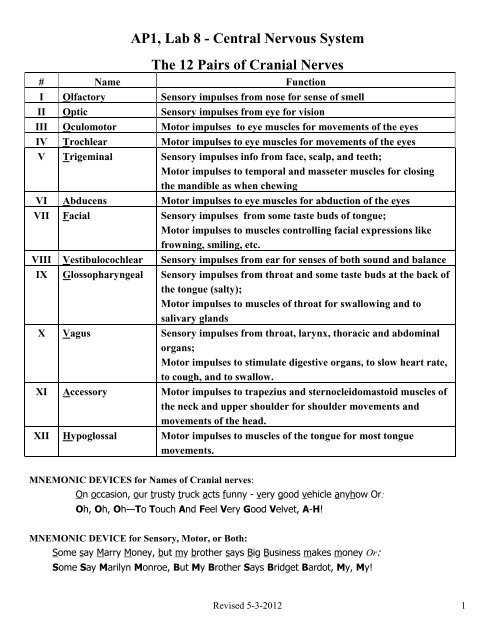
<strong>AP1</strong>, <strong>Lab</strong> 8 - Central Nervous System<br />
The 12 Pairs of Cranial Nerves<br />
# Name Function<br />
I Olfactory Sensory impulses from nose for sense of smell<br />
II Optic Sensory impulses from eye for vision<br />
III Oculomotor Motor impulses to eye muscles for movements of the eyes<br />
IV Trochlear Motor impulses to eye muscles for movements of the eyes<br />
V Trigeminal Sensory impulses info from face, scalp, and teeth;<br />
Motor impulses to temporal and masseter muscles for closing<br />
the mandible as when chewing<br />
VI Abducens Motor impulses to eye muscles for abduction of the eyes<br />
VII Facial Sensory impulses from some taste buds of tongue;<br />
Motor impulses to muscles controlling facial expressions like<br />
frowning, smiling, etc.<br />
VIII Vestibulocochlear Sensory impulses from ear for senses of both sound and balance<br />
IX Glossopharyngeal Sensory impulses from throat and some taste buds at the back of<br />
the tongue (salty);<br />
Motor impulses to muscles of throat for swallowing and to<br />
salivary glands<br />
X Vagus Sensory impulses from throat, larynx, thoracic and abdominal<br />
organs;<br />
Motor impulses to stimulate digestive organs, to slow heart rate,<br />
to cough, and to swallow.<br />
XI Accessory Motor impulses to trapezius and sternocleidomastoid muscles of<br />
the neck and upper shoulder for shoulder movements and<br />
movements of the head.<br />
XII Hypoglossal Motor impulses to muscles of the tongue for most tongue<br />
movements.<br />
MNEMONIC DEVICES for Names of Cranial nerves:<br />
On occasion, our trusty truck acts funny - very good vehicle anyhow Or:<br />
Oh, Oh, Oh—To Touch And Feel Very Good Velvet, A-H!<br />
MNEMONIC DEVICE for Sensory, Motor, or Both:<br />
Some say Marry Money, but my brother says Big Business makes money Or:<br />
Some Say Marilyn Monroe, But My Brother Says Bridget Bardot, My, My!<br />
Revised 5-3-2012 1
Visual learning: The figure below is especially useful to visual learners:<br />
There are 12 pairs of cranial nerves in humans.<br />
Their Roman numeral designations and<br />
names are:<br />
� I Olfactory<br />
� II Optic<br />
� III Oculomotor<br />
� IV Trochlear<br />
� V Trigeminal<br />
� VI Abducens<br />
� VII Facial<br />
� VIII<br />
Vestibulocochlear<br />
(auditory)<br />
� IX Glossopharyngeal<br />
� X Vagus<br />
� XI Accessory (spinal<br />
accessory)<br />
� XII Hypoglossal<br />
The cranial nerves mostly innervate structures in the head and neck.<br />
The cartoon codes for at least one major functional innervation of each cranial nerve. This drawing is<br />
based on one done by Beatrice Humphris, R.N. published in Nursing 1984.<br />
http://www.sci.uidaho.edu/med532/cranialnervebackgroundinfo.html<br />
Revised 5-3-2012 2
12 pairs of cranial nerves are associated with the ventral aspect of the brain.<br />
—The first 2 pairs attach to the forebrain, the rest originate from the brainstem.<br />
**Note: The Intermediate nerve included in the diagram above is actually the smaller root of the facial nerve (VII).<br />
Most images do not illustrate it separately. This one just happens to do so.<br />
Revised 5-3-2012 3
Test Your Cranial Nerves<br />
Now that you know the names and functions of the cranial nerves, let's test them. These tests will help you<br />
understand how the cranial nerves work. These tests are not meant to be a "clinical examination" of the cranial<br />
nerves.<br />
You will need to get a partner to help...both of you can serve as the experimenter (tester) and the subject. Record<br />
your observations of what your partner does and says.<br />
Olfactory Nerve (I): sensory for smell<br />
Gather some items with distinctive smells (for example, cloves, lemon, chocolate or coffee). Have your<br />
partner smell the items one at a time with each nostril. Have your partner record what the item is and the<br />
strength of the odor. Now you be the one who smells the items...have your partner use different smells<br />
for you.<br />
Optic Nerve (II): sensory for sight<br />
Use an eye chart (a "Snellen Chart") and have your partner try to read the lines at various distances away<br />
from the chart.<br />
Cranial nerves III, IV and VI are tested together. They each control muscles involved with eye movement.<br />
In addition, the oculomotor also functions in pupil dilation.<br />
Oculomotor Nerve (III), Trochlear Nerve (IV) and Abducens Nerve (VI): Motor<br />
These three nerves control eye movement and pupil diameter. Hold up a finger in front of your partner.<br />
Tell your partner to hold his or her head still and to follow your finger, then move your finger up and<br />
down, right and left. Do your partner's eyes follow your fingers?<br />
Check the pupillary response (oculomotor nerve): look at the diameter of your partner's eyes in dim light<br />
and also in bright light. Check for differences in the sizes of the right and left pupils.<br />
Trigeminal Nerve (V): sensory for touch/pain in face<br />
motor for chewing (mastication)<br />
The trigeminal nerve has both sensory and motor functions. To test the motor part of the nerve, tell your<br />
partner to close his or her jaws as if he or she was biting down on a piece of gum. Temporal and<br />
masseter muscles of the face should have equal strength<br />
To test the sensory part of the trigeminal nerve, lightly touch various parts of your partner's face with<br />
piece of cotton or a blunt object. Be careful not to touch your partner's eyes. Although much of the<br />
mouth and teeth are innervated by the trigeminal nerve, don't put anything into your subject's mouth.<br />
Facial Nerve (VII) Sensory for taste on anterior 2/3 of tongue and salivary glands<br />
Sensory to transmit information from ear to brain<br />
Motor for controlling muscles used in facial expression<br />
The motor part of the facial nerve can be tested by asking your partner to smile or frown or make funny<br />
faces. Check for symmetry.<br />
The sensory part of the facial nerve is responsible for taste on the front part of the tongue. You could try<br />
a few drops of sweet or salty water on this part of the tongue and see if your partner can taste it.<br />
Revised 5-3-2012 4
Vestibulocochlear Nerve (VIII): Sensory for sound and balance<br />
The vestibulocochlear nerve is a branching nerve. The cochlear nerve is responsible for hearing and the<br />
vestibular controls balance.<br />
Hearing: Have your partner cover one ear and close his or her eyes. Stand about 2 feet from the subject<br />
and whisper a word with 2 distinct syllables, like mailman, or football and observe for difficulties<br />
distinguishing words. You may also determine the distance at which he or she can hear the ticking of a<br />
clock or stopwatch.<br />
Balance: Test for balance by having the subject stand with his feet close together and eyes closed. The<br />
subject may sway a little, but should not fall.<br />
Cranial nerves IX and X can be tested together since they both serve to innervate the pharynx.<br />
Glossopharyngeal Nerve (IX): Sensory for taste on the posterior 1/3 of the tongue and conveying<br />
information from the tongue, tonsils and pharynx<br />
Motor for swallowing<br />
Have your partner drink some water and observe the swallowing reflex. Also the glossopharyngeal nerve<br />
is responsible for taste on the back part of the tongue. You could try a few drops of salty (or sugar) water<br />
on this part of the tongue and see if your partner can taste it.<br />
Vagus Nerve (X): Sensory from throat, larynx, thoracic and abdominal organs<br />
Motor for heart rate, swallowing, esophagus and stomach (digestion)<br />
Have the subject speak or cough. Her voice should be clear and strong. Cough should be strong. Subject<br />
should have a strong gag reflex.<br />
Spinal Accessory Nerve (XI): Motor to control the sternocleidomastoid and trapezius muscles involved in head<br />
movement<br />
Put your hands on your partner’s shoulders from the back. As you apply slight resistance, have him shrug<br />
his shoulders upward. Contraction should be symmetrical.<br />
Now place one hand on one side of your partner's jaw. Tell your partner to move his or her head toward<br />
your hand while you apply slight resistance. Repeat on the opposite side. Strength in the<br />
sternocleidomastoid should be symmetrical.<br />
Hypoglossal Nerve (XII): Motor for tongue movement<br />
Have your partner stick out his or her tongue and move it side to side. Check for symmetry.<br />
Revised 5-3-2012 5
DO YOU KNOW……?<br />
Which cranial nerve is the largest? _______________________________<br />
Which cranial nerves are responsible for eye movements? ______________<br />
What does "abducens" refer to? _________________________________<br />
Which cranial nerves carry gustatory (taste) information? ______________<br />
Which cranial nerve is the longest? _______________________________<br />
Which nerve or nerves are involved in:<br />
Rotating the head? _________________________________________<br />
Smelling a flower? _________________________________________<br />
Raising the eyelids, pupillary constriction? ________________________<br />
Slowing the heart rate? _____________________________________<br />
Increasing motility of the digestive tract? _______________________<br />
Bell’s Palsy? ______________________________________________<br />
Chewing food? ____________________________________________<br />
Listening to music? _________________________________________<br />
Seasickness? _____________________________________________<br />
Secretion of saliva? ________________________________________<br />
Sensation of taste? ________________________________________<br />
Rolling the eyes? (3 nerves)__________________________________<br />
Feeling a toothache? _______________________________________<br />
Reading a newspaper? ______________________________________<br />
Purely sensory in function? __________________________________<br />
Revised 5-3-2012 6
HOW THE BODY BLOCKS PAIN<br />
1. The Gate Control Theory:<br />
� Pain impulses are BLOCKED at the dorsal horns of the spinal cord and/or in the hypothalamus and/or<br />
midbrain by the release of natural opiates called ENDORPHINS and ENKEPHALINS. These (and possibly<br />
SEROTONIN) create IPSPs to cancel out EPSPs.<br />
� Fewer messages get through. The gate is partially “closed.”<br />
2. Pain messages aren’t really blocked at all but rather are “smothered” by pleasurable ones in the brain.<br />
� ENDORPHINS and ENKEPHALINS released in the brain inhibit the inhibition of the release of DOPAMINE.<br />
� DOPAMINE, a “feel-good” NT, floods the frontal lobes of the brain and smothers the unpleasant<br />
sensations with pleasant ones.<br />
3. In reality there is probably truth in both theories.<br />
HOW MEDICATIONS BLOCK PAIN:<br />
� ASPIRIN and IBUPROFEN inhibit prostaglandin activity thereby reducing inflammation and pain.<br />
� ACETAMINOPHEN (Tylenol) blocks pain but not inflammation by inhibiting activation of the COX<br />
enzyme.<br />
� NSAIDS are anti-inflammatory in a variety of ways; often as COX2 inhibitors<br />
� NARCOTICS such as morphine, Demerol, and heroin reduce pain by mimicking the effects of<br />
endorphins and enkephalins.<br />
Revised 5-3-2012 7
HOW A T.E.N.S. UNIT BLOCKS PAIN<br />
TENS = Transcutaneous Electrical Nerve Stimulation<br />
� Every day, ordinary afferent sensory impulses enter the spinal cord at the posterior horns of the gray<br />
matter. Here they must pass through synapses before traveling up to the brain via ascending tracts<br />
of the spinal cord. Pain messages follow the same pathway.<br />
� Applying continuous or repetitive stimulation through the skin of an appropriate area using TENS<br />
‘annoys’ the brain with a flood of sensory input. In response, the brain increases the frequency of<br />
impulses down the spinal cord to create IPSPs to “block” both the incoming messages from the<br />
skin as well as the pain messages.<br />
� Electrical stimulation of nerves using TENS also stimulates the release of endorphins so that fewer<br />
pain generating impulses reach the brain, so pain is not felt or at least is felt less.<br />
REFERRED PAIN<br />
� A painful sensation felt in a region of the body that is not the origin of the stimuli.<br />
� Usually, it’s a painful sensation of visceral origin felt in a somatic region of the body that is not the<br />
origin of the stimuli.<br />
Examples:<br />
1. Heart attack – the origin of the impulses is the heart muscle but the pain is often felt in the upper<br />
chest, left side of the mandible, and on the medial surface of L arm.<br />
2. Gallbladder and Liver – often felt at the top of the R shoulder.<br />
3. Kidney and Ureters – often felt in the lower back.<br />
� Happens because the visceral and somatic sensory neurons converge on the same ascending tracts of the<br />
spinal cord. When messages arrive on these tracts the brain can’t always distinguish between the two<br />
points of origin.<br />
PHANTOM PAIN<br />
� The sensation of pain experienced in a body part you no longer have – a missing foot, leg, arm, etc.<br />
� The brain receives no sensory information from the missing limb to inhibit pain sensations. If an action<br />
potential is initiated anywhere along the neuronal pathway from the severed limb, the idea of pain will be<br />
projected back to the missing limb even though the limb is not there.<br />
Revised 5-3-2012 8
Dissection of Cow Brain with Dura Mater<br />
� Use images from the dissection manuals in lab and your knowledge of the brain to identify the following.<br />
Recall the functions of those with an *.<br />
� Your group will be evaluated on the quality of your dissection. FOLLOW INSTRUCTIONS CAREFULLY.<br />
1. Glove up – your choice of latex or polyethylene.<br />
2. Safety – practice caution with the knives and other implements.<br />
3. Housekeeping (Your mother does not work here, kindly clean up after yourself) – Don’t put pieces of<br />
tissue in the sink. If you notice, there is no garbage disposal. At the end of lab, we will put brains back<br />
in the bag. Don’t get “slime” on the brain models. When you are finished, wash everything and return it<br />
where you found it.<br />
4. Open your bag and rinse away as much preservative as possible. Observe the dull white DURA MATER<br />
covering the surface of the brain.<br />
5. The two, large fatty masses dangling from the anterior and inferior surface are the tissues that were<br />
behind the eyes. Find the two OPTIC NERVES in these and then cut the fatty mass off.<br />
6. Use scissors to cut away the superior 90% of the DURA MATER. Leave the bottom portion intact as it will<br />
contain the pituitary gland. The ARACHNOID LAYER will come off with the dura mater leaving only the<br />
PIA MATER on the surface of the brain.<br />
7. Observe the LONGITUDINAL and TRANSVERSE FISSURES containing DURAL SINUSES as you remove the<br />
dura mater. What occurs at your dural sinuses? _______________<br />
1. Identify the following externally visible structures while the brain is still whole. No cutting necessary.<br />
CEREBRUM<br />
L and R CEREBRAL HEMISPHERES<br />
Various SULCI & GYRI<br />
OLFACTORY BULBS and OLFACTORY TRACTS. This is CRANIAL NERVE #1 carrying sensory<br />
messages to the brain for the sense of smell.<br />
Observe the delicate PIA MATER anchoring blood vessels to the cerebrum mostly in the sulci. These<br />
vessels are dark brown/black in color.<br />
CEREBELLUM<br />
OPTIC NERVES and OPTIC CHIASMA. Cut the optic nerves (cranial nerve #2) and other motor nerves as<br />
far out as you can clearly see them to get rid of those dangling lumps of fatty tissue that were<br />
located behind the eyes.<br />
Pituitary gland - usually appears as a nodule or “lump” approximately ¼ inch in diameter on the ventral<br />
side of the brain.)<br />
TRIGEMINAL NERVES - The lateral edges of the dura mater containing the pituitary gland also contain the<br />
large trigeminal nerves (cranial nerve #5). Cut the nerves near the dura mater to leave stumps on the<br />
lateral walls of the pons. Remove the dura mater containing any branches of this nerve but do not<br />
remove the pituitary gland.<br />
Revised 5-3-2012 9
OCULOMOTOR NERVES – Gently lift the pituitary gland from its anterior edge while looking for the<br />
oculomotor nerves (cranial nerve #3) to be raised with the dura mater and the pituitary gland. Go<br />
ahead and remove the dura mater and pituitary gland. The stumps of the oculomotor nerves will<br />
either be lying on the midbrain, projecting forward or they will have torn loose and be attached to the<br />
dura mater above the pituitary gland.<br />
BRAIN STEM<br />
Spinal cord – function?<br />
Medulla Oblongata – functions?<br />
Pons – functions?<br />
**Confirm identifications of everything up to this point with your instructor.<br />
2. Bisect the brain on the mid-sagittal plane along the longitudinal fissure all the way through the brain stem.<br />
Make one clean, straight cut all the way through. Try to cut as precisely in the middle as possible. Identify the<br />
following.<br />
Corpus Callosum and Fornix– recall function?<br />
Thalamus – recall functions?<br />
Hypothalamus – recall functions?<br />
Lateral ventricles - Use your metal probe to separate the corpus callosum and the fornix in order to see<br />
better into the lateral ventricles. The corpus callosum will be the roof and the fornix<br />
will be the floor of each ventricle.<br />
Third ventricle – Look above the thalamus but below the fornix.<br />
Choroid plexus - specialized capillaries for production of CSF. They are delicate and dark brown in color<br />
similar to the vessels on the surface of the cerebrum. Use your forceps to gently reach deep into the<br />
posterior portion of a lateral ventricle and pull some of this out. You might also find some in the 3 rd<br />
and 4 th ventricles.<br />
Cerebral aqueduct – passageway for CSF to flow from 3 rd to 4 th ventricle.<br />
Fourth ventricle – space between the cerebellum and spinal cord.<br />
3. Locate your thalamus again. Put the two hemispheres back together and perform a coronal section such that<br />
your cut passes through the thalamus. Hold the two posterior portions together and view them from the<br />
anterior side.<br />
Identify the following:<br />
Cerebral white matter<br />
Cerebral cortex - The term “cortex” refers to the outer layer of any structure.<br />
On the cow brain it’s the darker, more superficial layer that borders the white matter and follows<br />
the contour of the sulci. The majority of the synapses of the brain are found in the cortex.<br />
Corpus callosum<br />
Lateral ventricles<br />
Fornix<br />
Third ventricle<br />
Thalamus<br />
Revised 5-3-2012 10