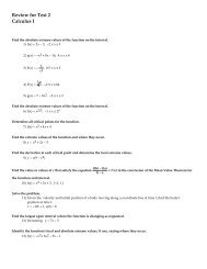
You also want an ePaper? Increase the reach of your titles
YUMPU automatically turns print PDFs into web optimized ePapers that Google loves.
OCULOMOTOR NERVES – Gently lift the pituitary gland from its anterior edge while looking for the<br />
oculomotor nerves (cranial nerve #3) to be raised with the dura mater and the pituitary gland. Go<br />
ahead and remove the dura mater and pituitary gland. The stumps of the oculomotor nerves will<br />
either be lying on the midbrain, projecting forward or they will have torn loose and be attached to the<br />
dura mater above the pituitary gland.<br />
BRAIN STEM<br />
Spinal cord – function?<br />
Medulla Oblongata – functions?<br />
Pons – functions?<br />
**Confirm identifications of everything up to this point with your instructor.<br />
2. Bisect the brain on the mid-sagittal plane along the longitudinal fissure all the way through the brain stem.<br />
Make one clean, straight cut all the way through. Try to cut as precisely in the middle as possible. Identify the<br />
following.<br />
Corpus Callosum and Fornix– recall function?<br />
Thalamus – recall functions?<br />
Hypothalamus – recall functions?<br />
Lateral ventricles - Use your metal probe to separate the corpus callosum and the fornix in order to see<br />
better into the lateral ventricles. The corpus callosum will be the roof and the fornix<br />
will be the floor of each ventricle.<br />
Third ventricle – Look above the thalamus but below the fornix.<br />
Choroid plexus - specialized capillaries for production of CSF. They are delicate and dark brown in color<br />
similar to the vessels on the surface of the cerebrum. Use your forceps to gently reach deep into the<br />
posterior portion of a lateral ventricle and pull some of this out. You might also find some in the 3 rd<br />
and 4 th ventricles.<br />
Cerebral aqueduct – passageway for CSF to flow from 3 rd to 4 th ventricle.<br />
Fourth ventricle – space between the cerebellum and spinal cord.<br />
3. Locate your thalamus again. Put the two hemispheres back together and perform a coronal section such that<br />
your cut passes through the thalamus. Hold the two posterior portions together and view them from the<br />
anterior side.<br />
Identify the following:<br />
Cerebral white matter<br />
Cerebral cortex - The term “cortex” refers to the outer layer of any structure.<br />
On the cow brain it’s the darker, more superficial layer that borders the white matter and follows<br />
the contour of the sulci. The majority of the synapses of the brain are found in the cortex.<br />
Corpus callosum<br />
Lateral ventricles<br />
Fornix<br />
Third ventricle<br />
Thalamus<br />
Revised 5-3-2012 10