Bond Strength of Self-etching Primer and Total-etch Adhesive ...
Bond Strength of Self-etching Primer and Total-etch Adhesive ...
Bond Strength of Self-etching Primer and Total-etch Adhesive ...
You also want an ePaper? Increase the reach of your titles
YUMPU automatically turns print PDFs into web optimized ePapers that Google loves.
JDC SCIENTIFIC ARTICLE<br />
<strong>Bond</strong> <strong>Strength</strong> <strong>of</strong> <strong>Self</strong>-<strong><strong>etch</strong>ing</strong> <strong>Primer</strong><br />
<strong>and</strong> <strong>Total</strong>-<strong>etch</strong> <strong>Adhesive</strong> Systems to<br />
Primary Dentin<br />
Carolina Paes Torres, DDS Silmara Aparecida Milori Corona, DDS, MS, PhD<br />
Renata Pereira Ramos, DDS, MS Regina Guenka Palma-Dibb, DDS, MS, PhD<br />
Maria Cristina Borsatto, DDS, MS, PhD<br />
ABSTRACT<br />
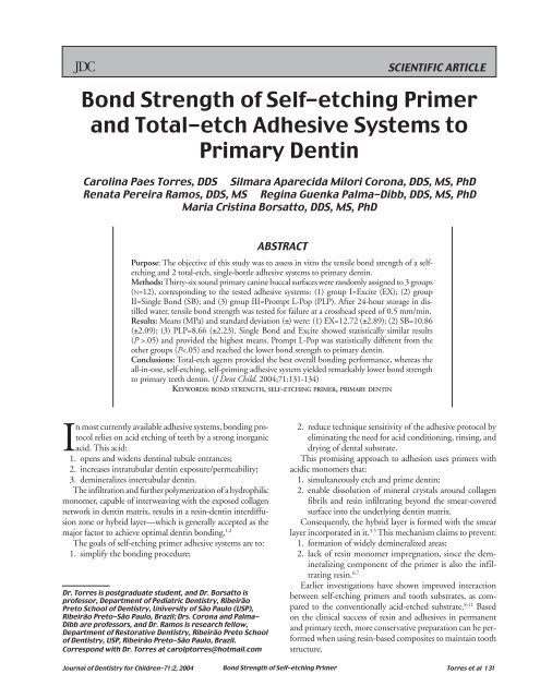
Purpose: The objective <strong>of</strong> this study was to assess in vitro the tensile bond strength <strong>of</strong> a self<strong><strong>etch</strong>ing</strong><br />
<strong>and</strong> 2 total-<strong>etch</strong>, single-bottle adhesive systems to primary dentin.<br />
Methods: Thirty-six sound primary canine buccal surfaces were r<strong>and</strong>omly assigned to 3 groups<br />
(N=12), corresponding to the tested adhesive systems: (1) group I=Excite (EX); (2) group<br />
II=Single <strong>Bond</strong> (SB); <strong>and</strong> (3) group III=Prompt L-Pop (PLP). After 24-hour storage in distilled<br />
water, tensile bond strength was tested for failure at a crosshead speed <strong>of</strong> 0.5 mm/min.<br />
Results: Means (MPa) <strong>and</strong> st<strong>and</strong>ard deviation (±) were: (1) EX=12.72 (±2.89); (2) SB=10.86<br />
(±2.09); (3) PLP=8.66 (±2.23). Single <strong>Bond</strong> <strong>and</strong> Excite showed statistically similar results<br />
(P >.05) <strong>and</strong> provided the highest means. Prompt L-Pop was statistically different from the<br />
other groups (P
Few articles investigate adhesive system bonding ability in<br />
primary teeth. 10-16 Due to an increasingly widespread use <strong>of</strong><br />
adhesive restorative systems, it seems relevant to assess behavior<br />
<strong>of</strong> such materials on primary tooth structure.<br />
The aim <strong>of</strong> this study was to assess the bond strength <strong>of</strong><br />
1 self-<strong><strong>etch</strong>ing</strong> <strong>and</strong> 2 total-<strong>etch</strong>, single-bottle adhesive systems<br />
to primary teeth dentin in vitro.<br />
METHODS<br />
Thirty-six sound primary canines exfoliated/extracted within<br />
a 6-month period <strong>and</strong> stored in 0.4% sodium azide solution<br />
at 4°C were selected <strong>and</strong> carefully cleaned with water/pumice<br />
slurry using dental prophylaxis cups. When necessary, roots<br />
were sectioned 2 mm below the amelocemental junction, <strong>and</strong><br />
crowns were embedded in polyester resin using polyvinyl chloride<br />
rings (2.1-cm diameter, 1.1-cm height).<br />
After resin polymerization, the rings were discarded <strong>and</strong><br />
the buccal surfaces <strong>of</strong> teeth were ground in a polishing<br />
machine (Politriz, Struers A/S, Copenhagen, DK-2610,<br />
Denmark) using water-cooled no. 320- to 400-grit silicon<br />
carbide (SiC) paper. This removed the overlying enamel <strong>and</strong><br />
exposed flat, smooth dentin surfaces.<br />
Additional grinding with no. 600-grit SiC paper was performed<br />
for 30 seconds to produce a st<strong>and</strong>ardized smear layer.<br />
To delimit the dentin bonding site, a piece <strong>of</strong> insulating tape<br />
with a 3-mm diameter central hole, created with a modified<br />
Ainsworth rubber-dam punch, was attached to the specimen<br />
surface. Limitation <strong>of</strong> the bonding site attempted to:<br />
1. define a fixed test surface so that the bond strengths recorded<br />
would be related solely to the evaluated area;<br />
2. warrant that the truncated resin composite cone would be<br />
further adhered precisely to the treated dentin surface, thus<br />
avoiding accidental adhesion to the surrounding enamel.<br />
The specimens were r<strong>and</strong>omly assigned to 3 groups <strong>of</strong> equal<br />
size (N=12), corresponding to the adhesive systems used:<br />
1. group I=Excite (EX), an ethanol-based, total-<strong>etch</strong> singlebottle<br />
bonding agent;<br />
2. group II=Single <strong>Bond</strong> (SB), an ethanol- <strong>and</strong> water-based<br />
total-<strong>etch</strong> adhesive system;<br />
3. group III=Prompt L-Pop (PLP), a self-<strong><strong>etch</strong>ing</strong> primer<br />
adhesive system.<br />
<strong>Adhesive</strong> systems for all groups were placed according<br />
to the manufacturers’ instructions. The agents were carefully<br />
applied onto the limited dentin surface with<br />
microbrush disposable brush tips. This avoided excess <strong>and</strong><br />
pooling <strong>of</strong> adhesive along the edges <strong>of</strong> the insulating tape<br />
that could compromise the distribution <strong>of</strong> tensions during<br />
the test <strong>and</strong> hence the validity <strong>of</strong> results.<br />
In group I, dentin surfaces were <strong>etch</strong>ed with a 35% phosphoric<br />
acid gel (Scotchbond Etchant, 3M/ESPE, ) for 10 seconds,<br />
rinsed thoroughly, <strong>and</strong> excess water was blotted with<br />
absorbing paper to keep the surface moist. Excite was applied<br />
to dental substrates with a light scrubbing motion for 10 seconds.<br />
Next, the remaining solvent was evaporated with a brief,<br />
mild air blast, <strong>and</strong> the bonding agent was light cured for 20<br />
seconds with a visible light curing unit using a 450 mW/cm 2<br />
output (XL 3000, 3M/ESPE).<br />
In group II, dentin sites were acid <strong>etch</strong>ed, rinsed <strong>and</strong> dried,<br />
as previously described. Then, 2 consecutive coats <strong>of</strong> Single<br />
<strong>Bond</strong> were applied to the <strong>etch</strong>ed surface, slightly thinned with<br />
a mild oil-free air stream, <strong>and</strong> light cured for 20 seconds.<br />
In group III, Prompt L-Pop components were mixed <strong>and</strong><br />
activated according to the manufacturer’s specifications, <strong>and</strong><br />
a uniform layer <strong>of</strong> the resulting mixture was applied onto dentin<br />
surfaces with a saturated microbrush, gently air thinned,<br />
left undisturbed for 15 seconds, <strong>and</strong> light cured.<br />
Once the bonding protocols were completed, specimens<br />
were individually fixed in a metallic clamping device (developed<br />
by Houston Biomaterial Research Center,) that allows<br />
the test dentin surface to remain parallel to a flat base. A splitbisected<br />
polytetrafluoroethylane jig was positioned on the<br />
tooth/resin block. This provided an inverted conical cavity<br />
with the smaller diameter coincident with the delimited bonding<br />
site (Ø 3 mm). A hybrid, light-cured composite resin (Filtek<br />
Z250, 3M/ESPE) was inserted into the jig in increments <strong>and</strong><br />
polymerized for 40 seconds each. As the cavity completely<br />
filled, the specimen was removed from the clamping device.<br />
The jig was opened <strong>and</strong> separated, leaving adhered to the<br />
delimited dentin site an inverted, truncated resin composite<br />
cone with a 6-mm diameter tapering to a 3-mm diameter<br />
<strong>and</strong> 4-mm height.<br />
After 24-hour storage in distilled water at 37°C, the coneshaped<br />
composite/polyester resin blocks were individually<br />
placed into an apparatus with an internal taper, corresponding<br />
to the resin cone’s shape. This configuration was loaded<br />
in tension to failure using a universal testing machine (Mod.<br />
MEM 2000,) at a crosshead speed <strong>of</strong> 0.5 mm/minute <strong>and</strong><br />
with 50 kgf load cell. This self-aligning system allows the<br />
forces to be applied perpendicular to the specimens’ surface.<br />
<strong>Bond</strong> strengths were recorded in kgf/cm <strong>and</strong> converted into<br />
MPA, <strong>and</strong> means were calculated. Sample distribution <strong>and</strong><br />
homogeneity were analyzed. Since a normal <strong>and</strong> homogeneous<br />
distribution was observed, data were submitted to<br />
one-way analysis <strong>of</strong> variance, using a factorial design with<br />
adhesive system as variable. Multiple comparisons were<br />
performed via Tukey test at 0.05 significance level.<br />
Fractured specimens were observed with a ×40 stereomicroscope<br />
to assess the failure modes, which were classified as<br />
adhesive, cohesive, or mixed.<br />
RESULTS<br />
Mean bond strengths <strong>and</strong> st<strong>and</strong>ard deviation to primary dentin<br />
are listed in Table 1.<br />
Table 1. <strong>Bond</strong> <strong>Strength</strong> Means (MPa) <strong>and</strong> St<strong>and</strong>ard<br />
Deviation (±) to Primary Dentin for the Experimental<br />
Groups*<br />
Groups<br />
Excite (I) Single <strong>Bond</strong> (II) Prompt L-Pop (III)<br />
12.72 (±4.89) ab 10.86 (±4.09) bc 8.66 (±2.23) c<br />
*Same letters indicate statistical similarity.<br />
132 Torres et al <strong>Bond</strong> <strong>Strength</strong> <strong>of</strong> <strong>Self</strong>-<strong><strong>etch</strong>ing</strong> <strong>Primer</strong> Journal <strong>of</strong> Dentistry for Children-71:2, 2004
The analysis <strong>of</strong> data revealed that, comparing the bonding<br />
agents assessed in this study, Single <strong>Bond</strong> <strong>and</strong> Excite total<strong>etch</strong><br />
adhesive systems showed statistically similar results (P>.05)<br />
<strong>and</strong> yielded the highest means to primary dentin. On the other<br />
h<strong>and</strong>, Prompt L-Pop self-<strong>etch</strong> primer reached a remarkably<br />
lower bond strength <strong>and</strong> was statistically different from the<br />
other groups (P
In spite <strong>of</strong> its expected bonding ability <strong>and</strong> advertised benefits,<br />
however, Prompt L-Pop did not perform as well as the<br />
total-<strong>etch</strong> adhesive systems. This leads to the assumption that<br />
an undermined clinical outcome may also be expected.<br />
Therefore, taking into account the outst<strong>and</strong>ing approach<br />
<strong>of</strong> adhesive dentistry in pediatric patients, it is m<strong>and</strong>atory that<br />
further studies be developed with the aim <strong>of</strong>:<br />
1. investigating<br />
a. bond strength;<br />
b. resin/dentin interface characteristics;<br />
c. the type <strong>of</strong> interaction occurring between the currently<br />
available self-<strong><strong>etch</strong>ing</strong> primer adhesive systems<br />
<strong>and</strong> the primary substrates;<br />
2. foreseeing, with some degree <strong>of</strong> reliability, the quality <strong>of</strong><br />
the adhesion obtained.<br />
CONCLUSIONS<br />
Based on the current study’s findings, <strong>and</strong> within the limitations<br />
<strong>of</strong> an in vitro investigation, it may be concluded that the<br />
tested total-<strong>etch</strong> agents provided the best overall bonding performance.<br />
The all-in-one, self-<strong><strong>etch</strong>ing</strong>, self-priming adhesive<br />
system, conversely, yielded a remarkably lower bond strength<br />
to primary teeth dentin.<br />
REFERENCES<br />
1. Nakabayashi N, Kojima K, Masuhara E. The promotion<br />
<strong>of</strong> adhesion by the infiltration <strong>of</strong> monomers into<br />
tooth substrates. J Biomed Mater Res. 1982;16:265-273.<br />
2. Nakabayashi N, Ashizawa M, Nakamura M. Identification<br />
<strong>of</strong> a resin-dentin hybrid layer in vital human dentin<br />
created in vivo: Durable bonding to vital dentin.<br />
Quintessence Int. 1992;23:135-141.<br />
3. Perdigão J, Lopes M, Lambrechts P, Leitão I, Van<br />
Meerbeek B, Vanherle G. Effect <strong>of</strong> a self-<strong><strong>etch</strong>ing</strong> primer<br />
on enamel shear bond strengths in SEM morphology.<br />
Am J Dent. 1997;10:141-146.<br />
4. Tay FR, Sano H, Carvalho RM, Pashley E, Pashley DH.<br />
An ultrastructural study <strong>of</strong> the influence <strong>of</strong> acidity <strong>of</strong><br />
self-<strong><strong>etch</strong>ing</strong> primers <strong>and</strong> smear layer thickness on bonding<br />
to intact dentin. J Adhes Dent. 2000;2:83-98.<br />
5. Tay FR, Pashley DH. Aggressiveness <strong>of</strong> contemporary<br />
self-<strong><strong>etch</strong>ing</strong> systems. I: Depth <strong>of</strong> penetration beyond<br />
dentin smear layers. Dent Mater. 2001;17:296-308.<br />
6. Ramos RP, Chimello DT, Chinelatti MA, Nonaka T,<br />
Pécora JD, Palma Dibb RG. Effect <strong>of</strong> Er:YAG laser on<br />
bond strength to dentin <strong>of</strong> a self-<strong><strong>etch</strong>ing</strong> primer <strong>and</strong> two<br />
single-bottle adhesive systems. Lasers Surg Med.<br />
2002;31:164-170.<br />
7. Sano H, Yoshikawa T, Pereira PNR, et al. Long-term<br />
durability <strong>of</strong> dentin bonds made with a self-<strong><strong>etch</strong>ing</strong><br />
primer, in vivo. J Dent Res. 1999;78:906-911.<br />
8. Watanabe I, Nakabayashi N, Pashley DH. <strong>Bond</strong>ing to<br />
ground dentin by a Phenyl-P self-<strong><strong>etch</strong>ing</strong> primer. J Dent<br />
Res. 1994;73:1212-1220.<br />
9. Miyasaka, K, Nakabayashi, N. Combination <strong>of</strong> EDTA<br />
conditioner <strong>and</strong> PHENYL-P/HEMA self-<strong><strong>etch</strong>ing</strong> primer<br />
for bonding to dentin. Dent Mater. 1999;15:153-157.<br />
10. Croll TP. <strong>Self</strong>-<strong><strong>etch</strong>ing</strong> adhesive system for resin bonding.<br />
J Dent Child. 2000;67:176-181.<br />
11. Fritz UB, Diedrich P, Finger WI. <strong>Self</strong>-<strong><strong>etch</strong>ing</strong> primer:<br />
An alternative to the conventional acid technique?<br />
J Or<strong>of</strong>ac Orthop. 2001;62:238-245.<br />
12. Elkins CJ, McCourt JW. <strong>Bond</strong> strength <strong>of</strong> dentinal<br />
adhesives in primary teeth. Quintessence Int. 1993;<br />
24:271-273.<br />
13. Truter MI, van der Vyver PJ, Nel JC. <strong>Bond</strong> strength <strong>of</strong><br />
composite resin bonded to deciduous <strong>and</strong> permanent<br />
dentin. J Dent Assoc S Afr. 1996;51:521-524.<br />
14. Fritz U, García-Godoy F, Finger WI. Enamel <strong>and</strong> dentin<br />
bond strength <strong>and</strong> bonding mechanism to dentin<br />
<strong>of</strong> Gluma CPS to primary teeth. J Dent Child. 1997;<br />
64:32-38.<br />
15. Pashley DH. Dentin: A dynamic substrate—a review.<br />
Scanning Microsc. 1989;3:161-176.<br />
16. Söderholm KJM. Correlation <strong>of</strong> in vivo <strong>and</strong> in vitro<br />
performance <strong>of</strong> adhesive restorative materials: A report<br />
<strong>of</strong> the ASCMD156 Task Group on test methods for<br />
the adhesion <strong>of</strong> restorative materials. Dent Mater.<br />
1991;7:74-83.<br />
17. Miyazaki M, Iwasaki K, Onose H. Adhesion <strong>of</strong> single<br />
application bonding systems to bovine enamel <strong>and</strong><br />
dentin. Oper Dent. 2002;27:88-94.<br />
18. Arrais CA, Giannini M. Morphology <strong>and</strong> thickness <strong>of</strong><br />
the diffusion <strong>of</strong> the resin through demineralized or<br />
unconditioned dental matrix. Pesqui Odontol Bras.<br />
2002;16:115-120.<br />
19. Lopes GC, Baratieri LN, Andrada MAC, Vieira LCC.<br />
Dental adhesion: Present state <strong>of</strong> the art <strong>and</strong> future<br />
perspectives. Quintessence Int. 2002;33:213-224.<br />
20. Perdigão J, Lopez M. Dentin bonding—questions for<br />
the new millenium. J Adhes Dent. 1999;1:191-209.<br />
21. Haller B. Recent developments in dentin bonding.<br />
Am J Dent. 2000;13:44-50.<br />
22. Van Meerbeek B, Vargas M, Inoue S, et al. <strong>Adhesive</strong>s<br />
<strong>and</strong> cements to promote preservation dentistry.<br />
Oper Dent. 2001;6:119-144.<br />
23. Agostini FG, Kaaden C, Powers JM. <strong>Bond</strong> strength <strong>of</strong><br />
self-<strong><strong>etch</strong>ing</strong> primers to enamel <strong>and</strong> dentin <strong>of</strong> primary<br />
teeth. Pediatr Dent. 2001;23:481-486.<br />
24. Miller J. Large tubules in dentine. J Dent Child.<br />
1981;48:269-271.<br />
25. Marshall GW, Inai N, Wu-Magidi IC. Dentin demineralization:<br />
Effects <strong>of</strong> dentin depth, pH <strong>and</strong> different acids.<br />
Dent Mater. 1997;13:338-347.<br />
26. Tay FR, Gwinnett AJ, Wei SHY. Micromorphological<br />
spectrum from over-drying to over-wetting acidconditioned<br />
dentin in water-free, acetone-based,<br />
single-bottle primer/adhesives. Dent Mater. 1996;<br />
12:236-244.<br />
27. Silva Telles PD, Aparecida M, Machado M, Nor JE. SEM<br />
study <strong>of</strong> a self-<strong><strong>etch</strong>ing</strong> primer adhesive system used for<br />
dentin bonding in primary <strong>and</strong> permanent teeth. Pediatr<br />
Dent. 2001;23:315-320.<br />
134 Torres et al <strong>Bond</strong> <strong>Strength</strong> <strong>of</strong> <strong>Self</strong>-<strong><strong>etch</strong>ing</strong> <strong>Primer</strong> Journal <strong>of</strong> Dentistry for Children-71:2, 2004


