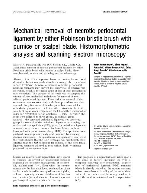
Mechanical removal of necrotic periodontal ligament by either ...
Mechanical removal of necrotic periodontal ligament by either ...
Mechanical removal of necrotic periodontal ligament by either ...

Create successful ePaper yourself
Turn your PDF publications into a flip-book with our unique Google optimized e-Paper software.
Dental Traumatology 2007; doi: 10.1111/j.1600-9657.2006.00472.x<br />
DENTAL TRAUMATOLOGY<br />
<strong>Mechanical</strong> <strong>removal</strong> <strong>of</strong> <strong>necrotic</strong> <strong>periodontal</strong><br />
<strong>ligament</strong> <strong>by</strong> <strong>either</strong> Robinson bristle brush with<br />
pumice or scalpel blade. Histomorphometric<br />
analysis and scanning electron microscopy<br />
Esper HR, Panzarini SR, Poi WR, Sonoda CK, Casatti CA.<br />
<strong>Mechanical</strong> <strong>removal</strong> <strong>of</strong> <strong>necrotic</strong> <strong>periodontal</strong> <strong>ligament</strong> <strong>by</strong> <strong>either</strong><br />
Robinson bristle brush with pumice or scalpel blade. Histomorphometric<br />
analysis and scanning electron microscopy.<br />
Abstract – One <strong>of</strong> the important factors accounting for successful<br />
delayed replantation <strong>of</strong> avulsed teeth is seemingly the type <strong>of</strong> root<br />
surface treatment. Removal <strong>of</strong> <strong>necrotic</strong> cemental <strong>periodontal</strong><br />
<strong>ligament</strong> remnants may prevent the occurrence <strong>of</strong> external root<br />
resorption, which is the major cause <strong>of</strong> loss <strong>of</strong> teeth replanted in<br />
such conditions. The purpose <strong>of</strong> this study was to compare the<br />
efficacy <strong>of</strong> two mechanical techniques for <strong>removal</strong> <strong>of</strong> rootadhered<br />
<strong>periodontal</strong> <strong>ligament</strong>. Preservation or <strong>removal</strong> <strong>of</strong> the<br />
cementum layer concomitantly with these procedures was also<br />
assessed. Forty-five roots <strong>of</strong> healthy premolars extracted for<br />
orthodontic purposes were selected. After extraction, the teeth<br />
were kept dry at room temperature for 1 h and then immersed in<br />
saline for rehydration for an additional 10 min. Thereafter, the<br />
roots were assigned to three groups, as follows: group 1<br />
(control) - the cemental <strong>periodontal</strong> <strong>ligament</strong> was preserved;<br />
group 2 - <strong>removal</strong> <strong>of</strong> the <strong>periodontal</strong> <strong>ligament</strong> <strong>by</strong> scraping root<br />
surface with a scalpel blade (SBS); group 3 - <strong>periodontal</strong> <strong>ligament</strong><br />
remnants were removed using a Robinson bristle brush at<br />
low-speed with pumice/water slurry (RBP). The specimens were<br />
analysed histomorphometrically and examined <strong>by</strong> scanning<br />
electron microscopy. The quantitative and qualitative analyses <strong>of</strong><br />
the results showed that the RBP technique was significantly more<br />
effective than the SBS technique for <strong>removal</strong> <strong>of</strong> the <strong>periodontal</strong><br />
<strong>ligament</strong> remnants adhered to root surface. Both techniques<br />
preserved the cementum layer.<br />
Helen Ramon Esper 1 ,Sônia Regina<br />
Panzarini 1 , Wilson Roberto Poi 1 , Celso<br />
Koogi Sonoda 1 , Cláudio Aparecido<br />
Casatti 2<br />
1 Discipline <strong>of</strong> Integrated Clinics, Department <strong>of</strong> Surgery and<br />
Integrated Clinics, Faculty <strong>of</strong> Dentistry <strong>of</strong> Araçatuba, UNESP,<br />
Araçatuba; 2 Discipline <strong>of</strong> Histology, Department <strong>of</strong> Basic<br />
Sciences, Faculty <strong>of</strong> Dentistry <strong>of</strong> Araçatuba, UNESP,<br />
Araçatuba, SP, Brazil<br />
Key words: delayed tooth replantation; <strong>periodontal</strong><br />
<strong>ligament</strong>; cementum<br />
Dra. Helen Ramon Esper, Departamento de Cirurgia e<br />
Clínica, Integrada, Faculdade de Odontologia do<br />
Campus de Araçatuba, UNESP, Rua José Bonifácio,<br />
1193, 1601-050 Araçatuba, SP, Brasil<br />
Tel.: +55 18 3636 3240<br />
Fax: +55 18 3636 3332<br />
e-mail: helen_esper@hotmail.com<br />
Accepted 1 November, 2005<br />
Studies on delayed tooth replantation have sought<br />
to elucidate the several yet unanswered questions<br />
regarding the treatment management <strong>of</strong> accidentally<br />
avulsed teeth (1–4). Even when the circumstances<br />
are far less than ideal, replantation <strong>of</strong> an<br />
avulsed tooth should be attempted because it yields,<br />
at least temporarily, the reestablishment <strong>of</strong> function<br />
and esthetics (5), and therefore has an important<br />
psychological impact on patient’s recovery.<br />
The prognosis <strong>of</strong> a replanted tooth relies upon a<br />
wide array <strong>of</strong> factors, including the type <strong>of</strong><br />
resorption occurring after replantation, degeneration<br />
<strong>of</strong> <strong>periodontal</strong> <strong>ligament</strong> (PDL) remnants<br />
(which is directly related to the trauma sustained<br />
and/or extra-alveolar handling <strong>of</strong> the root), desiccation<br />
<strong>of</strong> root surface and the storage medium in<br />
which the tooth is maintained until its replantation<br />
(6–9).<br />
Dental Traumatology 2007; 23: 333–339 Ó 2007 Blackwell Munksgaard 333
Esper et al.<br />
When an avulsed tooth is replanted with devitalized<br />
cemental <strong>periodontal</strong> <strong>ligament</strong> adhered to root<br />
surface, the granulation tissue may <strong>either</strong> be<br />
replaced <strong>by</strong> osseous tissue (10, 11) or trigger a<br />
resorption process (7, 12–14). In view <strong>of</strong> this, several<br />
techniques have been proposed for <strong>removal</strong> <strong>of</strong><br />
<strong>necrotic</strong> PDL remnants prior to delayed replantation<br />
(9, 10, 15–17) including mechanical <strong>removal</strong> <strong>by</strong><br />
scraping <strong>of</strong> the root surface with a scalpel blade (16),<br />
enzymatic techniques (18) or chemical <strong>removal</strong> <strong>by</strong><br />
immersion <strong>of</strong> the avulsed tooth in a sodium<br />
hypochlorite solution (15, 19–22). It has been<br />
advocated (13, 15, 20, 23) that whereas the use <strong>of</strong><br />
sodium hypochlorite does not seem to affect the<br />
cementum layer, scalpel blade scraping may result<br />
in <strong>removal</strong> <strong>of</strong> cementum together with the PDL<br />
remnants. The implications <strong>of</strong> such <strong>removal</strong> rely on<br />
the fact that the cementum layer seems to be more<br />
resistant to inflammatory resorption than dentin.<br />
Nevertheless, it has been reported that the roots<br />
<strong>of</strong> teeth treated with sodium hypochlorite before<br />
replantation appeared surrounded <strong>by</strong> a fibrous<br />
connective tissue with fibres organized parallel to<br />
the root surface without insertion areas (21). This<br />
fibrous capsule-like structure has been shown to<br />
compromise the stability and maintenance <strong>of</strong> the<br />
replanted tooth into the socket. The findings <strong>of</strong> a<br />
previous study investigating delayed rat teeth replantation<br />
after treatment <strong>of</strong> root surfaces with<br />
sodium hypochlorite at high concentrations (5%<br />
and 10%) showed premature exfoliation <strong>of</strong> the<br />
replanted teeth with no sign <strong>of</strong> root resorption (24).<br />
On account <strong>of</strong> these issues, the purpose <strong>of</strong> this<br />
study was to investigate, <strong>by</strong> means <strong>of</strong> histomorphometric<br />
analysis and scanning electron microscopy<br />
(SEM), the efficacy <strong>of</strong> two mechanical techniques<br />
for <strong>removal</strong> <strong>of</strong> root-adhered <strong>necrotic</strong> cemental<br />
<strong>periodontal</strong> <strong>ligament</strong>: Robinson bristle brush with<br />
pumice (RBP) or scalpel blade scraping (SBS).<br />
Preservation or <strong>removal</strong> <strong>of</strong> the cementum layer<br />
concomitantly with these procedures was also<br />
assessed.<br />
Material and methods<br />
Forty-five roots <strong>of</strong> noncarious, unrestored maxillary<br />
and mandibular premolars extracted for orthodontic<br />
reasons were utilized in this study. Immediately<br />
after extraction, the teeth were fixed <strong>by</strong> their<br />
crowns with wax on a flat base (Polidental Ind.<br />
e Com. Ltda, São Paulo, SP, Brazil) and left<br />
undisturbed at room temperature for 1 h. Thereafter,<br />
the teeth were individually immersed in disposable<br />
plastic recipients containing 50 ml <strong>of</strong> saline<br />
(Ariston Inds. Químs e Farms. Ltda, São Paulo, SP,<br />
Brazil), allowed for hydration for 10 min and<br />
assigned to three groups (n ¼ 15), as follows.<br />
Group 1 (Control): the <strong>periodontal</strong> <strong>ligament</strong> was<br />
preserved. Group 2 (SBS): the <strong>periodontal</strong> <strong>ligament</strong><br />
was removed gently <strong>by</strong> scraping root surfaces with a<br />
#15 scalpel blade (Medico International Trading<br />
Co. LTD., China) mounted on a #3 scalpel handle<br />
(J.O.N. Comércio de Produtos Odontológicos Ltda,<br />
São Paulo, SP, Brazil). Each root surface was<br />
scraped only once with the blade positioned<br />
perpendicularly to the long axis <strong>of</strong> the root and<br />
moved in a crown-apex direction. Group 3 (RBP):<br />
the <strong>periodontal</strong> <strong>ligament</strong> was removed using Robinson<br />
bristle brushes (Labor Dental Ltda, São Paulo,<br />
SP, Brazil) at low-speed and a polishing paste<br />
prepared with fine-grain pumice (Dental AG Ltda,<br />
São Paulo, SP, Brazil) and filtered water. Each root<br />
surface was polished in a crown-apex direction<br />
for 30 s and the roots were thoroughly rinsed<br />
with air-water spray to remove pumice residues<br />
completely.<br />
In all groups, two roots were processed for SEM<br />
examination. The other 13 roots were fixed in 10%<br />
neutral formalin for 24 h and then decalcified in<br />
EDTA solution (distilled water - 250 ml; EDTA -<br />
50 g; sodium hydroxide - 6.1 g) for 60 days. After<br />
decalcification, 10 roots were sectioned longitudinally<br />
and three roots were sectioned transversally at<br />
the middle third, and the fragments were included<br />
in paraffin. Semi-serial 6-lm-thick sections were<br />
obtained and stained with hematoxylin and eosin<br />
(H&E). All sections were examined histologically<br />
but only the longitudinal cuts were used for histomorphometric<br />
analysis.<br />
Histomorphometric analysis<br />
Ten longitudinal sections from different glass slides,<br />
each representative <strong>of</strong> one <strong>of</strong> the 10 teeth selected<br />
for histomorphometric analysis in each group, were<br />
chosen. To evaluate the entire root surface, the<br />
sections were further divided in smaller fragments<br />
and examined <strong>by</strong> an optical microscope with ·5<br />
magnification (Axiolab, Zeiss, Jena, Germany). The<br />
images were captured <strong>by</strong> a video camera (JVC<br />
TK-1270, Tokyo, Japan) and analysed using<br />
ImageLab 98 s<strong>of</strong>tware. In each fragment, the<br />
cementum and <strong>periodontal</strong> <strong>ligament</strong> perimeters<br />
were measured (in pixels) and the values were<br />
analysed statistically using one-way anova and the<br />
Tukey’s test.<br />
Scanning electron microscopy<br />
The teeth selected for SEM analysis were fixed in<br />
2.5% glutaraldehyde solution (ESM Electron Microscopy<br />
Sciences, Hatfield, PA, USA) and 4% paraformaldehyde<br />
(Sigma Chemical CO., St. Louis,<br />
MO, USA) in a 0.1 M sodium phosphate buffer at<br />
334 Dental Traumatology 2007; 23: 333–339 Ó 2007 Blackwell Munksgaard
Periodontal <strong>ligament</strong> <strong>removal</strong> <strong>by</strong> two mechanical techniques<br />
pH 7.4, for 12 h at 4°C. The specimens were<br />
washed in sodium phosphate buffer for 20 min and<br />
immersed in this solution for additional 12 h.<br />
Thereafter, the specimens were postfixed with 1%<br />
osmium tetroxide (ESM Electron Microscopy Sciences,<br />
Hatfield, PA, USA) for 2 h and received four<br />
20-minute washes with phosphate buffer solution.<br />
They were then dehydrated in a graded ethanol<br />
series (50, 70, 80, 90, 95 and 100%; 20 min each)<br />
and the critical point was obtained with liquid<br />
carbon dioxide (ESM Electron Microscopy Sciences,<br />
Hatfield, PA, USA). The specimens were<br />
mounted on metallic stubs with double-faced adhesive<br />
tape, sputter-coated with gold (Desk II, Denton<br />
Vacuum, Moorestown; NJ, USA) and kept in a<br />
vacuum chamber until examination with a scanning<br />
electron microscope (JSM 5410, JEOL, Tokyo,<br />
Japan).<br />
Fig. 2. Group 1 (Control) - Intact <strong>periodontal</strong> <strong>ligament</strong> (PL)<br />
and cementum (C). D ¼ Dentin. Longitudinal section. H&E<br />
(Original magnification ·160).<br />
Results<br />
Histomorphometric analysis<br />
The results were obtained after qualitative analysis<br />
<strong>of</strong> dentin, cementum and <strong>periodontal</strong> <strong>ligament</strong>.<br />
Group 1 (Control): in all specimens, cemental<br />
<strong>periodontal</strong> <strong>ligament</strong> was observed throughout the<br />
root surface. The cementum layer was intact and<br />
the insertion <strong>of</strong> PDL fibres could be clearly identified<br />
(Figs. 1 and 2).<br />
Group 2 (SBS): in both transversal (Fig. 3) and<br />
longitudinal sections, small areas with <strong>periodontal</strong><br />
<strong>ligament</strong> adhered to the cemental surface were<br />
observed. The cementum layer appeared preserved<br />
in all specimens (Fig. 4).<br />
Group 3 (RBP): both transversal (Fig. 5) and<br />
longitudinal (Fig. 6) sections exhibited small areas<br />
<strong>of</strong> <strong>periodontal</strong> <strong>ligament</strong> remnants adhered to the<br />
root surface. In this group, however, lesser<br />
amount <strong>of</strong> PDL fibres was noted. The cementum<br />
layer appeared preserved in all specimens<br />
(Fig. 6).<br />
Fig. 3. Group 2 (Scalpel blade scraping) - Small areas <strong>of</strong><br />
<strong>periodontal</strong> <strong>ligament</strong> (PL) adhered to cementum (C). D ¼ Dentin.<br />
Transversal section. H&E (Original magnification ·160).<br />
Fig. 4. Group 2 (Scalpel blade scraping) - Small areas <strong>of</strong><br />
<strong>periodontal</strong> <strong>ligament</strong> (PL) adhered to cementum (C). D ¼ Dentin.<br />
Transversal section. H&E (Original magnification ·160).<br />
Fig. 1. Group 1 (Control) - Intact <strong>periodontal</strong> <strong>ligament</strong> (PL)<br />
and cementum (C). D ¼ Dentin. Longitudinal section. H&E<br />
(Original magnification ·63).<br />
Scanning electron microscopy<br />
The middle third area <strong>of</strong> each specimen was<br />
observed at ·1000 magnification and the same<br />
structures, i.e., dentin, cementum and <strong>periodontal</strong><br />
<strong>ligament</strong>, were examined in all groups (Fig. 7). The<br />
SEM micrographs were consistent with the histomorphologic<br />
findings. In the control group, an<br />
extensive area <strong>of</strong> <strong>periodontal</strong> <strong>ligament</strong> fibres<br />
adhered to root surfaces was observed (Fig. 8). In<br />
Dental Traumatology 2007; 23: 333–339 Ó 2007 Blackwell Munksgaard 335
Esper et al.<br />
Fig. 5. Group 3 (Robinson bristle brush with pumice) - Small<br />
areas <strong>of</strong> <strong>periodontal</strong> <strong>ligament</strong> (PL) adhered to cemental surface<br />
(C). D ¼ Dentin. Transversal section. H&E (Original magnification<br />
·63).<br />
Fig. 8. Group 1 (control) - Extensive root area covered <strong>by</strong><br />
<strong>periodontal</strong> <strong>ligament</strong> (PL) fibres.<br />
Fig. 6. Group 3 (Robinson bristle brush with pumice) - Absence<br />
<strong>of</strong> <strong>periodontal</strong> <strong>ligament</strong> fibres and intact cementum (C).<br />
Transversal section. D ¼ Dentin. H&E (Original magnification<br />
·160).<br />
Fig. 9. Group 2 (Scalpel blade scraping) - Great amount <strong>of</strong><br />
<strong>periodontal</strong> <strong>ligament</strong> remnants (arrows). Preserved cementum<br />
layer (C).<br />
Fig. 10. Group 3 (Robinson bristle brush with pumice) -<br />
Smaller amount <strong>of</strong> <strong>periodontal</strong> <strong>ligament</strong> (PL) remnants.<br />
Preserved cementum layer (C). No exposed dentinal tubules<br />
can be identified.<br />
Fig. 7. Scanning electron micrograph <strong>of</strong> root surface. The<br />
demarcated area corresponds to the middle third.<br />
the SBS group, although the scraped root surface<br />
exhibited grooves caused <strong>by</strong> the scalpel blade, still a<br />
large amount <strong>of</strong> adhered <strong>periodontal</strong> <strong>ligament</strong><br />
remnants were observed. In the areas with absent<br />
<strong>periodontal</strong> <strong>ligament</strong>, an intact cementum lining<br />
was clearly identified without dentin exposure<br />
(Fig. 9). The RBP group showed considerably lesser<br />
amount <strong>of</strong> PDL remnants adhered to cementum<br />
surface and no exposure <strong>of</strong> dentinal tubules<br />
(Fig. 10).<br />
Statistical analysis<br />
One-way anova and the Tukey’s test showed highly<br />
significant difference between the surface treatments<br />
(P < 0.0001), the RBP technique presenting better<br />
results.<br />
336 Dental Traumatology 2007; 23: 333–339 Ó 2007 Blackwell Munksgaard
Periodontal <strong>ligament</strong> <strong>removal</strong> <strong>by</strong> two mechanical techniques<br />
Discussion<br />
The treatment guidelines for avulsed teeth recommend<br />
that first assistance be provided at the<br />
accident site immediately after sustaining the traumatic<br />
injury. The findings <strong>of</strong> a prospective study (9)<br />
evaluating the <strong>periodontal</strong> <strong>ligament</strong> healing in 400<br />
avulsed and replanted permanent teeth confirmed<br />
that immediate replantation provides the best<br />
prognosis for avulsed teeth and clearly has an<br />
overwhelming influence on the outcome <strong>of</strong> replantation.<br />
Nevertheless, delayed replantation is so far a<br />
clinical reality because trauma patients are not<br />
usually seen <strong>by</strong> a dentist readily enough to allow for<br />
an optimal treatment to be rendered. Clinical<br />
practise has shown that most avulsed teeth are<br />
replanted after long extra-alveolar times. Furthermore,<br />
in the majority <strong>of</strong> the cases, the first<br />
procedures are not correctly performed due to lack<br />
<strong>of</strong> knowledge <strong>of</strong> dental trauma management and<br />
replantation protocol (4, 25–27) and unawareness <strong>of</strong><br />
the best media for transportation <strong>of</strong> the avulsed<br />
tooth to the dental <strong>of</strong>fice. It is well documented that<br />
the survival rate <strong>of</strong> avulsed permanent teeth following<br />
replantation is affected primarily <strong>by</strong> the duration<br />
<strong>of</strong> the extra-buccal period and the nature <strong>of</strong> the<br />
storage conditions.<br />
Studies have shown that even after a short extraalveolar<br />
dry storage period (up to 30 min), only a<br />
small percentage <strong>of</strong> cells remain viable, because <strong>of</strong><br />
the rapid decrease in the proliferating capacity <strong>of</strong><br />
<strong>periodontal</strong> <strong>ligament</strong> cells (7, 28, 29). There is a<br />
consensus in the literature that the devitalized<br />
<strong>periodontal</strong> <strong>ligament</strong> should be removed when<br />
tooth replantation is delayed because the <strong>necrotic</strong><br />
remnants seem to stimulate the occurrence <strong>of</strong><br />
external root resorption (9, 10, 15, 16, 30, 31).<br />
According to the treatment guidelines for avulsed<br />
teeth replanted after an extended extra-alveolar<br />
period (31), <strong>removal</strong> <strong>of</strong> cemental PDL fibres should<br />
be the first step in root surface treatment prior to<br />
replantation.<br />
Several techniques have been described for the<br />
<strong>removal</strong> <strong>of</strong> <strong>necrotic</strong> <strong>periodontal</strong> <strong>ligament</strong>, including<br />
scaling <strong>of</strong> root surfaces with scalpel blades (16) and<br />
<strong>periodontal</strong> curettes (32–34). However, there are no<br />
reported studies attesting the efficacy <strong>of</strong> such<br />
methods with respect to not only the <strong>removal</strong> <strong>of</strong><br />
PDL remnants but also the preservation <strong>of</strong> the<br />
cementum layer, which is an important barrier<br />
against external root resorption (15, 23).<br />
In addition to mechanical procedures, chemical<br />
agents, such as sodium hypochlorite at different<br />
concentrations, have been indicated for <strong>removal</strong> <strong>of</strong><br />
<strong>necrotic</strong> <strong>periodontal</strong> <strong>ligament</strong> (19, 21, 22, 24). Even<br />
though it is an excellent solvent <strong>of</strong> organic material<br />
that efficiently removes PDL remnants (22)<br />
preserving the cementum layer, sodium hypochlorite<br />
is considered to be toxic for the <strong>periodontal</strong><br />
tissues (35), possibly because <strong>of</strong> its high pH.<br />
Experimental studies in dog and rat teeth using<br />
sodium hypochlorite for root surface treatment prior<br />
to replantation, revealed the formation <strong>of</strong> a connective<br />
tissue similar to a fibrous capsule that<br />
interfered with the maintenance <strong>of</strong> the replanted<br />
tooth into the alveolus (21, 24).<br />
This study was designed to investigate whether<br />
two mechanical techniques were able to remove<br />
root-adhered <strong>periodontal</strong> <strong>ligament</strong> remnants without<br />
causing any alterations to root surface pH, and<br />
preserve the cementum layer.<br />
Kenny et al. (36), in a study on the effect <strong>of</strong><br />
Emdogain in delayed replantation, evaluated the<br />
<strong>removal</strong> <strong>of</strong> root-adhered cemental <strong>periodontal</strong> <strong>ligament</strong><br />
using conventional rubber prophylaxis cups<br />
loaded with wet pumice. They advocated that this<br />
method produces minimal root surface damage and<br />
does not leave any organic residues. The methodology<br />
<strong>of</strong> the present study was designed based on<br />
these outcomes, however we used Robinson bristle<br />
brushes instead <strong>of</strong> rubber cups because the morphology<br />
<strong>of</strong> the brushes would yield a more effective<br />
<strong>removal</strong> <strong>of</strong> PDL remnants.<br />
Pumice is a siliceous material <strong>of</strong> volcanic origin<br />
that may be satisfactorily used both as an abrasive<br />
material and as a polishing agent, depending on the<br />
particle size (37). For this study, fine-grain pumice<br />
slurry was applied at low-speed for 30 s on each face<br />
<strong>of</strong> the roots to avoid unnecessary cementum abrasion,<br />
as <strong>removal</strong> <strong>of</strong> root-adhered PDL remnants<br />
could be observed macroscopically. The methodology<br />
adopted attempted to reproduce the clinical<br />
conditions as close as possible, i.e., the teeth were<br />
kept dry for 1 h and then hydrated <strong>by</strong> immersion in<br />
saline to facilitate the <strong>removal</strong> <strong>of</strong> <strong>necrotic</strong> cemental<br />
PDL. Pumice and Robinson bristle brushes mounted<br />
in a low-speed handpiece are routinely found in<br />
dental <strong>of</strong>fices.<br />
In this study, scalpel blade scraping was chosen<br />
amongst other mechanical techniques because it is a<br />
feasible procedure to be done at hospitals and<br />
emergency rooms, where first-aid care is usually<br />
provided in trauma cases, and was based on the<br />
methodology <strong>of</strong> a previous in vivo investigation (16).<br />
We evaluated the efficacy <strong>of</strong> a #15 blade to remove<br />
root-adhered PDL fibres (preserving the cementum<br />
layer), and compared its performance to that <strong>of</strong> a<br />
presumably less aggressive technique using Robinson<br />
bristle brushes and pumice. Control group<br />
served as a reference <strong>of</strong> the amount <strong>of</strong> <strong>periodontal</strong><br />
<strong>ligament</strong> present and the aspect <strong>of</strong> the cementum<br />
layer without any root surface treatment.<br />
The longitudinal root sections selected for<br />
histomorphometric analysis allowed for thorough<br />
Dental Traumatology 2007; 23: 333–339 Ó 2007 Blackwell Munksgaard 337
Esper et al.<br />
visualization <strong>of</strong> root extension, from the amelocemental<br />
junction to tooth apex. SEM examination <strong>of</strong><br />
the treated root surfaces at different magnifications<br />
(·35, ·100, ·500 and ·1000) revealed the presence<br />
<strong>of</strong> both cementum and <strong>periodontal</strong> <strong>ligament</strong> fibre<br />
remnants.<br />
The findings <strong>of</strong> this study showed that, unlike<br />
previously assumed, the cementum layer was preserved<br />
when the <strong>periodontal</strong> <strong>ligament</strong> fibres were<br />
removed <strong>by</strong> scraping with a scalpel blade. Although<br />
the SEM micrographs revealed the presence <strong>of</strong><br />
grooves produced <strong>by</strong> the blade in few areas, a<br />
continuous cementum lining was clearly identified<br />
in all specimens. The technique using Robinson<br />
brush plus pumice, in addition to preserve the<br />
cementum layer, was more effective in removing the<br />
<strong>periodontal</strong> <strong>ligament</strong> than the scalpel blade. Lesser<br />
amount <strong>of</strong> root-adhered PDL remnants was observed<br />
in the specimens <strong>of</strong> the RBP group. A<br />
noteworthy observation, however, was the limitation<br />
<strong>of</strong> both methods to detach <strong>periodontal</strong> <strong>ligament</strong><br />
fibres from root surface close to enamel.<br />
The outcome <strong>of</strong> this study indicates that for<br />
delayed tooth replantation requiring <strong>removal</strong> <strong>of</strong><br />
<strong>necrotic</strong> <strong>periodontal</strong> <strong>ligament</strong>, both scalpel blade<br />
scraping and Robinson bristle brush with pumice<br />
preserved the cementum layer while removing PDL<br />
remnants. However, brush/pumice technique<br />
yielded better efficacy in <strong>ligament</strong> <strong>removal</strong>.<br />
Conclusions<br />
The findings <strong>of</strong> the histomorphometric analysis and<br />
scanning electron microscopy examination revealed<br />
that:<br />
1 The cementum layer remained intact in the<br />
control and experimental groups alike.<br />
2 The use <strong>of</strong> Robinson bristle brush with pumice<br />
was more effective than scalpel blade for<br />
<strong>removal</strong> <strong>of</strong> root-adhered <strong>periodontal</strong> <strong>ligament</strong><br />
remnants.<br />
References<br />
1. Grossman LI, Ship II. Survival rate <strong>of</strong> replanted teeth. Oral<br />
Surg Oral Med Oral Pathol 1970;29:899–906.<br />
2. Kemp WB, Mourino AP. Accidental extraction and<br />
replantation <strong>of</strong> an immature permanent tooth. J Endod<br />
1977;3:240–1.<br />
3. Barrett EJ, Kenny DJ. Avulsed permanent teeth: a review <strong>of</strong><br />
the literature and treatment guidelines. Endod Dent<br />
Traumatol 1997;13:153–63.<br />
4. Panzarini SR, Saad-Neto M, Sonoda C, Poi WR, Perri de<br />
Carvalho AC. Avulsões dentárias em pacientes jovens e<br />
adultos na região de Araçatuba. Rev Assoc Paul Cir Dent<br />
2003;57:27–31.<br />
5. Duggal MS, Toumba KJ, Russell JL, Paterson SA.<br />
Replantation <strong>of</strong> avulsed permanent teeth with avital<br />
<strong>periodontal</strong> <strong>ligament</strong>s: a case report. Endod Dent Traumatol<br />
1994;10:282–5.<br />
6. Andreasen JO, Hjørting-Hansen E. Replantation <strong>of</strong> teeth.<br />
II. Histological study <strong>of</strong> 22 replanted anterior teeth in<br />
humans. Acta Odontol Scand 1966;24:286–306.<br />
7. Andreasen JO. Effect <strong>of</strong> extra-alveolar period and storage<br />
media upon <strong>periodontal</strong> and pulpal healing after replantation<br />
<strong>of</strong> mature permanent incisors in monkeys. Int J Oral<br />
Surg 1981a;10:43–53.<br />
8. Blomlöf L. Storage <strong>of</strong> human <strong>periodontal</strong> <strong>ligament</strong> cells in a<br />
combination <strong>of</strong> different media. J Dent Res 1981;60:1904–<br />
6.<br />
9. Andreasen JO, Borum M, Jacobsen HL, Andreasen FM.<br />
Replantation <strong>of</strong> 400 avulsed permanent incisors. 4. Factors<br />
related to <strong>periodontal</strong> <strong>ligament</strong> healing. Endod Dent<br />
Traumatol 1995;11:76–89.<br />
10. Andreasen JO. Analysis <strong>of</strong> pathogenesis and topography <strong>of</strong><br />
replacement root resorption (ankylosis) after replantation <strong>of</strong><br />
mature permanent incisors in monkeys. Sweed Dent J<br />
1980;4:231–40.<br />
11. Andreasen JO. Interrelation between alveolar bone and<br />
<strong>periodontal</strong> <strong>ligament</strong> repair after replantation <strong>of</strong> mature <strong>of</strong><br />
permanent incisors in monkeys. J Periodontal Res<br />
1981b;16:228–35.<br />
12. Andreasen JO. Relationship between cell damage in the<br />
<strong>periodontal</strong> <strong>ligament</strong> after replantation and subsequent<br />
development <strong>of</strong> root resorption: a time-related study in<br />
monkeys. Acta Odont Scand 1981c;39:15–25.<br />
13. Hammarström L, Lindskog S. General morphological<br />
aspects <strong>of</strong> resorption <strong>of</strong> teeth and alveolar bone. Int Endod<br />
J 1985;18:93–108.<br />
14. Hammarström L, Pierce A, Blomlöf L, Feiglin B, Lindskog<br />
S. Tooth avulsion and replantation: a review. Endod Dent<br />
Traumatol 1986;2:1–8.<br />
15. Lindskog S, Pierce AM, Blomlöf L, Hammarström L. The<br />
role <strong>of</strong> <strong>necrotic</strong> <strong>periodontal</strong> membrane in cementum resorption<br />
and ankylosis. Endod Dent Traumatol 1985;1:96–101.<br />
16. Okamoto T, Hanada E, Saad Neto M. Reimplante<br />
mediato de incisivo superior de rato sem e com <strong>ligament</strong>o<br />
<strong>periodontal</strong> cementário: estudo histológico. Rev Odontol<br />
UNESP 1986/87;15/16:53–64.<br />
17. Hammarström L, Lindskog S. Factors regulating and<br />
modifying dental resorption. Proc Finn Dent Soc<br />
1992;88:115–23.<br />
18. Nevins AJ, Porta LA, Borden BG, Lorenzo P. Replantation<br />
<strong>of</strong> enzimatically treated teeth in monkeys. Part 1. Oral Surg<br />
Oral Med Oral Pathol 1980;50:277–81.<br />
19. Percinoto C, Campos Russo M, Lima JE, , Andrioni JN,<br />
Benfati SV, Bertoz FA.Processo de reparo em dentes<br />
reimplantados após a remoção química das fibras periodontais<br />
radiculares. Rev Odontol UNESP 1988;17:73–81.<br />
20. Ehnevid H, Lindskog S, Jansson L, Blöml<strong>of</strong> L. Tissue<br />
formation on cementum surfaces in vivo. Sweed Dent J<br />
1993;17:1–8.<br />
21. Kanno CM, Saad Neto M, Oliveira JA, Escobar CAB,<br />
Saito CTMH. Efeito do hipoclorito de sódio sobre o<br />
<strong>ligament</strong>o <strong>periodontal</strong> de incisivos de ratos. Arq Odontol<br />
2001;37:35–43.<br />
22. Poi WR, Panzarini SR, Sonoda CK, Fernandes U, Mori<br />
GG. Influência do volume de hipoclorito de sódio a 1% na<br />
remoção do <strong>ligament</strong>o <strong>periodontal</strong> necrosado. Rev Assoc<br />
Paul Cir Dent 2001;55:286–90.<br />
23. Lindskog S, Hammarström L. Evidence in favor <strong>of</strong> an antiinvasion<br />
factor in cementum or <strong>periodontal</strong> membrane <strong>of</strong><br />
human teeth. Scand J Dent Res 1980;88:161–3.<br />
24. Sonoda CK, Poi WR, Okamoto T, Toyota E, Takeda RH.<br />
Reimplante imediato de dentes após o tratamento da raiz<br />
com solução de hipoclorito de sódio a 1%, 2,5%, 5% e<br />
10%. Rev Bras Odontol 2000;57:293–6.<br />
25. Hamilton FA, Hill RJ, Mackie IC. Investigation <strong>of</strong> lay<br />
knowledge <strong>of</strong> the management <strong>of</strong> avulsed permanent<br />
incisors. Endod Dent Traumatol 1997;13:19–23.<br />
338 Dental Traumatology 2007; 23: 333–339 Ó 2007 Blackwell Munksgaard
Periodontal <strong>ligament</strong> <strong>removal</strong> <strong>by</strong> two mechanical techniques<br />
26. Poi WR, Salineiro SL, Miziara FV, Miziara EV. A<br />
educação como forma de favorecer o prognóstico do<br />
reimplante dental. Rev Assoc Paul Cir Dent 1999;53:474–9.<br />
27. Chan AW, Wong TK, Cheung GS. Lay knowledge <strong>of</strong><br />
physical education teachers about management <strong>of</strong> dental<br />
trauma in Hong Kong. Dent Traumatol 2001;17:77–85.<br />
28. Lekic P, Kenny D, Moe HK, Barrett E, Mccullcch CAG.<br />
Relationship <strong>of</strong> clonogenic capacity to plating efficiency and<br />
vital dye staining <strong>of</strong> human <strong>periodontal</strong> <strong>ligament</strong> cells:<br />
implications for tooth replantation. J Periodontal Res<br />
1996;31:294–300.<br />
29. Kenny DJ, Barrett EJ. Recent developments in dental<br />
traumatology. Pediatr Dent 2001;23:464–8.<br />
30. Trope, M. Treatment <strong>of</strong> the avulsed tooth. Pediatr Dent<br />
2000;22:145–7.<br />
31. Flores MT, Andreasen JO, Bakland LK. Guidelines for the<br />
evaluation and management <strong>of</strong> traumatic dental injuries.<br />
Dent Traumatol 2001;17:193–6.<br />
32. Van Hassel HJ, Osvald RJ, Harrington GW. Replantation.<br />
2. The role <strong>of</strong> the <strong>periodontal</strong> <strong>ligament</strong>. J Endod<br />
1980;6:506–8.<br />
33. López NJ, Belvederessi M. Healing following implantation<br />
<strong>of</strong> healthy roots, with and without <strong>periodontal</strong> <strong>ligament</strong><br />
tissue in the oral mucosa. J Periodontol 1983;54:283–90.<br />
34. Nyman S, Houston F, Sarhed G, Lindhe J, Karring T.<br />
Healing following reimplantation <strong>of</strong> teeth subjected to root<br />
planning and citric acid treatment. J Clin Periodontol<br />
1985;12:294–305.<br />
35. Chang Y, Huang FM, Tai KW, Chou MY. The effect <strong>of</strong><br />
sodium hypochlorite and chlorhexidine on cultured human<br />
<strong>periodontal</strong> <strong>ligament</strong> cells. Oral Surg Oral Med Oral Pathol<br />
Oral Radiol Endod 2001;92:446–50.<br />
36. Kenny DJ, Barrett EJ, Johnson DH, Sigal MJ, Tenenbaum<br />
HC. Clinical Management <strong>of</strong> avulsed permanent incisors<br />
using Emdogain: initial report <strong>of</strong> an investigation. J Can<br />
Dent Assoc 2000;66:21–9.<br />
37. Skinner EW. Materiais dentários. Rio de Janeiro: Guanabara;<br />
1993. p. 334.<br />
Dental Traumatology 2007; 23: 333–339 Ó 2007 Blackwell Munksgaard 339