Mechanical removal of necrotic periodontal ligament by either ...
Mechanical removal of necrotic periodontal ligament by either ...
Mechanical removal of necrotic periodontal ligament by either ...

You also want an ePaper? Increase the reach of your titles
YUMPU automatically turns print PDFs into web optimized ePapers that Google loves.
Esper et al.<br />
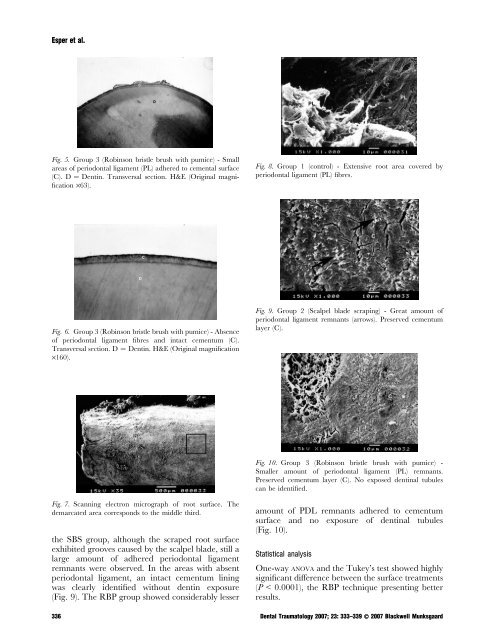
Fig. 5. Group 3 (Robinson bristle brush with pumice) - Small<br />
areas <strong>of</strong> <strong>periodontal</strong> <strong>ligament</strong> (PL) adhered to cemental surface<br />
(C). D ¼ Dentin. Transversal section. H&E (Original magnification<br />
·63).<br />
Fig. 8. Group 1 (control) - Extensive root area covered <strong>by</strong><br />
<strong>periodontal</strong> <strong>ligament</strong> (PL) fibres.<br />
Fig. 6. Group 3 (Robinson bristle brush with pumice) - Absence<br />
<strong>of</strong> <strong>periodontal</strong> <strong>ligament</strong> fibres and intact cementum (C).<br />
Transversal section. D ¼ Dentin. H&E (Original magnification<br />
·160).<br />
Fig. 9. Group 2 (Scalpel blade scraping) - Great amount <strong>of</strong><br />
<strong>periodontal</strong> <strong>ligament</strong> remnants (arrows). Preserved cementum<br />
layer (C).<br />
Fig. 10. Group 3 (Robinson bristle brush with pumice) -<br />
Smaller amount <strong>of</strong> <strong>periodontal</strong> <strong>ligament</strong> (PL) remnants.<br />
Preserved cementum layer (C). No exposed dentinal tubules<br />
can be identified.<br />
Fig. 7. Scanning electron micrograph <strong>of</strong> root surface. The<br />
demarcated area corresponds to the middle third.<br />
the SBS group, although the scraped root surface<br />
exhibited grooves caused <strong>by</strong> the scalpel blade, still a<br />
large amount <strong>of</strong> adhered <strong>periodontal</strong> <strong>ligament</strong><br />
remnants were observed. In the areas with absent<br />
<strong>periodontal</strong> <strong>ligament</strong>, an intact cementum lining<br />
was clearly identified without dentin exposure<br />
(Fig. 9). The RBP group showed considerably lesser<br />
amount <strong>of</strong> PDL remnants adhered to cementum<br />
surface and no exposure <strong>of</strong> dentinal tubules<br />
(Fig. 10).<br />
Statistical analysis<br />
One-way anova and the Tukey’s test showed highly<br />
significant difference between the surface treatments<br />
(P < 0.0001), the RBP technique presenting better<br />
results.<br />
336 Dental Traumatology 2007; 23: 333–339 Ó 2007 Blackwell Munksgaard