Clinical evaluation of low-level laser therapy and fluoride varnish for ...
Clinical evaluation of low-level laser therapy and fluoride varnish for ...
Clinical evaluation of low-level laser therapy and fluoride varnish for ...

You also want an ePaper? Increase the reach of your titles
YUMPU automatically turns print PDFs into web optimized ePapers that Google loves.
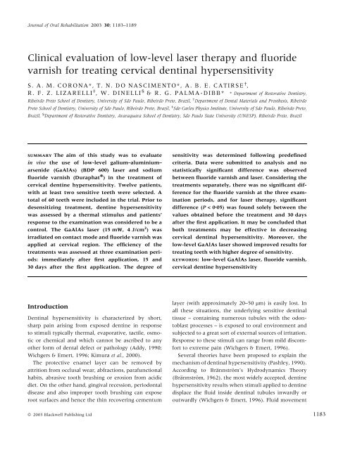
Journal <strong>of</strong> Oral Rehabilitation 2003 30; 1183–1189<br />
<strong>Clinical</strong> <strong>evaluation</strong> <strong>of</strong> <strong>low</strong>-<strong>level</strong> <strong>laser</strong> <strong>therapy</strong> <strong>and</strong> <strong>fluoride</strong><br />
<strong>varnish</strong> <strong>for</strong> treating cervical dentinal hypersensitivity<br />
S. A. M. CORONA*, T. N. DO NASCIMENTO*, A. B. E. CATIRSE † ,<br />
R. F. Z. LIZARELLI ‡ , W. DINELLI § & R. G. PALMA-DIBB* * Department <strong>of</strong> Restorative Dentistry,<br />
Ribeirão Preto School <strong>of</strong> Dentistry, University <strong>of</strong> São Paulo, Ribeirão Preto, Brazil, † Department <strong>of</strong> Dental Materials <strong>and</strong> Prosthesis, Ribeirão<br />
Preto School <strong>of</strong> Dentistry, University <strong>of</strong> São Paulo, Ribeirão Preto, Brazil, ‡ São Carlos Physics Institute, University <strong>of</strong> São Paulo, Ribeirão Preto,<br />
Brazil, § Department <strong>of</strong> Restorative Dentistry, Araraquara School <strong>of</strong> Dentistry, São Paulo State University (UNESP), Ribeirão Preto, Brazil<br />
SUMMARY The aim <strong>of</strong> this study was to evaluate<br />
in vivo the use <strong>of</strong> <strong>low</strong>-<strong>level</strong> galium–aluminium–<br />
arsenide (GaAlAs) (BDP 600) <strong>laser</strong> <strong>and</strong> sodium<br />
<strong>fluoride</strong> <strong>varnish</strong> (Duraphat Ò ) in the treatment <strong>of</strong><br />
cervical dentine hypersensitivity. Twelve patients,<br />
with at least two sensitive teeth were selected. A<br />
total <strong>of</strong> 60 teeth were included in the trial. Prior to<br />
desensitizing treatment, dentine hypersensitivity<br />
was assessed by a thermal stimulus <strong>and</strong> patients’<br />
response to the examination was considered to be a<br />
control. The GaAlAs <strong>laser</strong> (15 mW, 4 J/cm 2 ) was<br />
irradiated on contact mode <strong>and</strong> <strong>fluoride</strong> <strong>varnish</strong> was<br />
applied at cervical region. The efficiency <strong>of</strong> the<br />
treatments was assessed at three examination periods:<br />
immediately after first application, 15 <strong>and</strong><br />
30 days after the first application. The degree <strong>of</strong><br />
Introduction<br />
Dentinal hypersensitivity is characterized by short,<br />
sharp pain arising from exposed dentine in response<br />
to stimuli typically thermal, evaporative, tactile, osmotic<br />
or chemical <strong>and</strong> which cannot be ascribed to any<br />
other <strong>for</strong>m <strong>of</strong> dental defect or pathology (Addy, 1990;<br />
Wichgers & Emert, 1996; Kimura et al., 2000).<br />
The protective enamel layer can be removed by<br />
attrition from occlusal wear, abfractions, parafunctional<br />
habits, abrasive tooth brushing or erosion from acidic<br />
diet. On the other h<strong>and</strong>, gingival recession, periodontal<br />
disease <strong>and</strong> also improper tooth brushing can expose<br />
root surfaces <strong>and</strong> hence the thin recovering cementum<br />
sensitivity was determined fol<strong>low</strong>ing predefined<br />
criteria. Data were submitted to analysis <strong>and</strong> no<br />
statistically significant difference was observed<br />
between <strong>fluoride</strong> <strong>varnish</strong> <strong>and</strong> <strong>laser</strong>. Considering the<br />
treatments separately, there was no significant difference<br />
<strong>for</strong> the <strong>fluoride</strong> <strong>varnish</strong> at the three examination<br />
periods, <strong>and</strong> <strong>for</strong> <strong>laser</strong> <strong>therapy</strong>, significant<br />
difference (P
1184<br />
S. A. M. CORONA et al.<br />
then promotes a mechanical de<strong>for</strong>mation <strong>of</strong> nerve<br />
endings at the pulp/dentine interface (odontoblastic<br />
layer <strong>and</strong> subodontoblastic extract), which is transmitted<br />
as a painful sensation. There<strong>for</strong>e, it seems<br />
appropriate to assume that any substance or technique<br />
that reduces dentinal fluid movement or dentine<br />
permeability should decrease sensitivity (Pashley,<br />
1986).<br />
Considering the mechanism <strong>of</strong> action <strong>and</strong> treatment<br />
modalities, the desensitizing agents may be assigned to<br />
three main groups: the anti-inflammatory, the therapeutic<br />
tubule occlusive agents <strong>and</strong> those with effect on<br />
the depolarization <strong>of</strong> nerve endings. The products<br />
which promote partial or total closure <strong>of</strong> dentinal<br />
tubules, such as oxalates (Wichgers & Emert, 1996; Jain<br />
et al., 1997), resin bonding agents (Wichgers & Emert,<br />
1996; Li et al., 2000), <strong>for</strong>mulations containing potassium<br />
ions (Greenhill & Pashley, 1981; Muzzin &<br />
Johnson, 1989; Martinelli & Pereira, 2000; Orchardson<br />
& Gillam, 2000; Santiago & Pereira, 2000; Fitz,<br />
Markowitz & Napolitano, 2001) <strong>and</strong> abrasive dentifrices<br />
(Kodaka et al., 2001) are the most commonly used.<br />
These agents interfere with the hydrodynamic mechanism,<br />
as they act on the exposed sensitive area so as to<br />
reduce the number <strong>of</strong> open dentinal tubules or decrease<br />
their diameter thereby minimizing the movement <strong>of</strong><br />
dentinal fluid. The direct result is the relief <strong>of</strong> painful<br />
symptomatology.<br />
Sodium <strong>fluoride</strong> (NaF) has also been indicated <strong>for</strong><br />
treating dentine hypersensitivity <strong>and</strong> it is available in a<br />
variety <strong>of</strong> <strong>for</strong>ms. The use <strong>of</strong> fluoridating <strong>varnish</strong>es with<br />
sodium <strong>fluoride</strong> (in high concentrations) as the active<br />
ingredient has been advocated to increase time <strong>of</strong> action<br />
<strong>of</strong> NaF in contact with exposed dentin, thus aiming to<br />
enhance its effectiveness in decreasing dentine sensitivity<br />
(Gaffar, 1999; Lan, Liu & Lin, 1999). However, the<br />
attempt to provide tubule closure or narrowing is<br />
relatively short-lived because the <strong>varnish</strong> has a gradual<br />
therapeutic action (progressive in time) <strong>and</strong> can be<br />
removed during tooth brushing, be<strong>for</strong>e its desensitizing<br />
effect may be achieved (Lan et al., 1999).<br />
The advent <strong>of</strong> dental <strong>laser</strong>s has raised another<br />
possible treatment option <strong>for</strong> dentinal hypersensitivity<br />
<strong>and</strong> has become a research interest in the last decades.<br />
The <strong>laser</strong>s used <strong>for</strong> the treatment <strong>of</strong> sensitive teeth may<br />
be divided in to two groups. The middle output power<br />
<strong>laser</strong>s – Nd:YAG <strong>and</strong> CO 2 <strong>laser</strong>s <strong>and</strong> the <strong>low</strong>-<strong>level</strong> <strong>laser</strong>s<br />
– helium-neon (He–Ne) <strong>and</strong> gallium–aluminum–arsenide<br />
(GaAlAs) (diode) <strong>laser</strong>s.<br />
The <strong>low</strong>-<strong>level</strong> or ‘s<strong>of</strong>t’ <strong>laser</strong>s provide cold thermal <strong>low</strong><br />
energy wavelengths with little temperature increase <strong>of</strong><br />
Table 1. Degrees <strong>of</strong> cervical dentinal hypersensitivity<br />
Degree Sensitivity<br />
0 Without significant discom<strong>for</strong>t<br />
1 Discom<strong>for</strong>t with mild pain<br />
2 Sharp pain solely during the application<br />
<strong>of</strong> stimulus<br />
3 Sharp pain during the application <strong>of</strong> stimulus <strong>and</strong><br />
continuous after its removal<br />
complaints, were also excluded. After careful <strong>evaluation</strong>,<br />
12 individuals <strong>of</strong> both sexes, ranging from 20 to<br />
30 years <strong>of</strong> age were selected, with a total <strong>of</strong> 60 test<br />
teeth.<br />
Patients were required to be willing <strong>and</strong> able to<br />
return at specified study intervals <strong>for</strong> fol<strong>low</strong>-up examinations.<br />
The nature <strong>and</strong> objectives <strong>of</strong> the trial, as well<br />
as the possible discom<strong>for</strong>t <strong>and</strong> risks, were fully<br />
explained <strong>and</strong> all participants signed the appropriate,<br />
approved in<strong>for</strong>med consent documents.<br />
Be<strong>for</strong>e the treatments were accomplished, dentine<br />
hypersensitivity was assessed by a thermal stimulus <strong>and</strong><br />
patients’ response to this stimulation was considered to<br />
be a control. A cold air-blast from a three-way dental<br />
syringe was directed to the exposed area <strong>for</strong> 5 s under<br />
relative isolation.<br />
Data were st<strong>and</strong>ardized by scoring the teeth fol<strong>low</strong>ing<br />
the criteria proposed by Uchida et al. (1980), which<br />
establishes four degrees <strong>for</strong> sensitivity, depending on<br />
patient’s response to stimulation (Table 1).<br />
For each patient, half <strong>of</strong> sensitive teeth were painted<br />
with <strong>fluoride</strong> <strong>varnish</strong> <strong>and</strong> the other half were submitted<br />
to <strong>laser</strong> irradiation, which means that all subjects have<br />
been subjected to both types <strong>of</strong> desensitizing agents.<br />
Be<strong>for</strong>e desensitizing procedures, teeth surfaces were<br />
carefully cleaned with water/pumice slurry in dental<br />
prophylactic cups, rinsed <strong>and</strong> gently dried with absorbent<br />
paper. A relative isolation was obtained using<br />
cotton rolls <strong>and</strong> teeth were kept free from humidity<br />
with the aid <strong>of</strong> a high potency saliva ejector.<br />
Laser source (BDP 660)* was a <strong>low</strong>-<strong>level</strong> GaAlAs<br />
semiconductor diode <strong>laser</strong> doped with In, emitting a<br />
660 nm wavelength in the infrared spectrum. It consists<br />
<strong>of</strong> a class IIIb <strong>laser</strong> system (Makinson, 1986) with<br />
continuous wave <strong>and</strong> adjustable output energy ranging<br />
from 1 to 30 mW, when measured at the <strong>level</strong> <strong>of</strong> the<br />
diode <strong>laser</strong> itself, as the final useable output (from the<br />
*MM Optics LTDA, São Carlos, SP Brasil 13560-010.<br />
ª 2003 Blackwell Publishing Ltd, Journal <strong>of</strong> Oral Rehabilitation 30; 1183–1189<br />
T R E A T I N G C E R V I C A L D E N T I N A L H Y P E R S E N S I T I V I T Y 1185<br />
h<strong>and</strong>piece) will be less because <strong>of</strong> losses in the delivery<br />
system. The area <strong>of</strong> the active tip is 3.6 mm 2 <strong>and</strong> the<br />
h<strong>and</strong>piece is constituted by an optical fibres network.<br />
The sensitive teeth were irradiated on contact mode<br />
with the fol<strong>low</strong>ing parameters: 15 mW output power<br />
<strong>and</strong> 4 J cm )2 energy density. Laser beam was directed<br />
perpendicularly to tooth surface at three points: one<br />
apical <strong>and</strong> two cervical points (one mesio-buccal <strong>and</strong><br />
one disto-buccal). Each area was irradiated <strong>for</strong> 10 s<br />
(total <strong>of</strong> 30 s per tooth).<br />
Laser <strong>therapy</strong> was per<strong>for</strong>med within five sequential<br />
appointments with a 72-h interval between each one,<br />
as suggested by Groth (1993).<br />
For teeth treated by NaF <strong>varnish</strong> (Duraphat) † , five<br />
applications with a 5-day interval between each one<br />
were accomplished. The <strong>varnish</strong> was painted with a<br />
disposable brush at cervical region <strong>of</strong> both buccal <strong>and</strong><br />
lingual surfaces. The patients were instructed not to eat<br />
<strong>for</strong> 1 h fol<strong>low</strong>ing <strong>varnish</strong> application <strong>and</strong> to re-initiate<br />
tooth brushing solely after 12 h, thus enhancing the<br />
interaction <strong>of</strong> <strong>fluoride</strong> with tooth structure.<br />
The effectiveness <strong>of</strong> both therapies was assessed (by<br />
scoring patients’ response to thermal stimulation, fol<strong>low</strong>ing<br />
the predefined criteria) at three examination<br />
periods: immediately after the first application <strong>of</strong> the<br />
desensitizing agent, 15 <strong>and</strong> 30 days after the first<br />
application.<br />
Data obtained were submitted to statistical analysis<br />
using Friedman test (P
1186<br />
S. A. M. CORONA et al.<br />
Degree<br />
presented sharp pain during thermal stimulation <strong>and</strong><br />
continuous discom<strong>for</strong>t after the removal <strong>of</strong> stimulus<br />
(degree 3). At the second <strong>evaluation</strong> (15 days after the<br />
first application), there was an observed increase in the<br />
number <strong>of</strong> teeth with no report <strong>of</strong> significant discom<strong>for</strong>t<br />
(degree 0). Thirty days after the treatment was initiated,<br />
little decline in the relief/absence <strong>of</strong> painful symptomatology<br />
was noticed. However, no tooth showed<br />
recurrent sensitivity at degree 3.<br />
The results <strong>of</strong> <strong>fluoride</strong> <strong>varnish</strong> treatment also disclosed<br />
that be<strong>for</strong>e desensitizing <strong>therapy</strong>, all test teeth<br />
presented dentinal hypersensitivity, at different degrees<br />
(Table 3). Immediately after the first application, 11<br />
teeth did not present significant discom<strong>for</strong>t (degree 0)<br />
<strong>and</strong> three teeth continued to presented sharp pain<br />
during cold air-blasting <strong>and</strong> even after the removal <strong>of</strong><br />
stimulus (degree 3). Fifteen days after the first application,<br />
there was an observed increase in the number <strong>of</strong><br />
teeth without significant discom<strong>for</strong>t (degree 0) <strong>and</strong> a<br />
decrease in the amount <strong>of</strong> teeth with the most severe<br />
degree <strong>of</strong> sensitivity. Nevertheless, at the last recall<br />
(30 days after the treatment was initiated), one tooth<br />
remained with persistent sharp pain (degree 3).<br />
The overall analysis <strong>of</strong> scores, per<strong>for</strong>med 30 days<br />
after the first application <strong>of</strong> GaAlAs <strong>laser</strong>, showed that<br />
70% <strong>of</strong> teeth which scored 1, 54% <strong>of</strong> teeth which<br />
scored 2 <strong>and</strong> 14% <strong>of</strong> teeth which scored 3, did not<br />
present a significant discom<strong>for</strong>t. Additionally, 39% <strong>of</strong><br />
teeth which scored 2 <strong>and</strong> 71% <strong>of</strong> teeth which scored 3<br />
showed discom<strong>for</strong>t with mild pain, while 14% <strong>of</strong> teeth<br />
which scored 3 showed sharp pain during the application<br />
<strong>of</strong> thermal stimuli. Considering the same exam-<br />
Degree<br />
Be<strong>for</strong>e<br />
treatment<br />
Be<strong>for</strong>e<br />
treatment<br />
Immediately after<br />
the first application<br />
Immediately after<br />
the first application<br />
15 days after the<br />
first application<br />
15 days after<br />
the first application<br />
30 days after the<br />
first application<br />
0 0 13 18 16<br />
1 10 8 6 12<br />
2 13 9 6 2<br />
3 7 0 0 0<br />
ination period (30-day recall), the analysis <strong>of</strong> scores<br />
after <strong>fluoride</strong> <strong>varnish</strong> <strong>therapy</strong> disclosed that that 64% <strong>of</strong><br />
teeth which scored 1, 62% <strong>of</strong> teeth which scored 2 <strong>and</strong><br />
17% <strong>of</strong> teeth which scored 3, showed no significant<br />
discom<strong>for</strong>t while 25% <strong>of</strong> teeth which scored 2 <strong>and</strong> 33%<br />
<strong>of</strong> teeth scored 3 showed discom<strong>for</strong>t with mild pain.<br />
Discussion<br />
Despite the great variety <strong>of</strong> available therapeutic<br />
methods, dentinal hypersensitivity still remains a chronic<br />
dental problem with a difficult treatment conduct<br />
<strong>and</strong> an uncertain prognostic. A possible elimination <strong>of</strong><br />
painful symptomatology resulting from the dentine<br />
hypersensitivity mechanism, seems to be directly related<br />
to the interruption <strong>of</strong> stimuli transmission to the<br />
nerve endings <strong>of</strong> odontoblast processes by reducing the<br />
fluid movement inside the dentinal canalicules,<br />
through the narrowing or occlusion <strong>of</strong> tubules openings<br />
(Brännström, 1986). However, other treatment modalities<br />
have been proposed, such as ‘s<strong>of</strong>t’ <strong>laser</strong> <strong>therapy</strong>.<br />
Reduction in sensitivity to thermal <strong>and</strong> tactile stimuli<br />
has been widely reported using <strong>and</strong> He–Ne <strong>and</strong> GaAlAs<br />
<strong>laser</strong>s (Matsumoto et al., 1985; Nakamura et al., 1986;<br />
Yamaguchi et al., 1990; Gerschman et al., 1994; Walsh,<br />
1997; Coleton, 1998; Kimura et al., 2000). Accordingly,<br />
in the present investigation, <strong>laser</strong> <strong>therapy</strong> promoted a<br />
considerable decrease in sensitivity, after 30 days <strong>of</strong> the<br />
first application.<br />
Although <strong>low</strong>-<strong>level</strong> <strong>laser</strong>s <strong>and</strong> <strong>fluoride</strong> <strong>varnish</strong>es<br />
present distinct modes <strong>of</strong> action, in the present study<br />
both treatments provided a significant overall relief in<br />
30 days after the<br />
first application<br />
0 0 11 15 16<br />
1 11 6 8 9<br />
2 13 10 5 4<br />
3 6 3 2 1<br />
Table 2. GaAlAs <strong>low</strong>-<strong>level</strong> <strong>laser</strong><br />
<strong>therapy</strong>. Degree <strong>of</strong> dentine sensitivity<br />
at three examination periods<br />
Table 3. Sodium <strong>fluoride</strong> <strong>varnish</strong><br />
(Duraphat Ò ) <strong>therapy</strong>. Degree <strong>of</strong><br />
dentine sensitivity at three<br />
examination periods<br />
ª 2003 Blackwell Publishing Ltd, Journal <strong>of</strong> Oral Rehabilitation 30; 1183–1189
dentine hypersensitivity (particularly <strong>for</strong> the sharpest<br />
pains) <strong>and</strong> showed similar per<strong>for</strong>mance.<br />
In the conducted research, it was observed that teeth,<br />
which presented exacerbated sharp pain during airblasting<br />
<strong>and</strong> continuous discom<strong>for</strong>t after the removal <strong>of</strong><br />
thermal stimulation be<strong>for</strong>e desensitizing treatments<br />
were accomplished, showed an accentuated decrease<br />
<strong>of</strong> painful sensation immediately after the first application<br />
<strong>of</strong> <strong>low</strong>-<strong>level</strong> <strong>laser</strong> <strong>and</strong> even 1 month after initial<br />
irradiation. On the contrary, the <strong>fluoride</strong> <strong>varnish</strong><br />
provided a gradual decrease in sharp pain <strong>and</strong>, one<br />
patient reported the persistence <strong>of</strong> painful symptoms<br />
(sensitivity) at the 30-day recall.<br />
The gradual action <strong>of</strong> NaF <strong>varnish</strong> may be attributed<br />
to the reaction that occurs between NaF <strong>and</strong> calcium<br />
ions <strong>of</strong> dentinal fluid <strong>and</strong> that leads to <strong>for</strong>mation <strong>of</strong><br />
calcium <strong>fluoride</strong> (CaF 2) crystals, which are deposited on<br />
the dentinal tubules openings. As the crystal size <strong>of</strong><br />
CaF 2 is small (about 0Æ05 micrometers), a single application<br />
<strong>of</strong> NaF would not be effective in narrowing the<br />
diameter <strong>of</strong> dentinal tubules <strong>and</strong> multiple applications<br />
should be required. Also, the patency <strong>of</strong> sensitive<br />
dentine interferes with the action <strong>of</strong> therapeutic tubule<br />
occlusive agents <strong>and</strong> dem<strong>and</strong>s a longer treatment, as<br />
the number <strong>and</strong> width <strong>of</strong> dentinal tubules in hypersensitive-exposed<br />
areas have been shown to be higher<br />
than in normal dentine (Absi, Addy & Adams, 1987;<br />
Liu, Lin & Lan, 1997; Lan et al., 1999).<br />
On the other h<strong>and</strong>, the effect <strong>of</strong> GaAlAs <strong>low</strong>-<strong>level</strong><br />
<strong>laser</strong> relies upon <strong>laser</strong>-induced changes in neural<br />
transmission networks within the dental pulp<br />
(depressed nerve transmission), rather than alterations<br />
in the exposed dentine surface, as observed with other<br />
treatment modalities (Walsh, 1997). The faster desensitizing<br />
effect <strong>of</strong> <strong>laser</strong> <strong>therapy</strong> observed in the conducted<br />
research may be attributed to that mechanism.<br />
Moreover, besides the immediate analgesic effect, the<br />
<strong>laser</strong> <strong>therapy</strong> – if used within the correct parameters –<br />
may stimulate the normal physiological cellular functions.<br />
There<strong>for</strong>e, at subsequent appointments, the<br />
pulpal tissue would be less injured <strong>and</strong> inflamed <strong>and</strong><br />
the <strong>laser</strong> would stimulate the production <strong>of</strong> sclerotic<br />
dentin, thus promoting the internal obliteration <strong>of</strong><br />
dentinal tubules.<br />
Studies on other <strong>laser</strong> systems (Liu et al., 1997; Lan<br />
et al., 1999; Kimura et al., 2000) showed that the mode<br />
<strong>of</strong> action <strong>and</strong> the effects on dentine hypersensitivity<br />
<strong>therapy</strong> are different <strong>for</strong> each type <strong>of</strong> <strong>laser</strong>. The<br />
morphological analysis <strong>of</strong> dentine surface using scan-<br />
ª 2003 Blackwell Publishing Ltd, Journal <strong>of</strong> Oral Rehabilitation 30; 1183–1189<br />
T R E A T I N G C E R V I C A L D E N T I N A L H Y P E R S E N S I T I V I T Y 1187<br />
ning electron microscopy revealed that Nd:YAG <strong>laser</strong><br />
irradiation causes melting <strong>and</strong> fusion <strong>of</strong> dentine <strong>and</strong><br />
there<strong>for</strong>e the closure <strong>of</strong> exposed dentinal tubules (Lan<br />
& Liu, 1995). The combined effect <strong>of</strong> Nd:YAG <strong>laser</strong><br />
irradiation <strong>and</strong> sodium <strong>fluoride</strong> <strong>varnish</strong> on dentine<br />
hypersensitivity was also investigated <strong>and</strong> the closure <strong>of</strong><br />
90% <strong>of</strong> dentinal tubules was observed. The use <strong>of</strong><br />
<strong>fluoride</strong> <strong>varnish</strong> alone resulted in partially opened<br />
dentinal tubules <strong>and</strong> it was noticed the removal <strong>of</strong><br />
desensitizing agent during tooth brushing was noticed<br />
(Lan et al., 1999).<br />
Regardless <strong>of</strong> the method <strong>and</strong> materials employed, the<br />
<strong>evaluation</strong> <strong>of</strong> treatments <strong>for</strong> dentine hypersensitivity is<br />
not simple. In estimating treatment effects on hypersensitive<br />
teeth, investigations may be h<strong>and</strong>icapped by a<br />
the difficulty to assess patient’s response objectively <strong>and</strong><br />
are dependent upon the patient’s interpretation, which<br />
is in turn subjected to suggestion.<br />
Furthermore, placebo effect has been described in<br />
clinical dentine hypersensitivity trials (Wilder-Smith,<br />
1988). In the present investigation, the possibility <strong>of</strong> a<br />
placebo effect must also be taken into account, especially<br />
as patients’ reports were strongly positive immediately<br />
after the first application <strong>of</strong> GaAlAs <strong>laser</strong>, whereas<br />
normally one would expect the cumulative effect <strong>of</strong><br />
any <strong>therapy</strong> to provide a gradual lessening <strong>of</strong> painful<br />
sensation from visit to visit. However, it must be<br />
emphasized that <strong>for</strong> teeth on which <strong>laser</strong> <strong>therapy</strong> was<br />
accomplished, a considerable decrease in sensitivity was<br />
observed at the second <strong>evaluation</strong> (15 days after the<br />
first application) <strong>and</strong> a continued desensitizing effect<br />
was noticed at the last recall.<br />
Indeed, the desensitizing agents provide a significant<br />
relief in sharp painful symptomatology <strong>and</strong> can be<br />
reapplied, in case <strong>of</strong> recurrent discom<strong>for</strong>t, as they consist<br />
<strong>of</strong> non-invasive methods. Nevertheless, the ideal treatment<br />
technique, one which should not irritate the pulp,<br />
nor cause pain, should be easy <strong>and</strong> practical to per<strong>for</strong>m,<br />
effective <strong>for</strong> a long period, as well as accessible to the<br />
great majority <strong>of</strong> population, is still to be developed.<br />
There is a need <strong>for</strong> a more systematic approach to the<br />
clinical management <strong>of</strong> cervical dentine hypersensitivity,<br />
particularly in determining the long-tem effect <strong>of</strong><br />
the currently available desensitizing agents. For more<br />
effective treatment, further investigation is required to<br />
increase the underst<strong>and</strong>ing <strong>of</strong> the mechanisms <strong>and</strong><br />
aetiology <strong>of</strong> dentinal pain. The findings revealed by<br />
both laboratory <strong>and</strong> clinical research are extremely<br />
important to support the development or improvement
1188<br />
S. A. M. CORONA et al.<br />
<strong>of</strong> therapies that may acutely contribute to the treatment<br />
<strong>of</strong> dentinal hypersensitivity sufferers.<br />
Conclusions<br />
Based on the findings <strong>of</strong> this clinical <strong>evaluation</strong>, it may<br />
be concluded that <strong>low</strong>-<strong>level</strong> GaAlAs <strong>laser</strong> <strong>and</strong> NaF<br />
<strong>varnish</strong> showed similar overall per<strong>for</strong>mance <strong>and</strong> provided<br />
a decrease in cervical dentine hypersensitivity.<br />
Low-<strong>level</strong> <strong>laser</strong> showed improved results <strong>for</strong> treating<br />
teeth with higher degree <strong>of</strong> sensitivity.<br />
References<br />
Absi, E.G., Addy, M.&Adams, D. (1987) Dentin hypersensitivity.<br />
A study <strong>of</strong> the patency <strong>of</strong> dentinal tubules in sensitive <strong>and</strong> nonsensitive<br />
cervical dentin. Journal <strong>of</strong> <strong>Clinical</strong> Periodontology, 14,<br />
280.<br />
Addy, M. (1990) Etiology <strong>and</strong> clinical implications <strong>of</strong> dentine<br />
hypersensitivity. Dental Clinics <strong>of</strong> North America, 34, 503.<br />
Brännström, M.A. (1962) A hydrodynamic mechanism in the<br />
transmission <strong>of</strong> pain producing stimuli trough the dentine. In:<br />
Sensory Mechanism in Dentine (ed. D. J. Anderson), p. 73–9.<br />
Pergamon Press, Ox<strong>for</strong>d.<br />
Brännström, M.A. (1986) The hydrodynamic theory <strong>of</strong> dentinal<br />
pain: sensation in preparations, caries <strong>and</strong> dentinal crack.<br />
Journal <strong>of</strong> Endodontics, 12, 453.<br />
Coleton, S. (1998) Sensitivity <strong>and</strong> <strong>laser</strong> treatment. Journal <strong>of</strong><br />
American Dental Association, 129, 1200.<br />
Fitz, B., Markowitz, K.&Napolitano, N. (2001) Tooth hypersensitivity<br />
treatment: enhanced potassium delivery from tubule<br />
occluding composition. Journal <strong>of</strong> Dental Research, 80 Abstract <strong>of</strong><br />
Papers (special issue), p.191. Abstract no. 1242.<br />
Fukuoka, M., Yokoi, T., Fukuda, S., Usuki, M., Matsuo, S.,<br />
Taniguchi, K.&Kimura, K. (1988) Effects <strong>of</strong> GaAlAs <strong>laser</strong> diode<br />
in the treatment <strong>of</strong> hypersensitive dentin. Fukuoka Shikka<br />
Daigaku Gakkai Zasshi 15, 42.<br />
Gaffar, A. (1999) Treating hypersensitivity with <strong>fluoride</strong> <strong>varnish</strong><br />
Compendium <strong>of</strong> Continuing Education in Dentistry. 20 (1 Spec No),<br />
27, quiz 35.<br />
Gerschman, J.A., Ruben, J.&Gebart-Eaglemont, J. (1994) Low<strong>level</strong><br />
<strong>laser</strong> <strong>therapy</strong> <strong>for</strong> dentinal tooth hypersensitivity. Australian<br />
Dental Journal, 39, 353.<br />
Greenhill, J.D. & Pashley, D.H. (1981) The effects <strong>of</strong> desensitizing<br />
agents on the hydraulic condutante <strong>of</strong> human dentin in vitro.<br />
Journal <strong>of</strong> Dental Research 60, 686.<br />
Groth, E.B. (1993) Contribution <strong>for</strong> the study <strong>of</strong> use <strong>of</strong> <strong>low</strong>-<strong>level</strong><br />
GaAlAs <strong>laser</strong> in the treatment <strong>of</strong> dentinal hypersensitivity.<br />
Thesis. 60 p. School <strong>of</strong> Dentistry, University <strong>of</strong> São Paulo.<br />
Hoji, T. (1990) Effects <strong>of</strong> s<strong>of</strong>t <strong>laser</strong> irradiation on dentinal pain.<br />
Gifu Shikka Gakkai Zasshi 17, 534.<br />
Jain, P., Vargas, M.A., Denehy, G.E. & Boyer, D.B. (1997) Dentin<br />
desensitizing agents: SEM <strong>and</strong> X-ray microanalysis assessment.<br />
American Journal <strong>of</strong> Dentistry 10, 21.<br />
Kimura, Y., Wilder-Smith, P., Yonaga, K.&Matsumoto, K. (2000)<br />
Treatment <strong>of</strong> dentine hypersensitivity by <strong>laser</strong>s: a review.<br />
Journal <strong>of</strong> <strong>Clinical</strong> Periodontal 27, 715.<br />
Kodaka, T., Kuroiwa, M., Kuroiwa, M., Okumura, J., Mori, R.,<br />
Hirasawa, S.&Kobori, M. (2001) Effects <strong>of</strong> brushing with a<br />
dentifrice <strong>for</strong> sensitive teeth on tubule occlusion <strong>and</strong> abrasion <strong>of</strong><br />
dentin. Journal <strong>of</strong> Electron Microscopy 50, 57.<br />
Lan, W.H. & Liu, H.C. (1995) Sealing <strong>of</strong> human dentinal tubules<br />
by Nd:YAG <strong>laser</strong>. Journal <strong>of</strong> <strong>Clinical</strong> Laser Medicine & Surgery 13,<br />
329.<br />
Lan, W.H., Liu, H.C. & Lin, C.P. (1999) The combined occluding<br />
effect <strong>of</strong> sodium <strong>fluoride</strong> <strong>varnish</strong> <strong>and</strong> Nd:YAG <strong>laser</strong> irradiation<br />
on human dentinal tubules. Journal <strong>of</strong> Endodontics 25, 424.<br />
Li, D., Turner, S., Rached, R.N., Powers, J.M. & Chan, J. (2000)<br />
Effect <strong>of</strong> desensitizers <strong>and</strong> bonding agent on human dentin.<br />
Journal <strong>of</strong> Dental Research 79 Abstract <strong>of</strong> Papers (special issue),<br />
p. 276. Abstract no. 1059.<br />
Liu, H.C., Lin, C.P. & Lan, W.H. (1997) Sealing depth <strong>of</strong> Nd:YAG<br />
<strong>laser</strong> on human dentinal tubules. Journal <strong>of</strong> Endodontics 23, 691.<br />
Makinson, O.F. (1986) S<strong>of</strong>t <strong>laser</strong>s <strong>and</strong> dentistry. Current note n.<br />
63. Australian Dental Journal 31, 139.<br />
Martinelli, A.C.B.F. & Pereira, J.C. (2000) Treating dentin<br />
hypersensitivity with potassium oxalate: a one-year <strong>evaluation</strong>.<br />
Journal <strong>of</strong> Dental Research 79 Abstract <strong>of</strong> Papers (special issue),<br />
p. 361. Abstract no. 1740.<br />
Matsumoto, K., Funai, H., Wakabayashi, H.&Oyoama, T. (1985)<br />
Study on the treatment <strong>of</strong> hypersensitive dentine by GaAlAs<br />
<strong>laser</strong> diode. Japanese Journal <strong>of</strong> Conservative Dentistry 28, 766.<br />
Muzzin, K.B. & Johnson, R. (1989) Effects <strong>of</strong> potassium oxalate on<br />
dentin hypersensitivity in vivo. Journal <strong>of</strong> Periodontology 60, 151.<br />
Nakamura, N., Saito, H., Uematsu, N., Morimoto, I.&Takeda, G.<br />
(1986) Pain relieving effects <strong>of</strong> s<strong>of</strong>t <strong>laser</strong> (He-Ne) irradiation on<br />
various oral lesions. Japanese Journal <strong>of</strong> Conservative Dentistry 29,<br />
1548.<br />
Orchardson, R.&Gillam, D.G. (2000) The efficacy <strong>of</strong> potassium<br />
salts as agents <strong>for</strong> treating dentin hypersensitivity. Journal <strong>of</strong><br />
Or<strong>of</strong>acial Pain 14, 9.<br />
Pashley, D.H. (1986) Dentin permeability, dentin sensitivity <strong>and</strong><br />
treatment through tubule occlusion. Journal <strong>of</strong> Endodontics 12,<br />
465.<br />
Pashley, D.H. (1990) Mechanisms <strong>of</strong> dentin sensitivity. Dental<br />
Clinics <strong>of</strong> North America 34, 449.<br />
Santiago, S.L. & Pereira, J.C. (2000) Time dependence <strong>of</strong> the<br />
effects <strong>of</strong> dentin desensitizing agents Journal <strong>of</strong> Dental Research 79<br />
Abstract <strong>of</strong> Papers (special issue), p. 276. Abstract no. 1058.<br />
Uchida, A., Wakano, Y., Fukuyama, O., Miki, T., Iwayama, Y.&<br />
Okada, H. (1980) Controlled clinical <strong>evaluation</strong> <strong>of</strong> 10% strontium<br />
chloride dentifrice in treatment <strong>of</strong> dentin hypersensitivity<br />
fol<strong>low</strong>ing periodontal surgery. Journal <strong>of</strong> Periodontology 51, 578.<br />
Walsh, L.J. (1997) The current status <strong>of</strong> <strong>low</strong>-<strong>level</strong> <strong>laser</strong> <strong>therapy</strong> in<br />
dentistry. Part 2. Hard tissue application. Australian Dental<br />
Journal 42, 302.<br />
Wichgers, T.G. & Emert, R.L. (1996) Dentin hypersensitivity.<br />
General Dentistry 44, 225.<br />
Wilder-Smith, P. (1988) The s<strong>of</strong>t <strong>laser</strong>: therapeutic tool or popular<br />
placebo? Oral Surgery Oral Medicine Oral Pathology 68, 271.<br />
ª 2003 Blackwell Publishing Ltd, Journal <strong>of</strong> Oral Rehabilitation 30; 1183–1189
Yamaguchi, M., Ito, M., Miwata, T., Horiba, N., Matsumoto, T.,<br />
Nakamura, H. & Fukaya, M. (1990) <strong>Clinical</strong> study on the<br />
treatment <strong>of</strong> hypersensitivity dentin by GaAlAs <strong>laser</strong> diode<br />
using double blind test. Aichi Gakuin Daigaku Shigakki Shi 28,<br />
703.<br />
ª 2003 Blackwell Publishing Ltd, Journal <strong>of</strong> Oral Rehabilitation 30; 1183–1189<br />
T R E A T I N G C E R V I C A L D E N T I N A L H Y P E R S E N S I T I V I T Y 1189<br />
Correspondence: Silmara Aparecida Milori Corona, Faculdade de<br />
Odontologia de Ribeirão Preto, Departamento de Odontologia Restauradora,<br />
Universidade de São Paulo, Av. do Café S/N, Monte Alegre,<br />
CEP: 14040-904, Ribeirão Preto, SP – Brazil.<br />
E-mail: nelsoncorona@uol.com.br