The Theory of the Microscope - Leica Microsystems
The Theory of the Microscope - Leica Microsystems
The Theory of the Microscope - Leica Microsystems

Create successful ePaper yourself
Turn your PDF publications into a flip-book with our unique Google optimized e-Paper software.
12<br />
11.0 Binocular Observation<br />
Thus far we have considered only <strong>the</strong> monocular microscope. For prolonged use, <strong>the</strong><br />
inclined binocular form is preferred since it gives a more natural and restful condition <strong>of</strong><br />
observation. Figure 15 shows a cutaway view <strong>of</strong> <strong>the</strong> optical system <strong>of</strong> a binocular microscope.<br />
Binocular vision is attained by <strong>the</strong> use <strong>of</strong> a beam-dividing prism and three mirrors.<br />
This system divides <strong>the</strong> light equally, sending half to <strong>the</strong> left eye and half to <strong>the</strong> right. <strong>The</strong><br />
coating on <strong>the</strong> mirrors is enhanced aluminum, having a multiple-film transparent coating<br />
on top <strong>of</strong> <strong>the</strong> aluminum to increase <strong>the</strong> reflectivity and protect <strong>the</strong> aluminum. Coverglass<br />
seals are used to keep dust from entering <strong>the</strong> binocular body.<br />
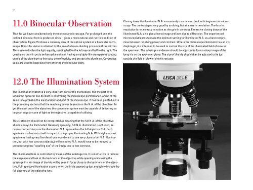
12.0 <strong>The</strong> Illumination System<br />
<strong>The</strong> illumination system is a very important part <strong>of</strong> <strong>the</strong> microscope. It is <strong>the</strong> part with<br />
which <strong>the</strong> operator can do most in controlling <strong>the</strong> microscope performance, and is at <strong>the</strong><br />
same time probably <strong>the</strong> least understood part <strong>of</strong> <strong>the</strong> microscope. It has been pointed out in<br />
<strong>the</strong> preceding sections that <strong>the</strong> resolving power depends on <strong>the</strong> N.A. <strong>of</strong> <strong>the</strong> objective. To<br />
get <strong>the</strong> most out <strong>of</strong> <strong>the</strong> objective, <strong>the</strong> condenser system must be capable <strong>of</strong> delivering as<br />
large an angular cone <strong>of</strong> light as <strong>the</strong> objective is capable <strong>of</strong> utilizing.<br />
This statement should not be interpreted as meaning that <strong>the</strong> full N.A. <strong>of</strong> <strong>the</strong> objective<br />
should always be illuminated. Generally speaking, full N.A. illumination is not used, because<br />
contrast drops as <strong>the</strong> illuminated N.A. approaches <strong>the</strong> full objective N.A. Each<br />
specimen is a law unto itself in regard to <strong>the</strong> proper illuminating N.A. With high contrast<br />
specimens having very fine detail one would want to use very close to full N.A. illumination,<br />
but with low contrast objects,<strong>the</strong> illuminated N.A. would have to be reduced to<br />
prevent complete “washing out” <strong>of</strong> <strong>the</strong> image due to low contrast.<br />
<strong>The</strong> illuminated N.A. is controlled by means <strong>of</strong> <strong>the</strong> substage iris. It is instructive to remove<br />
<strong>the</strong> eyepiece and look at <strong>the</strong> back lens <strong>of</strong> <strong>the</strong> objective while opening and closing <strong>the</strong><br />
substage iris. An image <strong>of</strong> <strong>the</strong> iris will be seen in focus close to <strong>the</strong> back lens <strong>of</strong> <strong>the</strong> objective.<br />
Full-aperture illumination occurs when <strong>the</strong> iris is opened up just enough to include <strong>the</strong><br />
full aperture <strong>of</strong> <strong>the</strong> objective lens.<br />
Closing down <strong>the</strong> illuminated N.A. excessively is a common fault with beginners in microscopy.<br />
<strong>The</strong> contrast gets very good by so doing, but at a loss in resolution. <strong>The</strong> loss in<br />
resolution is not so easy to notice as <strong>the</strong> gain in contrast. Excessive closing down <strong>of</strong> <strong>the</strong><br />
illuminated N.A. also gives rise to image artifacts due to diffraction. <strong>The</strong> experienced<br />
microscopist learns to make <strong>the</strong> optimum setting for illuminated N.A. as a best compromise<br />
between resolving power and contrast. Where <strong>the</strong> microscope illuminator has an iris<br />
diaphragm, it is intended to be used to control <strong>the</strong> size <strong>of</strong> <strong>the</strong> illuminated field <strong>of</strong> view on<br />
<strong>the</strong> specimen. <strong>The</strong> substage condenser should be adjusted to form a sharp image <strong>of</strong> <strong>the</strong><br />
lamp iris on <strong>the</strong> specimen plane. <strong>The</strong> size <strong>of</strong> <strong>the</strong> iris should <strong>the</strong>n be adjusted to lie just<br />
outside <strong>the</strong> field <strong>of</strong> view <strong>of</strong> <strong>the</strong> microscope.