The Theory of the Microscope - Leica Microsystems
The Theory of the Microscope - Leica Microsystems
The Theory of the Microscope - Leica Microsystems

You also want an ePaper? Increase the reach of your titles
YUMPU automatically turns print PDFs into web optimized ePapers that Google loves.
4<br />
Introduction<br />
<strong>The</strong> aim <strong>of</strong> this booklet is to provide <strong>the</strong> microscopist with a basic explanation <strong>of</strong> <strong>the</strong><br />
<strong>the</strong>ory <strong>of</strong> <strong>the</strong> microscope sufficient to enable him to understand <strong>the</strong> reasons behind<br />
accepted microscope techniques. It is felt that such an understanding will not only add to<br />
his interest in using <strong>the</strong> microscope, but will help him to work his way out <strong>of</strong> possible<br />
problems that may arise later on when detailed instructions, originally grasped, may have<br />
been forgotten. Where possible, ma<strong>the</strong>matical formulae have been avoided in favor <strong>of</strong><br />
physical or pictorial explanations, as it is felt that such explanations are more easily<br />
grasped and better retained than explanations involving ma<strong>the</strong>matics.<br />
2.0 What's in a <strong>Microscope</strong>?<br />
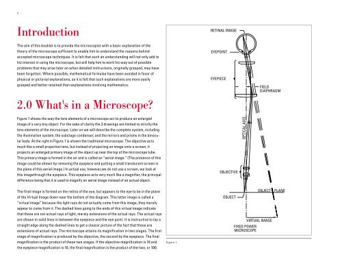
Figure 1 shows <strong>the</strong> way <strong>the</strong> lens elements <strong>of</strong> a microscope act to produce an enlarged<br />
image <strong>of</strong> a very tiny object. For <strong>the</strong> sake <strong>of</strong> clarity <strong>the</strong> 3 drawings are limited to strictly <strong>the</strong><br />
lens elements <strong>of</strong> <strong>the</strong> microscope. Later on we will describe <strong>the</strong> complete system, including<br />
<strong>the</strong> illumination system, <strong>the</strong> substage condenser, and <strong>the</strong> mirrors and prisms in <strong>the</strong> binocular<br />
body. At <strong>the</strong> right in Figure 1 is shown <strong>the</strong> traditional microscope. <strong>The</strong> objective acts<br />
much like a small projection lens, but instead <strong>of</strong> projecting an image onto a screen, it<br />
projects an enlarged primary image <strong>of</strong> <strong>the</strong> object up near <strong>the</strong> top <strong>of</strong> <strong>the</strong> microscope tube.<br />
This primary image is formed in <strong>the</strong> air and is called an “aerial image.” (<strong>The</strong> presence <strong>of</strong> this<br />
image could be shown by removing <strong>the</strong> eyepiece and putting a small translucent screen in<br />
<strong>the</strong> plane <strong>of</strong> this aerial image.) In actual use, however,we do not use a screen, we look at<br />
this imagethrough <strong>the</strong> eyepiece. This eyepiece acts very much like a magnifier, <strong>the</strong> principal<br />
difference being that it is used to magnify an aerial image instead <strong>of</strong> an actual object.<br />
<strong>The</strong> final image is formed on <strong>the</strong> retina <strong>of</strong> <strong>the</strong> eye, but appears to <strong>the</strong> eye to be in <strong>the</strong> plane<br />
<strong>of</strong> <strong>the</strong> Virtual Image down near <strong>the</strong> bottom <strong>of</strong> <strong>the</strong> diagram. This latter image is called a<br />
“virtual image” because <strong>the</strong> light rays do not actually come from this image, <strong>the</strong>y merely<br />
appear to come from it. <strong>The</strong> dashed lines going to <strong>the</strong> ends <strong>of</strong> this virtual image indicate<br />
that <strong>the</strong>se are not actual rays <strong>of</strong> light, merely extensions <strong>of</strong> <strong>the</strong> actual rays. <strong>The</strong> actual rays<br />
are shown in solid lines in between <strong>the</strong> eyepiece and <strong>the</strong> eye point. It is instructive to lay a<br />
straight edge along <strong>the</strong> dashed lines to get a clearer picture <strong>of</strong> <strong>the</strong> fact that <strong>the</strong>se are<br />
extensions <strong>of</strong> actual rays. <strong>The</strong> microscope attains its magnification in two stages. <strong>The</strong> first<br />
stage <strong>of</strong> magnification is produced by <strong>the</strong> objective, <strong>the</strong> second by <strong>the</strong> eyepiece. <strong>The</strong> final<br />
magnification is <strong>the</strong> product <strong>of</strong> <strong>the</strong>se two stages. If <strong>the</strong> objective magnification is 10 and<br />
<strong>the</strong> eyepiece magnification is 10, <strong>the</strong> final magnification is <strong>the</strong> product <strong>of</strong> <strong>the</strong> two, or 100.<br />
Figure 1<br />
RETINAL IMAGE<br />
EYEPOINT<br />
EYEPIECE<br />
OBJECTIVE<br />
OBJECT<br />
OPTICAL AXIS<br />
FIXED POWER<br />
MICROSCOPE<br />
FIELD<br />
DIAPHRAGM<br />
OBJECT PLANE<br />
VIRTUAL IMAGE