Chapter 3 Puberty and Biological Foundations - The McGraw-Hill ...
Chapter 3 Puberty and Biological Foundations - The McGraw-Hill ...
Chapter 3 Puberty and Biological Foundations - The McGraw-Hill ...

You also want an ePaper? Increase the reach of your titles
YUMPU automatically turns print PDFs into web optimized ePapers that Google loves.
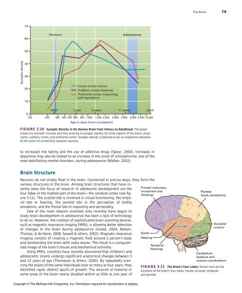
Synaptic density<br />
70<br />
60<br />
50<br />
40<br />
30<br />
20<br />
10<br />
birth 1 year 3 years 11 years adult<br />
0<br />
100 200 300 400 500 600 800 1,000 1,500 2,000 3,000 4,000 6,000 8,000 10,000<br />
FIGURE 3.10 Synaptic Density in the Human Brain from Infancy to Adulthood. <strong>The</strong> graph<br />
shows the dramatic increase <strong>and</strong> then pruning in synaptic density for three regions of the brain: visual<br />
cortex, auditory cortex, <strong>and</strong> prefrontal cortex. Synaptic density is believed to be an important indication<br />
of the extent of connectivity between neurons.<br />
to increased risk taking <strong>and</strong> the use of addictive drugs (Spear, 2000). Increases in<br />
dopamine may also be related to an increase in the onset of schizophrenia, one of the<br />
most debilitating mental disorders, during adolescence (Walker, 2002).<br />
Brain Structure<br />
Newborn<br />
Visual cortex (vision)<br />
Auditory cortex (hearing)<br />
Prefrontal cortex (reasoning,<br />
self-regulation)<br />
Age in days (from conception)<br />
Adolescence<br />
Neurons do not simply float in the brain. Connected in precise ways, they form the<br />
various structures in the brain. Among brain structures that have recently<br />
been the focus of research in adolescent development are the<br />
four lobes in the highest part of the brain—the cerebral cortex (see figure<br />
3.11). <strong>The</strong> occipital lobe is involved in visual functioning, the temporal<br />
lobe in hearing, the parietal lobe in the perception of bodily<br />
sensations, <strong>and</strong> the frontal lobe in reasoning <strong>and</strong> personality.<br />
One of the main reasons scientists only recently have begun to<br />
study brain development in adolescence has been a lack of technology<br />
to do so. However, the creation of sophisticated brain scanning devices,<br />
such as magnetic resonance imaging (MRI), is allowing better detection<br />
of changes in the brain during adolescence (Giedd, 2004; Nelson,<br />
Thomas, & de Hann, 2006; Sowell & others, 2002). Magnetic resonance<br />
imaging consists of creating a magnetic field around a person’s body<br />
<strong>and</strong> bombarding the brain with radio waves. <strong>The</strong> result is a computerized<br />
image of the brain’s tissues <strong>and</strong> biochemical activities.<br />
Using MRIs, scientists have recently discovered that children’s <strong>and</strong><br />
adolescents’ brains undergo significant anatomical changes between 3<br />
<strong>and</strong> 15 years of age (Thompson & others, 2000). By repeatedly scanning<br />
the brains of the same individuals over as many as four years, they<br />
identified rapid, distinct spurts of growth. <strong>The</strong> amount of material in<br />
some areas of the brain nearly doubled within as little as one year of<br />
Copyright © <strong>The</strong> <strong>McGraw</strong>-<strong>Hill</strong> Companies, Inc. Permission required for reproduction or display.<br />
Frontal (voluntary<br />
movement <strong>and</strong><br />
thinking)<br />
Smell<br />
Hearing<br />
Temporal<br />
(hearing)<br />
<strong>The</strong> Brain 79<br />
Parietal<br />
(body sensations)<br />
Occipital<br />
(vision)<br />
Cerebellum<br />
(balance <strong>and</strong><br />
muscle coordination)<br />
FIGURE 3.11 <strong>The</strong> Brain’s Four Lobes. Shown here are the<br />
locations of the brain’s four lobes: frontal, occipital, temporal,<br />
<strong>and</strong> parietal.