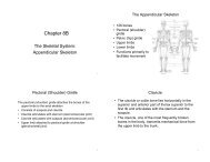
Chapter 8A The Skeletal System: The Axial Skeleton
Chapter 8A The Skeletal System: The Axial Skeleton
Chapter 8A The Skeletal System: The Axial Skeleton

You also want an ePaper? Increase the reach of your titles
YUMPU automatically turns print PDFs into web optimized ePapers that Google loves.
• 5 basic types of bones:<br />
– long = compact bone<br />
– short = spongy except<br />
surface<br />
– flat = plates of compact<br />
enclosing spongy<br />
– irregular = variable<br />
– sesamoid = develop in<br />
tendons or ligaments<br />
(patella)<br />
• Sutural bones = in joint<br />
between skull bones (not<br />
named)<br />
<strong>Chapter</strong> <strong>8A</strong><br />
<strong>The</strong> <strong>Skeletal</strong> <strong>System</strong>:<br />
<strong>The</strong> <strong>Axial</strong> <strong>Skeleton</strong><br />
Types of Bones<br />
1<br />
3<br />
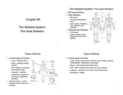
<strong>The</strong> <strong>Skeletal</strong> <strong>System</strong>: <strong>The</strong> <strong>Axial</strong> <strong>Skeleton</strong><br />
• 206 named bones<br />
• <strong>Axial</strong> <strong>Skeleton</strong><br />
– 80 bones<br />
– lie along longitudinal<br />
axis<br />
– skull, hyoid, vertebrae,<br />
ribs, sternum, ear<br />
ossicles<br />
• Appendicular <strong>Skeleton</strong><br />
– 126 bones<br />
– upper & lower limbs<br />
and pelvic & pectoral<br />
girdles<br />
Types of Bones<br />
• 5 basic types of bones:<br />
– Long: femur, tibia, fibula, humerus, ulna, radius, clavicle,<br />
metacarpals, metatarsals, phalanges<br />
– Short: most carpal and tarsal bones<br />
– Flat: some cranial bones, sternum, ribs, scapula<br />
– Irregular: vertabrae, os coxae, most facial bones<br />
– Sesamoid: patella<br />
2<br />
4
BONE SURFACE MARKINGS<br />
• <strong>The</strong>re are two major types of surface markings<br />
– Depressions and openings participate in joints or allow<br />
the passage of soft tissue (e.g., nerves, blood vessels)<br />
– Processes are projections or outgrowths that either help<br />
form joints or serve as attachment points for connective<br />
tissue (e.g., ligaments, tendons)<br />
SKULL<br />
• <strong>The</strong> skull, composed of 22 bones, consists of the 8<br />
cranial bones (cranium) and the 14 facial bones<br />
(face)<br />
• General Features<br />
– <strong>The</strong> skull forms the large cranial cavity and smaller<br />
cavities, including the nasal cavity and orbits (eye<br />
sockets).<br />
– Certain skull bones contain mucous membrane lined<br />
cavities called paranasal sinuses.<br />
– <strong>The</strong> only moveable bone of the skull, other than the ear<br />
ossicles within the temporal bones, is the mandible.<br />
– Immovable joints called sutures hold the skull bones<br />
together.<br />
5<br />
7<br />
Bone Surface Markings<br />
• Foramen = opening<br />
• Fossa = shallow depression<br />
• Sulcus = groove<br />
• Meatus = tubelike passageway or<br />
canal<br />
• Condyle = large, round<br />
protuberance<br />
• Facet = smooth flat articular<br />
surface<br />
• Trochanter = very large projection<br />
• Tubercle = small, rounded<br />
projection<br />
• Tuberosity = large, rounded,<br />
roughened projection<br />
• Protect brain & house ear ossicles<br />
• Muscle attachment for jaw, neck & facial muscles<br />
<strong>The</strong> 8 Cranial<br />
Bones<br />
•Frontal<br />
•Parietal (2)<br />
•Temporal (2)<br />
•Occipital<br />
•Sphenoid<br />
•Ethmoid<br />
6<br />
8
Temporal<br />
Bones<br />
• Temporal<br />
– zygomatic process forms part of arch<br />
– external auditory meatus<br />
– internal auditory meatus (VIII)<br />
– mastoid process<br />
– styloid process<br />
– mandibular fossa (TMJ)<br />
Sphenoid<br />
Bone<br />
• Base of skull<br />
• Pterygoid processes are attachment sites<br />
for jaw muscles<br />
9<br />
11<br />
Temporal &<br />
Occipital Bones<br />
• Temporal<br />
– carotid foramen<br />
(carotid artery)<br />
– jugular foramen<br />
(jugular vein)<br />
• Occipital<br />
– foramen magnum<br />
– occipital condyles<br />
– external occipital<br />
protuberance attachment<br />
for ligamentum nuchae<br />
Sphenoid in Anterior View<br />
• Greater and lesser wings<br />
• Optic foramen - optic (II) nerve<br />
10<br />
12
Sphenoid<br />
from<br />
Superior<br />
View<br />
• Lesser wing & greater wing<br />
• Sella turcica holds pituitary gland<br />
Ethmoid Bone<br />
• Perpendicular plate is upper part of nasal septum<br />
• Superior & middle nasal conchae<br />
– filters & warms air<br />
13<br />
15<br />
Ethmoid Bone<br />
• Forms part of the anterior portion of the cranial floor, the<br />
medial wall of the orbits, the superior portion of the nasal<br />
septum, and most of the superior side walls of the nasal<br />
cavity<br />
• Cribiform plate (roof of nasal cavity) and olfactory foramina<br />
14 Facial Bones<br />
Nasal (2) Maxillae (2) Zygomatic (2)<br />
Mandible (1) Lacrimal (2) Palatine (2)<br />
Inferior nasal conchae (2) Vomer (1)<br />
14<br />
16
Maxillary Bones<br />
• Floor of orbit, floor of nasal cavity or hard palate<br />
• Alveolar processes hold upper teeth<br />
• Cleft palate is lack of union of maxillary bones<br />
Lacrimal & Inferior Nasal Conchae<br />
• Lacrimal bones<br />
– part of medial wall of orbit<br />
Inferior Nasal Conchae<br />
– lacrimal fossa houses lacrimal sac<br />
• Inferior nasal concha (not part of ethmoid)<br />
17<br />
19<br />
Zygomatic Bones<br />
• Cheekbones<br />
• Lateral wall of orbit along with sphenoid<br />
• Part of zygomatic arch<br />
Mandible<br />
• Condylar process (part of temporomandibular joint)<br />
• Coronoid process (attachment of chewing muscles)<br />
• Mandibular & mental foramen<br />
18<br />
20
TMJ<br />
• <strong>The</strong> mandible articulates with the temporal bone to<br />
form the temporomandibular joint (TMJ)<br />
• TMJ is the only movable joint in the skull<br />
• Temporomandibular joint (TMJ) syndrome is<br />
dysfunction of this joint<br />
Nasal Septum<br />
• Divides nasal cavity into left and right sides<br />
• Formed by vomer, perpendicular plate of ethmoid<br />
and septal cartilage<br />
• Deviated septum does not run along the midline<br />
– developmental abnormality or trauma<br />
21<br />
23<br />
Palatine & Vomer<br />
• Palatine<br />
– L-shaped : one end is back part of hard palate,<br />
other end is a small part of orbit<br />
• Vomer<br />
– posterior part of nasal septum<br />
<strong>The</strong> Orbits (Eye Sockets)<br />
• <strong>The</strong> orbits contain the eyeballs and associated<br />
structures and are formed by seven bones of the skull<br />
22<br />
24
Sutures<br />
• Sutures are immovable joints found only between skull bones<br />
and hold skull bones together.<br />
• Sutures include the coronal, sagittal, lambdoid, and squamous<br />
sutures<br />
Fontanels<br />
• Fontanels are connective tissue membranes between the<br />
cranial bones of fetuses and infants. <strong>The</strong>y remain unossified<br />
at birth but close early in a child’s life<br />
• “Soft spots”<br />
•Fontanels have two<br />
major functions:<br />
–<strong>The</strong>y enable the fetal skull<br />
change shape as it passes<br />
through the birth canal<br />
–<strong>The</strong>y permit rapid growth<br />
of the brain during infancy<br />
25<br />
27<br />
Paranasal Sinuses<br />
• Paired cavities in ethmoid, sphenoid, frontal and maxillary<br />
• Lined with mucous membranes and open into nasal cavity<br />
• Resonating chambers for voice, lighten the skull<br />
• Sinusitis is inflammation of the membrane (allergy)<br />
Hyoid Bone<br />
– U-shaped single<br />
bone<br />
– Articulates with no<br />
other bone of the<br />
body<br />
– Suspended by<br />
ligament and<br />
muscle from skull<br />
– Supports the<br />
tongue & provides<br />
attachment for<br />
tongue, neck and<br />
pharyngeal<br />
muscles<br />
26<br />
28
Vertebral Column<br />
• Backbone or spine built of<br />
26 vertebrae<br />
• Five vertebral regions<br />
– cervical vertebrae (7) in the<br />
neck<br />
– thoracic vertebrae (12) in the<br />
thorax<br />
– lumbar vertebrae (5) in the<br />
low back region<br />
– sacrum (5 fused)<br />
– coccyx (4 fused)<br />
Intervertebral Discs<br />
• Between adjacent vertebrae<br />
• Fibrocartilagenous ring (annulus fibrosus) with a pulpy center<br />
(nucleus pulposus)<br />
• Form strong joints<br />
• Permit various movements of the vertebral column<br />
• Absorb vertical shock<br />
29<br />
31<br />
Normal Curves of the Vertebral Column<br />
• Primary curves<br />
– thoracic and sacral are formed during fetal development<br />
• Secondary curves<br />
– cervical is formed when infant raises head at 4 months<br />
– lumbar forms when infant sits up & begins to walk at 1<br />
year<br />
Typical Vertebrae • Body<br />
– weight bearing<br />
• Vertebral arch<br />
– pedicles<br />
– laminae<br />
• Vertebral foramen<br />
• Seven processes<br />
– 2 transverse<br />
– 1 spinous<br />
– 4 articular<br />
• Vertebral notches<br />
30<br />
32
Intervertebral Foramen & Spinal Canal<br />
• Spinal canal (vertebral cavity) is all vertebral foramen<br />
together<br />
• Intervertebral foramen are 2 vertebral notches together<br />
Typical<br />
Cervical<br />
Vertebrae<br />
(C3-C7)<br />
• Smaller bodies but larger spinal canal<br />
• Transverse processes<br />
– shorter, with transverse foramen for vertebral artery<br />
• Spinous processes of C2 to C6 often bifurcated<br />
• 1st and 2nd cervical vertebrae are unique - atlas & axis<br />
33<br />
35<br />
Cervical Region<br />
• <strong>The</strong>re are 7 cervical vertebrae<br />
– <strong>The</strong> first cervical vertebra is the atlas and supports the<br />
skull<br />
– <strong>The</strong> second cervical vertebra is the axis, which permits<br />
side-to-side rotation of the head<br />
– <strong>The</strong> third to sixth correspond to the structural patterns of<br />
the typical cervical vertebrae<br />
– <strong>The</strong> seventh called the vertebra prominens is somewhat<br />
different<br />
Atlas &<br />
Axis (C1-<br />
C2)<br />
• Atlas -- ring of bone, superior facets for occipital<br />
condyles<br />
– nodding movement at atlanto-occipital joint signifies “yes”<br />
• Axis -- dens or odontoid process is body of atlas<br />
– pivotal movement at atlanto-axial joint signifies “no”<br />
34<br />
36
Thoracic Vertebrae<br />
(T1-T12)<br />
• <strong>The</strong>re are 12 thoracic<br />
vertebrae<br />
• <strong>The</strong>se vertebrae articulate<br />
with the 12 pairs of ribs<br />
• Larger and stronger bodies<br />
• Longer transverse & spinous<br />
processes<br />
• Facets on body for head of<br />
rib<br />
• Facets on transverse<br />
processes (T1-T10) for<br />
tubercle of rib<br />
Sacrum<br />
• Union of 5 vertebrae (S1-S5) by age 30<br />
• Sacral ala is fused transverse processes<br />
• Winglike alae articulate with the hipbones<br />
• Four pairs of sacral foramina allow passage of<br />
nerves and blood vessels<br />
37<br />
39<br />
Lumbar Vertebrae<br />
(L1-L5)<br />
• <strong>The</strong>re are 5 lumbar<br />
vertebrae<br />
• Largest and<br />
strongest vertebrae<br />
• Short thick spinous<br />
& transverse<br />
processes<br />
Coccyx<br />
• Union of 4 vertebrae (Co1-Co4) by age 30<br />
• Caudal anesthesia (epidural block) during<br />
delivery<br />
– into sacral hiatus anesthetizes sacral & coccygeal<br />
nerves<br />
38<br />
40
• <strong>The</strong> term thorax refers to<br />
the entire chest<br />
• <strong>The</strong> skeletal part of the<br />
thorax (a bony cage)<br />
consists of the sternum,<br />
costal cartilages, ribs, and<br />
the bodies of the thoracic<br />
vertebrae<br />
• <strong>The</strong> thoracic cage<br />
encloses and protects the<br />
organs in the thoracic and<br />
superior abdominal<br />
cavities<br />
• Provides support for the<br />
bones of the shoulder<br />
girdle and upper limbs<br />
THORAX<br />
Ribs<br />
• <strong>The</strong> 12 pairs of ribs give structural support to the<br />
sides of the thoracic cavity<br />
– Ribs 1-7 are called true ribs<br />
– Ribs 8-12 are called false ribs (with the last two false ribs<br />
called floating ribs)<br />
– Rib fractures are the most common types of chest<br />
injuries<br />
41<br />
43<br />
Sternum • 3 parts fuse by<br />
age 25:<br />
Rib Articulation<br />
• Manubrium<br />
– suprasternal<br />
angle<br />
– 1st & 2nd ribs<br />
• Body<br />
– sternal angle<br />
– costal<br />
cartilages of<br />
ribs 2-10<br />
• Xiphoid<br />
process<br />
• Tubercle articulates with transverse process<br />
• Head articulates with vertebral bodies<br />
• Neck is between the head and tubercle<br />
• Body<br />
42<br />
44
• Protrusion of the<br />
nucleus pulposus<br />
• Most commonly in<br />
lumbar region<br />
• Pressure on spinal<br />
nerves causes<br />
pain<br />
Herniated (Slipped) Disc<br />
45<br />
Clinical Problems<br />
• Abnormal curves of the spine<br />
– scoliosis (lateral bending of the column)<br />
– kyphosis (exaggerated thoracic curve)<br />
– lordosis (exaggerated lumbar curve)<br />
• Spina bifida is a congenital defect<br />
– failure of the vertebral laminae to unite<br />
– nervous tissue is unprotected<br />
– paralysis<br />
46