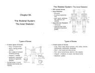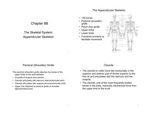
Chapter 8B: Skeletal System: Appendicular Skeleton
Chapter 8B: Skeletal System: Appendicular Skeleton
Chapter 8B: Skeletal System: Appendicular Skeleton

You also want an ePaper? Increase the reach of your titles
YUMPU automatically turns print PDFs into web optimized ePapers that Google loves.
<strong>Chapter</strong> <strong>8B</strong><br />
The <strong>Skeletal</strong> <strong>System</strong>:<br />
<strong>Appendicular</strong> <strong>Skeleton</strong><br />
Pectoral (Shoulder) Girdle<br />
The pectoral (shoulder) girdle attaches the bones of the<br />
upper limbs to the axial skeleton<br />
• Consists of scapula and clavicle<br />
• Clavicle articulates with sternum (sternoclavicular joint)<br />
• Clavicle articulates with scapula (acromioclavicular joint)<br />
• Upper limb attached to pectoral girdle at shoulder<br />
(glenohumeral joint)<br />
1<br />
3<br />
• 126 bones<br />
• Pectoral (shoulder)<br />
girdle<br />
• Pelvic (hip) girdle<br />
• Upper limbs<br />
• Lower limbs<br />
• Functions primarily to<br />
facilitate movement<br />
The <strong>Appendicular</strong> <strong>Skeleton</strong><br />
Clavicle<br />
• The clavicle or collar bone lies horizontally in the<br />
superior and anterior part of thorax superior to the<br />
first rib and articulates with the sternum and the<br />
scapula<br />
• The clavicle, one of the most frequently broken<br />
bones in the body, transmits mechanical force from<br />
the upper limb to the trunk.<br />
2<br />
4
Clavicle (collarbone)<br />
• S-shaped bone with two curves<br />
• Extends from sternum to scapula above 1st rib<br />
• Fracture site is junction of curves<br />
• Ligaments attached to clavicle stabilize its position<br />
Anterior Surface of Scapula<br />
• Subscapular fossa filled with muscle<br />
• Coracoid process for muscle attachment<br />
5<br />
7<br />
Scapula<br />
• The scapula or shoulder blade articulates with the<br />
clavicle and the humerus<br />
• The scapulae articulate with other bones anteriorly,<br />
but are held in place posteriorly only by complex<br />
shoulder and back musculature<br />
Posterior Surface of Scapula<br />
• Triangular flat bone found in upper back region<br />
• Scapular spine ends as acromion process<br />
– a sharp ridge widening to a flat process<br />
• Glenoid cavity forms shoulder joint with head of humerus<br />
• Supraspinous & infraspinous fossa for muscular<br />
attachments<br />
6<br />
8
Upper Extremity<br />
• Each upper limb = 30 bones<br />
– humerus within the arm<br />
– ulna & radius within the forearm<br />
– carpal bones within the wrist<br />
– metacarpal bones within the palm<br />
– phalanges in the fingers<br />
• Joints<br />
– shoulder (glenohumeral), elbow,<br />
wrist, metacarpophalangeal,<br />
interphalangeal<br />
Humerus --- Proximal End<br />
• Part of shoulder joint<br />
• Head & anatomical neck<br />
• Greater & lesser tubercles for muscle<br />
attachments<br />
• Intertubercular<br />
sulcus or<br />
groove<br />
• Surgical neck is<br />
fracture site<br />
• Deltoid tuberosity<br />
• Shaft<br />
9<br />
11<br />
Humerus<br />
• The humerus is the longest and largest bone of the<br />
upper limb<br />
• It articulates proximally with the scapula and distally<br />
at the elbow with both the radius and ulna.<br />
Humerus --- Distal End<br />
anterior and posterior<br />
• Forms elbow joint with<br />
ulna and radius<br />
• Capitulum<br />
– articulates with head of radius<br />
• Trochlea<br />
– articulation with ulna<br />
• Olecranon fossa<br />
– posterior depression for<br />
olecranon process of ulna<br />
• Medial & lateral epicondyles<br />
– attachment of forearm<br />
muscles<br />
10<br />
12
Ulna and Radius<br />
• The ulna is located on the medial aspect of the<br />
forearm<br />
• The radius is located on the lateral aspect of the<br />
forearm<br />
• The radius and ulna articulate with the humerus at<br />
the elbow joint, with each other, and with three<br />
carpal bones<br />
Elbow Joint<br />
• Articulation of humerus with ulna and radius<br />
• Ulna articulates with trochlea of humerus<br />
• Radius articulates with capitulum of humerus<br />
• Interosseous membrane between ulna & radius provides site<br />
for muscle attachment<br />
13<br />
15<br />
Ulna & Radius --- Proximal End<br />
• Ulna (on little finger side)<br />
– trochlear notch articulates with<br />
humerus & radial notch with radius<br />
– olecranon process forms point of elbow<br />
• Radius (on thumb side)<br />
– head articulates with capitulum of<br />
humerus & radial notch of ulna<br />
– radial tuberosity for muscle attachment<br />
Ulna and Radius - Distal End<br />
• Ulna - styloid process<br />
– head separated from wrist joint by fibrocartilage<br />
disc<br />
• Radius - styloid process<br />
– forms wrist joint with scaphoid, lunate & triquetrum<br />
– forms distal radioulnar joint with head of ulna<br />
14<br />
16
Carpals, Metacarpal, and Phalanges<br />
• The eight carpal bones, bound together by ligaments,<br />
comprise the wrist<br />
• Five metacarpal bones are contained in the palm of each<br />
hand<br />
• Each hand contains 14 phalanges, three in each finger and<br />
two in each thumb<br />
Metacarpals and Phalanges<br />
• Metacarpals<br />
– 5 total----#1 proximal to<br />
thumb<br />
– knuckles<br />
(metacarpophalangeal joints)<br />
• Phalanges<br />
– 14 total: each is called<br />
phalanx<br />
– proximal, middle, distal on<br />
each finger, except thumb<br />
17<br />
19<br />
Hand<br />
8 Carpal Bones (wrist)<br />
• Proximal row - lateral to medial<br />
– scaphoid - boat shaped<br />
– lunate - moon shaped<br />
– triquetrum - 3 corners<br />
– pisiform - pea shaped<br />
• Distal row - lateral to medial<br />
– trapezium - four sided<br />
– trapezoid - four sided<br />
– capitate - large head<br />
– hamate - hooked process<br />
• Carpal tunnel - tunnel of bone &<br />
flexor retinaculum<br />
18<br />
20
PELVIC (HIP) GIRDLE<br />
• The pelvic (hip) girdle consists of two hipbones (os<br />
coxa or coxal bones) and provides a strong and<br />
stable support for the lower extremities, on which<br />
the weight of the body is carried<br />
• Each hipbone is composed of three separate bones<br />
at birth: the ilium, ischium, and pubis<br />
• These bones eventually fuse at a depression called<br />
the acetabulum, which forms the socket for the hip<br />
joint<br />
The Ilium<br />
• The larger of the three components of the hip bone<br />
and articulates (fuses) with the ischium and pubis<br />
• Bone marrow aspiration or bone marrow biopsy are<br />
frequently performed on the iliac crest in adults.<br />
• The ischium is the inferior, posterior portion of the<br />
hip bone<br />
• The pubis is the anterior and inferior part of the hip<br />
bone<br />
21<br />
23<br />
Pelvic Girdle and Hip Bones<br />
• Pelvic girdle = two hipbones united at pubic symphysis<br />
– articulate posteriorly with sacrum at sacroiliac joints<br />
• Each hip bone = ilium, pubis, and ischium<br />
– fuse after birth at acetabulum<br />
• Bony pelvis = 2 hip bones, sacrum and coccyx<br />
Ilium<br />
• Iliac crest and iliac spines for muscle attachment<br />
• Iliac fossa for muscle attachment<br />
• Sacroiliac joint at auricular surface & iliac tuberosity<br />
• Greater sciatic notch for sciatic nerve<br />
22<br />
24
Ischium and Pubis<br />
• Ischium<br />
– ischial tuberosity<br />
• Pubis<br />
– pubic symphysis is pad of<br />
fibrocartilage between 2 pubic<br />
bones<br />
• Obturator foramen<br />
• Acetabulum<br />
Female and Male <strong>Skeleton</strong>s<br />
• Male skeleton<br />
– larger and heavier<br />
– larger articular surfaces<br />
– larger muscle attachments<br />
• Female pelvis<br />
– wider & shallower<br />
– larger pelvic inlet & outlet<br />
– more space in true pelvis<br />
– pubic arch >90 degrees<br />
25<br />
27<br />
Pelvis<br />
Female Male<br />
• Pelvis = sacrum, coccyx<br />
& 2 hip bones<br />
• Pelvic brim<br />
– sacral promontory to<br />
symphysis pubis<br />
– separates false from<br />
true pelvis<br />
– false pelvis holds<br />
only abdominal<br />
organs<br />
26<br />
28
COMPARISON OF PECTORAL AND PELVIC<br />
GIRDLES<br />
• The pectoral girdle does not directly articulate with<br />
the vertebral column; the pelvic girdle does.<br />
• The pectoral girdle sockets are shallow and<br />
maximize movement; those of the pelvic girdle are<br />
deeper and allow less movement.<br />
• The structure of the pectoral girdle offers more<br />
movement than strength; the pelvic girdle, more<br />
strength than movement.<br />
Femur<br />
• The femur or thighbone is the largest, heaviest,<br />
and strongest bone of the body<br />
• It articulates with the hip bone and the tibia.<br />
– head articulates with acetabulum (attached by<br />
ligament of head of femur)<br />
– medial & lateral condyles articulate with tibia<br />
• neck is common fracture site<br />
• greater & lesser trochanters, linea aspera, &<br />
gluteal tuberosity -- muscle attachments<br />
• patellar surface is visible anteriorly between<br />
condyles<br />
29<br />
31<br />
Lower Extremity<br />
• Each lower limb = 30 bones<br />
– femur and patella within the<br />
thigh<br />
– tibia & fibula within the leg<br />
– tarsal bones in the foot<br />
– metatarsals within the forefoot<br />
– phalanges in the toes<br />
• Joints<br />
– hip, knee, ankle<br />
– proximal & distal tibiofibular<br />
– metatarsophalangeal<br />
Femur<br />
30<br />
32
Patella<br />
• triangular sesamoid bone<br />
• increases leverage of quadriceps femoris tendon<br />
Fibula<br />
• not part of knee joint<br />
• muscle attachment only<br />
• lateral malleolus at ankle<br />
Tibia and Fibula<br />
33<br />
35<br />
Tibia and Fibula<br />
Tibia<br />
• medial & larger bone<br />
of leg<br />
• weight-bearing bone<br />
• lateral & medial<br />
condyles<br />
• tibial tuberosity for<br />
patellar ligament<br />
• proximal tibiofibular<br />
joint<br />
• medial malleolus at<br />
ankle<br />
Tarsals, Metatarsals, and Phalanges<br />
• Seven tarsal bones constitute the ankle and share<br />
the weight associated with walking<br />
• Five metatarsal bones are contained in the foot<br />
• Fractures of the metatarsals are common among<br />
dancers, especially ballet dancers.<br />
• The arrangement of phalanges in the toes is the<br />
same as that described for the fingers and thumb<br />
above - fourteen bones in each foot<br />
34<br />
36
Tarsus<br />
• Proximal region of<br />
foot (contains 7 tarsal<br />
bones)<br />
• Talus = ankle bone<br />
(articulates with tibia<br />
& fibula)<br />
• Calcaneus - heel<br />
bone<br />
• Cuboid, navicular & 3<br />
cuneiforms<br />
Arches of the Foot<br />
• Function<br />
– distribute body weight over foot<br />
– yield & spring back when weight is lifted<br />
• Longitudinal arches along each side of foot<br />
• Transverse arch across midfoot region<br />
– navicular, cuneiforms & bases of metatarsals<br />
37<br />
39<br />
Metatarsus and Phalanges<br />
• Flatfoot<br />
– weakened ligaments<br />
allow bones of<br />
medial arch to drop<br />
• Clawfoot<br />
– medial arch is too<br />
elevated<br />
• Hip fracture<br />
– 1/2 million/year in US<br />
– osteoporosis<br />
Clinical Problems<br />
• Metatarsus<br />
– midregion of the foot<br />
– 5 metatarsals (1 is most<br />
medial)<br />
– each with base, shaft and<br />
head<br />
• Phalanges<br />
– distal portion of the foot<br />
– similar in number and<br />
arrangement to the hand<br />
– big toe is hallux<br />
38<br />
40