Arterial vascularisation of the anterior perforated ... - APAMED Central
Arterial vascularisation of the anterior perforated ... - APAMED Central
Arterial vascularisation of the anterior perforated ... - APAMED Central

Create successful ePaper yourself
Turn your PDF publications into a flip-book with our unique Google optimized e-Paper software.
2a 2b<br />
METHODS<br />
The arteries <strong>of</strong> <strong>the</strong> APS were investigated in 60 cerebral<br />
hemispheres from 30 adult cadaveric brains. Brains<br />
having signs <strong>of</strong> central nervous system trauma or<br />
disease were excluded. First, <strong>the</strong> internal carotid arteries<br />
were cannulated and perfused with coloured latex in<br />
fresh brains, after which <strong>the</strong> brains were embalmed in<br />
10% formaline solution for fixation. Dissections were<br />
performed using microsurgical instruments and an OPMI<br />
99 surgical microscope (Carl Zeiss, Göttingen, Germany).<br />
The number <strong>of</strong> arteries <strong>of</strong> <strong>the</strong> APS was recorded, and<br />
<strong>the</strong>ir origins were investigated. The Statistical Package<br />
for <strong>the</strong> Social Sciences version 15.0 (SPSS Inc, Chicago,<br />
IL, USA) was used for statistical analyses. A p-value <<br />
0.05 was considered to be statistically significant. Overall<br />
measurements were evaluated with <strong>the</strong> Wilcoxon signed-<br />
rank test.<br />
RESULTS<br />
TB<br />
MCA<br />
ON<br />
ICA<br />
OT<br />
In all <strong>the</strong> 60 specimens, <strong>the</strong> internal carotid artery<br />
(ICA) bifurcated into <strong>the</strong> MCA and <strong>anterior</strong> cerebral<br />
artery (ACA) below <strong>the</strong> central portion <strong>of</strong> <strong>the</strong> APS. The<br />
branches <strong>of</strong> <strong>the</strong> MCA that penetrated <strong>the</strong> APS (lateral<br />
lenticulostriate arteries [LLAs]) were investigated. In<br />
all hemispheres, <strong>the</strong> average number <strong>of</strong> LLAs that arose<br />
from <strong>the</strong> M1 segment <strong>of</strong> <strong>the</strong> MCA was seven (range 4–11)<br />
(Figs. 1 & 2a). The number <strong>of</strong> LLAs on <strong>the</strong> left and right<br />
cerebral hemispheres was 6–11 and 4–10, respectively.<br />
No statistically significant difference was observed<br />
between <strong>the</strong> two sides with <strong>the</strong> Wilcoxon signed-rank<br />
test (p = 0.764 left, p > 0.05 right). We observed early<br />
Singapore Med J 2011; 52(6) : 411<br />
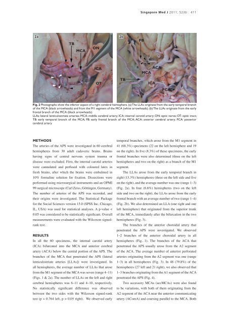
Fig. 2 Photographs show <strong>the</strong> inferior aspect <strong>of</strong> a right cerebral hemisphere. (a) The LLAs originate from <strong>the</strong> early temporal branch<br />
<strong>of</strong> <strong>the</strong> MCA (black arrowheads) and from <strong>the</strong> M1 segment <strong>of</strong> <strong>the</strong> MCA (white arrowheads). (b) The LLAs originate from <strong>the</strong> early<br />
frontal branch <strong>of</strong> <strong>the</strong> MCA (black arrowheads).<br />
LLAs: lateral lenticulostriate arteries; MCA: middle cerebral artery; ICA: internal carotid artery; ON: optic nerve; OT: optic tract;<br />
TB: early temporal branch <strong>of</strong> <strong>the</strong> MCA; FB: early frontal branch <strong>of</strong> <strong>the</strong> MCA; ACA: <strong>anterior</strong> cerebral artery; PCA: posterior<br />
cerebral artery<br />
TB<br />
temporal branches, which arose from <strong>the</strong> M1 segment in<br />
41 (68.3%) specimens (22 on <strong>the</strong> left hemisphere and 19<br />
on <strong>the</strong> right). In five (8.3%) <strong>of</strong> <strong>the</strong>se specimens, <strong>the</strong> early<br />
frontal branches were also determined (three on <strong>the</strong> left<br />
hemispheres and two on <strong>the</strong> right) as a branch <strong>of</strong> <strong>the</strong> M1<br />
segment.<br />
FB<br />
The LLAs arose from <strong>the</strong> early temporal branch in<br />
eight (13.3%) hemispheres (three on <strong>the</strong> left side and five<br />
on <strong>the</strong> right), and <strong>the</strong> average number was one (range 1–3)<br />
(Fig. 2a). In four (6.6%) hemispheres (two on <strong>the</strong> left<br />
side and two on <strong>the</strong> right), <strong>the</strong> LLAs arose from <strong>the</strong> early<br />
frontal branch with an average number <strong>of</strong> two (range 1–4)<br />
(Fig. 2b). We also determined an LLA (one right and one<br />
left hemisphere) that originated from <strong>the</strong> superior trunk<br />
<strong>of</strong> <strong>the</strong> MCA, immediately after <strong>the</strong> bifurcation in <strong>the</strong> two<br />
hemispheres (Fig. 3).<br />
ACA<br />
MCA<br />
ICA<br />
The branches <strong>of</strong> <strong>the</strong> <strong>anterior</strong> choroidal artery that<br />
penetrated <strong>the</strong> APS were investigated. We observed<br />
1–2 branches <strong>of</strong> <strong>the</strong> <strong>anterior</strong> choroidal artery in all<br />
hemispheres (Fig. 1). The branches <strong>of</strong> <strong>the</strong> ACA that<br />
penetrated <strong>the</strong> APS usually arose from <strong>the</strong> A2 segment<br />
<strong>of</strong> <strong>the</strong> ACA. The average number <strong>of</strong> <strong>anterior</strong> <strong>perforated</strong><br />
arteries originating from <strong>the</strong> A2 segment was one (range<br />
1–3) in all hemispheres (Fig. 3). In 48 (79.8%) <strong>of</strong> <strong>the</strong><br />
hemispheres (27 left and 21 right), we also observed that<br />
1–3 branches originating from <strong>the</strong> A1 segment <strong>of</strong> <strong>the</strong> ACA<br />
penetrated <strong>the</strong> APS (Fig. 4).<br />
PCA<br />
Two accessory MCAs (accMCAs) were also found<br />
to be variations, with both <strong>of</strong> <strong>the</strong>m originating from <strong>the</strong><br />
A2 segment <strong>of</strong> <strong>the</strong> ACA near <strong>the</strong> <strong>anterior</strong> communicating<br />
artery (AComA) and coursing parallel to <strong>the</strong> MCA. Both