Arterial vascularisation of the anterior perforated ... - APAMED Central
Arterial vascularisation of the anterior perforated ... - APAMED Central
Arterial vascularisation of the anterior perforated ... - APAMED Central

Create successful ePaper yourself
Turn your PDF publications into a flip-book with our unique Google optimized e-Paper software.
Department <strong>of</strong><br />
Anatomy,<br />
Ankara University<br />
School <strong>of</strong> Medicine,<br />
Morfoloji Binasi,<br />
Sihhiye,<br />
Ankara 06100,<br />
Turkey<br />
Sen T, MD<br />
Senior Consultant<br />
Esmer AF, MD<br />
Senior Consultant<br />
Acar HI, MD<br />
Associate Pr<strong>of</strong>essor<br />
Karahan ST, MD<br />
Pr<strong>of</strong>essor<br />
Tuccar E, MD, PhD<br />
Pr<strong>of</strong>essor<br />
Correspondence to:<br />
Dr Ali Firat Esmer<br />
Tel: (90) 312 3105001<br />
Fax: (90) 312 3105001<br />
Email: alife76@yahoo.<br />
com<br />
Original Article<br />
Singapore Med J 2011; 52(6) : 410<br />
<strong>Arterial</strong> <strong>vascularisation</strong> <strong>of</strong> <strong>the</strong> <strong>anterior</strong><br />
<strong>perforated</strong> substance<br />
Sen T, Esmer A F, Acar H I, Karahan S T, Tuccar E<br />
ABSTRACT<br />
Introduction: The arteries <strong>of</strong> <strong>the</strong> <strong>anterior</strong><br />
<strong>perforated</strong> substance (APS) are important due to<br />
<strong>the</strong>ir role in supplying blood to important internal<br />
structures such as <strong>the</strong> internal capsule, putamen<br />
and caudate nucleus. The purpose <strong>of</strong> this study was<br />
to investigate in detail <strong>the</strong> arteries <strong>of</strong> <strong>the</strong> APS.<br />
Methods: The arteries <strong>of</strong> <strong>the</strong> APS were investigated<br />
in 60 cerebral hemispheres from 30 adult cadaveric<br />
brains. The internal carotid arteries were<br />
cannulated and perfused with coloured latex. The<br />
branches <strong>of</strong> <strong>the</strong> middle cerebral artery (MCA)<br />
penetrating <strong>the</strong> APS were investigated. These<br />
arteries, known as <strong>the</strong> lateral lenticulostriate<br />
arteries and originating from <strong>the</strong> M1 segment,<br />
early temporal and early frontal branches <strong>of</strong> <strong>the</strong><br />
MCA, were recorded.<br />
Results: The branches <strong>of</strong> <strong>the</strong> <strong>anterior</strong> choroidal<br />
artery, which reached <strong>the</strong> APS, were seen in all<br />
specimens. We found one to three branches that<br />
arose from <strong>the</strong> A2 segment <strong>of</strong> <strong>the</strong> <strong>anterior</strong> cerebral<br />
artery (ACA) to <strong>the</strong> APS in all hemispheres, and<br />
one to three branches that originated from <strong>the</strong><br />
A1 segment <strong>of</strong> <strong>the</strong> ACA in 48 hemispheres. In<br />
addition, two accessory MCAs that originated<br />
from <strong>the</strong> A2 segment <strong>of</strong> <strong>the</strong> ACA were recorded<br />
as variations, and perforating branches to <strong>the</strong> APS<br />
were observed.<br />
Conclusion: Serious complications like motor<br />
deficits can occur as a result <strong>of</strong> injury to <strong>the</strong> arteries<br />
<strong>of</strong> <strong>the</strong> APS. Hence, neurosurgeons performing<br />
operations such as aneurysm or insular tumour<br />
surgeries must be aware <strong>of</strong> <strong>the</strong> importance <strong>of</strong><br />
preserving <strong>the</strong>se arteries.<br />
Keywords: anatomy, <strong>anterior</strong> cerebral artery,<br />
<strong>anterior</strong> <strong>perforated</strong> substance, middle cerebral<br />
artery<br />
Singapore Med J 2011; 52(6): 410-414<br />
OC<br />
ON<br />
OT<br />
AccMCA<br />
INTRODUCTION<br />
ICA<br />
A1 M1<br />
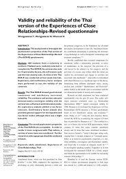
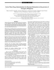
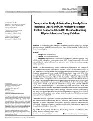
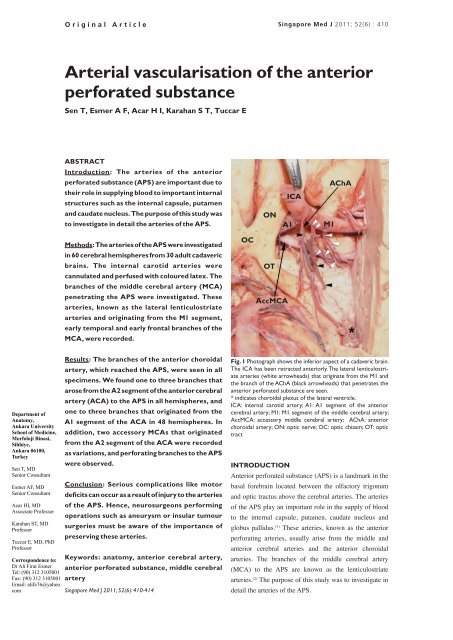
Fig. 1 Photograph shows <strong>the</strong> inferior aspect <strong>of</strong> a cadaveric brain.<br />
The ICA has been retracted <strong>anterior</strong>ly. The lateral lenticulostriate<br />
arteries (white arrowheads) that originate from <strong>the</strong> M1 and<br />
<strong>the</strong> branch <strong>of</strong> <strong>the</strong> AChA (black arrowheads) that penetrates <strong>the</strong><br />
<strong>anterior</strong> <strong>perforated</strong> substance are seen.<br />
* indicates choroidal plexus <strong>of</strong> <strong>the</strong> lateral ventricle.<br />
ICA: internal carotid artery; A1: A1 segment <strong>of</strong> <strong>the</strong> <strong>anterior</strong><br />
cerebral artery; M1: M1 segment <strong>of</strong> <strong>the</strong> middle cerebral artery;<br />
AccMCA: accessory middle cerebral artery; AChA: <strong>anterior</strong><br />
choroidal artery; ON: optic nerve; OC: optic chiasm; OT: optic<br />
tract<br />
Anterior <strong>perforated</strong> substance (APS) is a landmark in <strong>the</strong><br />
basal forebrain located between <strong>the</strong> olfactory trigonum<br />
and optic tractus above <strong>the</strong> cerebral arteries. The arteries<br />
<strong>of</strong> <strong>the</strong> APS play an important role in <strong>the</strong> supply <strong>of</strong> blood<br />
to <strong>the</strong> internal capsule, putamen, caudate nucleus and<br />
globus pallidus. (1) These arteries, known as <strong>the</strong> <strong>anterior</strong><br />
perforating arteries, usually arise from <strong>the</strong> middle and<br />
<strong>anterior</strong> cerebral arteries and <strong>the</strong> <strong>anterior</strong> choroidal<br />
arteries. The branches <strong>of</strong> <strong>the</strong> middle cerebral artery<br />
(MCA) to <strong>the</strong> APS are known as <strong>the</strong> lenticulostriate<br />
arteries. (2) The purpose <strong>of</strong> this study was to investigate in<br />
detail <strong>the</strong> arteries <strong>of</strong> <strong>the</strong> APS.<br />
AChA
2a 2b<br />
METHODS<br />
The arteries <strong>of</strong> <strong>the</strong> APS were investigated in 60 cerebral<br />
hemispheres from 30 adult cadaveric brains. Brains<br />
having signs <strong>of</strong> central nervous system trauma or<br />
disease were excluded. First, <strong>the</strong> internal carotid arteries<br />
were cannulated and perfused with coloured latex in<br />
fresh brains, after which <strong>the</strong> brains were embalmed in<br />
10% formaline solution for fixation. Dissections were<br />
performed using microsurgical instruments and an OPMI<br />
99 surgical microscope (Carl Zeiss, Göttingen, Germany).<br />
The number <strong>of</strong> arteries <strong>of</strong> <strong>the</strong> APS was recorded, and<br />
<strong>the</strong>ir origins were investigated. The Statistical Package<br />
for <strong>the</strong> Social Sciences version 15.0 (SPSS Inc, Chicago,<br />
IL, USA) was used for statistical analyses. A p-value <<br />
0.05 was considered to be statistically significant. Overall<br />
measurements were evaluated with <strong>the</strong> Wilcoxon signed-<br />
rank test.<br />
RESULTS<br />
TB<br />
MCA<br />
ON<br />
ICA<br />
OT<br />
In all <strong>the</strong> 60 specimens, <strong>the</strong> internal carotid artery<br />
(ICA) bifurcated into <strong>the</strong> MCA and <strong>anterior</strong> cerebral<br />
artery (ACA) below <strong>the</strong> central portion <strong>of</strong> <strong>the</strong> APS. The<br />
branches <strong>of</strong> <strong>the</strong> MCA that penetrated <strong>the</strong> APS (lateral<br />
lenticulostriate arteries [LLAs]) were investigated. In<br />
all hemispheres, <strong>the</strong> average number <strong>of</strong> LLAs that arose<br />
from <strong>the</strong> M1 segment <strong>of</strong> <strong>the</strong> MCA was seven (range 4–11)<br />
(Figs. 1 & 2a). The number <strong>of</strong> LLAs on <strong>the</strong> left and right<br />
cerebral hemispheres was 6–11 and 4–10, respectively.<br />
No statistically significant difference was observed<br />
between <strong>the</strong> two sides with <strong>the</strong> Wilcoxon signed-rank<br />
test (p = 0.764 left, p > 0.05 right). We observed early<br />
Singapore Med J 2011; 52(6) : 411<br />
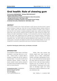
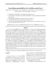
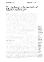
Fig. 2 Photographs show <strong>the</strong> inferior aspect <strong>of</strong> a right cerebral hemisphere. (a) The LLAs originate from <strong>the</strong> early temporal branch<br />
<strong>of</strong> <strong>the</strong> MCA (black arrowheads) and from <strong>the</strong> M1 segment <strong>of</strong> <strong>the</strong> MCA (white arrowheads). (b) The LLAs originate from <strong>the</strong> early<br />
frontal branch <strong>of</strong> <strong>the</strong> MCA (black arrowheads).<br />
LLAs: lateral lenticulostriate arteries; MCA: middle cerebral artery; ICA: internal carotid artery; ON: optic nerve; OT: optic tract;<br />
TB: early temporal branch <strong>of</strong> <strong>the</strong> MCA; FB: early frontal branch <strong>of</strong> <strong>the</strong> MCA; ACA: <strong>anterior</strong> cerebral artery; PCA: posterior<br />
cerebral artery<br />
TB<br />
temporal branches, which arose from <strong>the</strong> M1 segment in<br />
41 (68.3%) specimens (22 on <strong>the</strong> left hemisphere and 19<br />
on <strong>the</strong> right). In five (8.3%) <strong>of</strong> <strong>the</strong>se specimens, <strong>the</strong> early<br />
frontal branches were also determined (three on <strong>the</strong> left<br />
hemispheres and two on <strong>the</strong> right) as a branch <strong>of</strong> <strong>the</strong> M1<br />
segment.<br />
FB<br />
The LLAs arose from <strong>the</strong> early temporal branch in<br />
eight (13.3%) hemispheres (three on <strong>the</strong> left side and five<br />
on <strong>the</strong> right), and <strong>the</strong> average number was one (range 1–3)<br />
(Fig. 2a). In four (6.6%) hemispheres (two on <strong>the</strong> left<br />
side and two on <strong>the</strong> right), <strong>the</strong> LLAs arose from <strong>the</strong> early<br />
frontal branch with an average number <strong>of</strong> two (range 1–4)<br />
(Fig. 2b). We also determined an LLA (one right and one<br />
left hemisphere) that originated from <strong>the</strong> superior trunk<br />
<strong>of</strong> <strong>the</strong> MCA, immediately after <strong>the</strong> bifurcation in <strong>the</strong> two<br />
hemispheres (Fig. 3).<br />
ACA<br />
MCA<br />
ICA<br />
The branches <strong>of</strong> <strong>the</strong> <strong>anterior</strong> choroidal artery that<br />
penetrated <strong>the</strong> APS were investigated. We observed<br />
1–2 branches <strong>of</strong> <strong>the</strong> <strong>anterior</strong> choroidal artery in all<br />
hemispheres (Fig. 1). The branches <strong>of</strong> <strong>the</strong> ACA that<br />
penetrated <strong>the</strong> APS usually arose from <strong>the</strong> A2 segment<br />
<strong>of</strong> <strong>the</strong> ACA. The average number <strong>of</strong> <strong>anterior</strong> <strong>perforated</strong><br />
arteries originating from <strong>the</strong> A2 segment was one (range<br />
1–3) in all hemispheres (Fig. 3). In 48 (79.8%) <strong>of</strong> <strong>the</strong><br />
hemispheres (27 left and 21 right), we also observed that<br />
1–3 branches originating from <strong>the</strong> A1 segment <strong>of</strong> <strong>the</strong> ACA<br />
penetrated <strong>the</strong> APS (Fig. 4).<br />
PCA<br />
Two accessory MCAs (accMCAs) were also found<br />
to be variations, with both <strong>of</strong> <strong>the</strong>m originating from <strong>the</strong><br />
A2 segment <strong>of</strong> <strong>the</strong> ACA near <strong>the</strong> <strong>anterior</strong> communicating<br />
artery (AComA) and coursing parallel to <strong>the</strong> MCA. Both
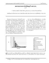
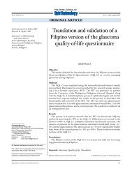
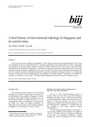
Fig. 3 Photograph shows bifurcation <strong>of</strong> <strong>the</strong> ICA. The ICA is<br />
dissected to separate <strong>the</strong> MCA and ACA, after which <strong>the</strong>y<br />
are respectively retracted laterally and medially to expose<br />
<strong>the</strong> arteries <strong>of</strong> <strong>the</strong> <strong>anterior</strong> <strong>perforated</strong> substance. The lateral<br />
lenticulostriate artery originates from <strong>the</strong> ST trunk <strong>of</strong> <strong>the</strong> MCA<br />
(black arrowheads), while <strong>the</strong> <strong>anterior</strong> perforating artery arises<br />
from <strong>the</strong> A2 segment <strong>of</strong> <strong>the</strong> ACA (while arrowheads).<br />
ICA: internal carotid artery; ACA: <strong>anterior</strong> cerebral artery;<br />
MCA: middle cerebral artery; TB: early temporal branch <strong>of</strong><br />
<strong>the</strong> MCA; IT: inferior trunk <strong>of</strong> <strong>the</strong> MCA; ST: superior trunk <strong>of</strong><br />
<strong>the</strong> MCA; OC: optic chiasm; OT: optic tract; A1: A1 segment<br />
<strong>of</strong> <strong>the</strong> ACA; A2: A2 segment <strong>of</strong> <strong>the</strong> ACA; AComA: <strong>anterior</strong><br />
communicating artery<br />
accMCAs were found in <strong>the</strong> left cerebral hemispheres,<br />
and gave rise to three and four perforating branches <strong>of</strong> <strong>the</strong><br />
APS (Fig. 4).<br />
DISCUSSION<br />
The MCA is divided into four major segments: (a) The<br />
M1 (sphenoidal) segment extends from <strong>the</strong> terminal<br />
bifurcation <strong>of</strong> <strong>the</strong> ICA to <strong>the</strong> main MCA bifurcation,<br />
which is usually located at <strong>the</strong> level <strong>of</strong> <strong>the</strong> limen insulae;<br />
(b) The M2 (insular) segment, which includes <strong>the</strong><br />
superior and inferior trunks <strong>of</strong> <strong>the</strong> MCA, extends from<br />
<strong>the</strong> main bifurcation to <strong>the</strong> peri-insular sulci; (c) The M3<br />
(opercular) segment extends from <strong>the</strong> peri-insular sulci to<br />
<strong>the</strong> cortical surface <strong>of</strong> <strong>the</strong> sylvian fissure; and (d) The M4<br />
(cortical) segment is located on <strong>the</strong> parasylvian surface <strong>of</strong><br />
<strong>the</strong> brain and spreads over <strong>the</strong> cortical surface. (3)<br />
According to Fischer, (4) <strong>the</strong> ACA is divided into five<br />
major segments. The A1 segment (proximal ACA) extends<br />
from <strong>the</strong> terminal bifurcation <strong>of</strong> <strong>the</strong> ICA to <strong>the</strong> AComA.<br />
The A1 segment ends and <strong>the</strong> A2 segment begins at <strong>the</strong><br />
level <strong>of</strong> <strong>the</strong> AComA. The A2 segment extends to <strong>the</strong><br />
region between <strong>the</strong> rostrum and <strong>the</strong> genu <strong>of</strong> <strong>the</strong> corpus<br />
callosum (CC). The A3 segment curves around <strong>the</strong> genu<br />
<strong>of</strong> CC and ends at <strong>the</strong> rostral part <strong>of</strong> <strong>the</strong> body <strong>of</strong> <strong>the</strong> CC.<br />
The A4 and A5 segments follow <strong>the</strong> superior surface <strong>of</strong><br />
<strong>the</strong> CC. The A4 segment extends from <strong>the</strong> point at which<br />
<strong>the</strong> artery turns sharply to <strong>the</strong> posterior on <strong>the</strong> genu <strong>of</strong> <strong>the</strong><br />
Singapore Med J 2011; 52(6) : 412<br />
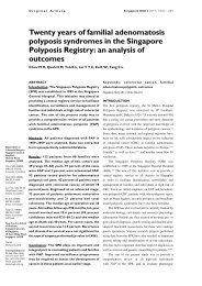
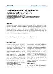
Fig. 4 Photograph shows <strong>the</strong> <strong>anterior</strong> choroidal artery and LLAs<br />
arising from <strong>the</strong> MCA, which are removed for a better aspect <strong>of</strong><br />
<strong>the</strong> AccMCA. The AccMCA is retracted <strong>anterior</strong>ly with a suture<br />
to show <strong>the</strong> LLAs that originated from it. The LLAs arise from<br />
<strong>the</strong> AccMCA (black arrowheads) and <strong>the</strong> <strong>anterior</strong> perforating<br />
artery arises from <strong>the</strong> A1 segment <strong>of</strong> <strong>the</strong> ACA (*).<br />
LLAs: lateral lenticulostriate arteries; ON: optic nerve; OC: optic<br />
chiasm; OT: optic tract; ICA: internal carotid artery; ACA: <strong>anterior</strong><br />
cerebral artery; MCA: middle cerebral artery; AccMCA:<br />
accessory middle cerebral artery<br />
CC to <strong>the</strong> line where <strong>the</strong> CC intersects laterally with <strong>the</strong><br />
coronal suture. The A5 segment extends from <strong>the</strong> point<br />
at which <strong>the</strong> CC intersects with <strong>the</strong> coronal suture to <strong>the</strong><br />
splenium <strong>of</strong> <strong>the</strong> CC. (4) In this study, we focused on <strong>the</strong><br />
perforating branches <strong>of</strong> <strong>the</strong> A1 and proximal A2 segments<br />
<strong>of</strong> <strong>the</strong> ACA.<br />
The microsurgical anatomy <strong>of</strong> <strong>the</strong> LLAs has been<br />
examined in detail in many studies. (1,3,5-7) Türe et al<br />
found 1–15 (average 7.75) LLAs, while Marinkovíc et al<br />
observed 3–18 (average nine) LLAs that originated from<br />
<strong>the</strong> M1 segment <strong>of</strong> <strong>the</strong> MCA. (1,5) Similar findings were<br />
noted in our study as well. The cortical arteries arising from<br />
<strong>the</strong> main trunk <strong>of</strong> <strong>the</strong> MCA before bifurcation are called<br />
‘early branches’. (8) Türe et al reported LLAs originating<br />
from <strong>the</strong> early frontal branch <strong>of</strong> <strong>the</strong> M1 in nine (22.5%)<br />
hemispheres, (1) while we observed this in four (6.6%)<br />
hemispheres and LLAs from <strong>the</strong> early temporal branch in<br />
eight (13.3%) hemispheres. Umansky et al reported 5.7%<br />
<strong>of</strong> perforating arteries arising from <strong>the</strong> early branches <strong>of</strong><br />
<strong>the</strong> MCA, (9) while Marinkovíc et al found one or more<br />
LLAs originating from <strong>the</strong> early branches in a quarter <strong>of</strong><br />
<strong>the</strong> hemispheres. (5) Also, nearly one in five LLAs arose<br />
from an early branch in <strong>the</strong> study <strong>of</strong> Tanrıover et al. (10)<br />
When early cortical branches exist as a branch <strong>of</strong> <strong>the</strong><br />
MCA, <strong>the</strong> LLAs have a high possibility <strong>of</strong> originating<br />
from <strong>the</strong>se arteries. Yasargil’s studies found that <strong>the</strong><br />
frequency and site <strong>of</strong> origin <strong>of</strong> <strong>the</strong> early branches as well
as <strong>the</strong> course and number <strong>of</strong> LLAs arising from <strong>the</strong>m have<br />
practical application in transsylvian approaches, which<br />
require exposure <strong>of</strong> part or all <strong>of</strong> <strong>the</strong> insula. (11,12)<br />
Chyatte and Porterfield reported that LLAs arising<br />
from <strong>the</strong> M1 trunk never supply <strong>the</strong> frontal lobe, and<br />
<strong>the</strong>refore, it is easier to retract <strong>the</strong> frontal lobe away from<br />
<strong>the</strong> MCA during surgery. (13) Moreover, Tanrıover et al<br />
also reported in <strong>the</strong>ir study that an early frontal branch<br />
arising from <strong>the</strong> M1 segment may exist in more than 30%<br />
<strong>of</strong> cases, and on average, this gives rise to more LLAs per<br />
vessel than <strong>the</strong> early temporal branch. They also added<br />
that if a large, proximal early frontal branch is seen to<br />
give rise to several LLAs on an angiogram, consideration<br />
should be given to <strong>the</strong> initial retraction <strong>of</strong> <strong>the</strong> temporal<br />
lobe ra<strong>the</strong>r than <strong>the</strong> frontal lobe, away from <strong>the</strong> MCA. (10)<br />
Hence, surgeons must be aware <strong>of</strong> <strong>the</strong> possibility <strong>of</strong> such a<br />
situation arising during surgery. Türe et al recorded LLAs<br />
originating from <strong>the</strong> superior or inferior trunk <strong>of</strong> <strong>the</strong> M2<br />
segment, which are located near <strong>the</strong> main bifurcation <strong>of</strong><br />
<strong>the</strong> MCA, in three (7.5%) hemispheres. (1) However, we<br />
observed that <strong>the</strong> LLAs originated from <strong>the</strong> superior<br />
trunk <strong>of</strong> <strong>the</strong> MCA in only two (3.3%) hemispheres, but<br />
none from <strong>the</strong> inferior trunk. In addition, it has been noted<br />
that <strong>the</strong> LLAs usually arise from <strong>the</strong> pre-bifurcation trunk<br />
<strong>of</strong> <strong>the</strong> MCA in previous studies, and our results were<br />
concordant with this finding. (1,11,12)<br />
The LLAs that penetrate <strong>the</strong> APS play an important<br />
role in <strong>the</strong> supply <strong>of</strong> blood to <strong>the</strong> internal capsule,<br />
putamen, caudate nucleus and globus pallidus. (1,14) The<br />
APS lies just medial to <strong>the</strong> limen insulae and serves as<br />
an important surgical landmark. In <strong>the</strong>ir study, Tanrıover<br />
et al considered <strong>the</strong> point <strong>of</strong> entrance <strong>of</strong> <strong>the</strong> most lateral<br />
LLA to be <strong>the</strong> lateral limit <strong>of</strong> <strong>the</strong> APS, and referred to it<br />
as <strong>the</strong> “limen recess”, which is between <strong>the</strong> medial border<br />
<strong>of</strong> <strong>the</strong> limen insulae and <strong>the</strong> point <strong>of</strong> entrance <strong>of</strong> <strong>the</strong> most<br />
lateral LLA. (15) The limen recess is devoid <strong>of</strong> important<br />
perforating arteries and may be used as <strong>the</strong> medial limit<br />
<strong>of</strong> dissection during insular tumour surgery. Thus, an<br />
awareness <strong>of</strong> <strong>the</strong> location <strong>of</strong> <strong>the</strong> most lateral LLA may be<br />
helpful during insular tumour surgery, as motor deficits,<br />
such as hemiparesis due to obliteration <strong>of</strong> <strong>the</strong>se perforating<br />
arteries, constitute a significant number <strong>of</strong> complications<br />
that occur following such surgeries. (15-17) It is well known<br />
that aneurysms on <strong>the</strong> MCA are not unusual. The orifices<br />
<strong>of</strong> LLAs can be affected by <strong>the</strong>se aneurysms, and thus, <strong>the</strong><br />
surgical procedures used for <strong>the</strong> treatment <strong>of</strong> aneurysms<br />
may damage <strong>the</strong> LLAs. (12,18)<br />
In our study, <strong>the</strong> <strong>anterior</strong> perforating arteries<br />
arose from <strong>the</strong> <strong>anterior</strong> choroidal artery in all 60<br />
hemispheres. However, Rosner et al noted that <strong>the</strong>se<br />
arteries arose from <strong>the</strong> main trunk or superior branch<br />
Singapore Med J 2011; 52(6) : 413<br />
<strong>of</strong> <strong>the</strong> <strong>anterior</strong> choroidal artery and enter <strong>the</strong> brain<br />
through <strong>the</strong> APS. In addition, <strong>the</strong>y also reported that<br />
<strong>the</strong> <strong>anterior</strong> perforating arteries arose from <strong>the</strong> ICA, (2)<br />
which differs from our findings. Previous studies have<br />
found that <strong>the</strong> arteries <strong>of</strong> APS originate from <strong>the</strong> A1<br />
or A2 segments <strong>of</strong> <strong>the</strong> ACA; (2,19,20) <strong>the</strong>se findings were<br />
also observed in our study. As mentioned earlier, like<br />
<strong>the</strong> LLAs that originate from <strong>the</strong> MCA, <strong>the</strong>se arteries<br />
have an important role to play in <strong>the</strong> supply <strong>of</strong> blood to<br />
some important internal structures such as <strong>the</strong> internal<br />
capsule or nuclei basales. (2,12,21)<br />
The accMCA usually originates from <strong>the</strong> ACA,<br />
particularly from its A1 or <strong>the</strong> proximal part <strong>of</strong> <strong>the</strong> A2<br />
segments, and courses parallel to <strong>the</strong> MCA. This artery<br />
supplies <strong>the</strong> orbit<strong>of</strong>rontal and prefrontal regions <strong>of</strong> <strong>the</strong><br />
territory <strong>of</strong> <strong>the</strong> MCA. (10,22,23) Knowledge <strong>of</strong> <strong>the</strong> accMCA<br />
is important for <strong>the</strong> surgical treatment <strong>of</strong> cerebral<br />
aneurysms and for understanding <strong>the</strong> collateral blood<br />
supply in cerebral ischaemia. (24,25) The incidence <strong>of</strong><br />
accMCA has been reported to be 0.4%–4% (approximately<br />
3%). (10-12,22,23) In our study, this variation was seen in two<br />
(3.3%) hemispheres. The perforating branches <strong>of</strong> <strong>the</strong><br />
accMCA to <strong>the</strong> APS have been mentioned in previous<br />
studies, (3,10,11,22,23,26) and this finding was observed<br />
in both cases <strong>of</strong> accMCA in our study. Therefore,<br />
when <strong>the</strong> accMCA appears as a variation, <strong>the</strong> high<br />
possibility <strong>of</strong> its branches perforating to <strong>the</strong> APS must<br />
be considered during surgery.<br />
In conclusion, this study highlights <strong>the</strong> complex<br />
arterial <strong>vascularisation</strong> <strong>of</strong> <strong>the</strong> APS. The arteries <strong>of</strong> APS<br />
are important due to <strong>the</strong>ir role in supplying blood to<br />
important structures such as <strong>the</strong> internal capsule, putamen,<br />
caudate nucleus and globus pallidus. Neurosurgeons must<br />
be aware <strong>of</strong> <strong>the</strong> importance <strong>of</strong> preserving <strong>the</strong>se arteries<br />
and should also keep in mind that damage to <strong>the</strong>se<br />
arteries during aneurysm or insular tumour surgery can<br />
cause serious motor complications such as dyskinesias,<br />
hemiparesis or hemiplagias.<br />
REFERENCES<br />
1. Türe U, Yaşargil MG, Al-Mefty O, Yaşargil DC. Arteries <strong>of</strong> <strong>the</strong><br />
insula. J Neurosurg 2000; 92:676-87.<br />
2. Rosner SS, Rhoton AL Jr, Ono M, Barry M. Microsurgical<br />
anatomy <strong>of</strong> <strong>the</strong> <strong>anterior</strong> perforating arteries. J Neurosurg 1984;<br />
61:468-85.<br />
3. Gibo H, Carver CC, Rhoton AL Jr, Lenkey C, Mitchell RJ.<br />
Microsurgical anatomy <strong>of</strong> <strong>the</strong> middle cerebral artery. J Neurosurg<br />
1981; 54:151-69.<br />
4. Fischer E. [Positional variations <strong>of</strong> <strong>the</strong> <strong>anterior</strong> cerebral artery<br />
in <strong>the</strong> vascular picture]. 1938; 3:300-12. German.<br />
5. Marinković SV, Kovacević MS, Marinković JM. Perforating<br />
branches <strong>of</strong> <strong>the</strong> middle cerebral artery. Microsurgical anatomy <strong>of</strong><br />
<strong>the</strong>ir extracerebral segments. J Neurosurg 1985; 63:266-71.<br />
6. Marinkovic SV, Milisavljevic MM, Kovacevic MS, Stevic ZD.<br />
Perforating branches <strong>of</strong> <strong>the</strong> middle cerebral artery. Microanatomy
and clinical significance <strong>of</strong> <strong>the</strong>ir intracerebral segments. Stroke<br />
1985; 16:1022-9.<br />
7. Umansky F, Juarez SM, Dujovny M, et al. Microsurgical<br />
anatomy <strong>of</strong> <strong>the</strong> proximal segments <strong>of</strong> <strong>the</strong> middle cerebral artery. J<br />
Neurosurg 1984; 61:458-67.<br />
8. Crompton MR. The pathology <strong>of</strong> ruptured middle-cerebral<br />
aneurysms, with special reference to <strong>the</strong> differences between <strong>the</strong><br />
sexes. Lancet 1962; 2:421-5.<br />
9. Umansky F, Gomes FB, Dujovny M, et al. The perforating<br />
branches <strong>of</strong> <strong>the</strong> middle cerebral artery. A microanatomical study.<br />
J Neurosurg 1985; 62:261-8.<br />
10. Tanrıover N, Kawashima M, Rhoton AL Jr, Ulm AJ, Mericle<br />
RA. Microsurgical anatomy <strong>of</strong> <strong>the</strong> early branches <strong>of</strong> <strong>the</strong> middle<br />
cerebral artery: morphometric analysis and classification with<br />
angiographic correlation. J Neurosurg 2003; 98:1277-90.<br />
11. Y asargil MG. Microneurosurgery, Vol I. Stuttgart: George Thieme<br />
Verlag, 1984.<br />
12. Yasargil MG. Microneurosurgery, Vol II. Stuttgart: George<br />
Thieme Verlag, 1984.<br />
13. Chyatte D, Porterfield R. Nuances <strong>of</strong> middle cerebral artery<br />
aneurysm microsurgery. Neurosurgery 2001; 48:339-46.<br />
14. Marinkovic S, Gibo H, Milisavljevic M, Cetkovic M. Anatomic<br />
and clinical correlations <strong>of</strong> <strong>the</strong> lenticulostriate arteries. Clin Anat<br />
2001; 14:190-5.<br />
15. Tanriover N, Rhoton AL Jr, Kawashima M, Ulm AJ, Yasuda A.<br />
Microsurgical anatomy <strong>of</strong> <strong>the</strong> insula and <strong>the</strong> sylvian fissure. J<br />
Neurosurg 2004; 100:891-922.<br />
16. Lang FF, Olansen NE, DeMonte F, et al. Surgical resection <strong>of</strong><br />
intrinsic insular tumors: complication avoidance. J Neurosurg<br />
2001; 95:638-50.<br />
Singapore Med J 2011; 52(6) : 414<br />
17. Zentner J, Meyer B, Stangl A, Schramm J. Intrinsic tumors <strong>of</strong><br />
<strong>the</strong> insula: a prospective study <strong>of</strong> 30 patients. J Neurosurg 1996;<br />
85:263-71.<br />
18. Rhoton AL Jr. Anatomy <strong>of</strong> saccular aneurysms. Surg Neurol 1980;<br />
14:59-66.<br />
19. Gomes FB, Dujovny M, Umansky F, et al. Microanatomy <strong>of</strong> <strong>the</strong><br />
<strong>anterior</strong> cerebral artery. Surg Neurol 1986; 26:129-41.<br />
20. Perlmutter D, Rhoton AL Jr. Microsurgical anatomy <strong>of</strong> <strong>the</strong> <strong>anterior</strong><br />
cerebral-<strong>anterior</strong> communicating-recurrent artery complex. J<br />
Neurosurg 1976; 45:259-72.<br />
21. Marinkovic S, Gibo H, Brigante L, Milisavljevic M, Donzelli R.<br />
Arteries <strong>of</strong> <strong>the</strong> Brain and Spinal Cord. Anatomic Features and<br />
Clinical Significance. Avellino: De Angelis, 1997: 201-25.<br />
22. Takahashi S, Hoshino F, Uemura K, Takahashi A, Sakamoto<br />
K. Accessory middle cerebral artery: is it a variant form <strong>of</strong> <strong>the</strong><br />
recurrent artery <strong>of</strong> Heubner? AJNR Am J Neuroradiol 1989;<br />
10:563-8.<br />
23. Umansky F, Dujovny M, Ausman JI, Diaz FG, Mirchandani<br />
HG. Anomalies and variations <strong>of</strong> <strong>the</strong> middle cerebral artery: a<br />
microanatomical study. Neurosurgery 1988; 22:1023-7.<br />
24. Komiyama M, Nishikawa M, Yasui T. The accessory middle<br />
cerebral artery as a collateral blood supply. AJNR A J Neuroradiol<br />
1997; 18:587-90.<br />
25. Kwak R, Kuwahara K, Niizuma H, Suzuki J. [Anomalies <strong>of</strong> <strong>the</strong><br />
middle cerebral artery with intracranial saccular aneurysms:<br />
duplication and fenestration]. No Shinkei Geka. 1979; 7:691-6.<br />
Japanese.<br />
26. Komiyama M, Nakajima H, Nishikawa M, Yasui T. Middle<br />
cerebral artery variations: duplicated and accessory arteries.<br />
AJNR Am J Neuroradiol 1998; 19:45-9.