Study Of Femoral Neck Anteversion 7 Study Of Femoral Neck ... - njirm
Study Of Femoral Neck Anteversion 7 Study Of Femoral Neck ... - njirm
Study Of Femoral Neck Anteversion 7 Study Of Femoral Neck ... - njirm

Create successful ePaper yourself
Turn your PDF publications into a flip-book with our unique Google optimized e-Paper software.
<strong>Study</strong> <strong>Of</strong> <strong>Femoral</strong> <strong>Neck</strong> <strong>Anteversion</strong> 7<br />
<strong>Study</strong> <strong>Of</strong> <strong>Femoral</strong> <strong>Neck</strong> <strong>Anteversion</strong> <strong>Of</strong> Adult Dry Femora In Gujarat Region<br />
Dr. Ankur Zalawadia*, Dr. Srushti Ruparelia*, Dr. Shaival Shah*, Dr. Dhara Parekh**, Dr. Shailesh Patel***,<br />
Dr. S. P. Rathod****, Dr. S. V. Patel*****<br />
*Assistant Professor, **Tutor, ***Associate Professor, ****Professor and Head, Department of Anatomy, Medical College, Bhavnagar;<br />
*****Professor and Head, Department of Anatomy, GMERS, Medical College, Patan<br />
Abstract Abstract : The purpose of this study was to estimate the average angle of femoral neck anteversion in an Indian population. Unpaired 92<br />
dry femurs, 50 of female (27 right and 23 left) and 42 of male (22 right and 20 left) devoid of any gross pathology were used to<br />
measure the femoral neck angle (FNA) from Department of Anatomy, Govt. Medical College, Bhavnagar in year 2005. They were evaluated<br />
by the Kingsley Olmsted method, and the data were statistically analyzed. In female femoral neck anteversion range form –8.3° to +30.4º<br />
with a mean of 16.4º on left and 10.5º on right sides. In male femoral neck anteversion range from –13.7° to +25.6º with a mean of<br />
14.3º on right and 7.2º on left sides. The female femora showed about 2.7° more anteversion than the male femora. The average left-sided<br />
femora showed about 6.4° more anteversion than the right-sided femora.<br />
Key Key words : <strong>Femoral</strong> neck anteversion, <strong>Neck</strong> head axis, Transcondylar plane, Femur, Angle<br />
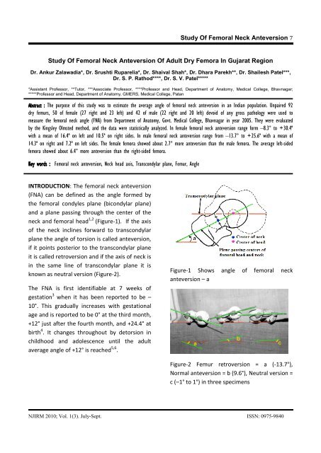
INTRODUCTION: The femoral neck anteversion<br />
(FNA) can be defined as the angle formed by<br />
the femoral condyles plane (bicondylar plane)<br />
and a plane passing through the center of the<br />
neck and femoral head 1,2 (Figure-1). If the axis<br />
of the neck inclines forward to transcondylar<br />
plane the angle of torsion is called anteversion,<br />
if it points posterior to the transcondylar plane<br />
it is called retroversion and if the axis of neck is<br />
in the same line of transcondylar plane it is<br />
known as neutral version (Figure-2).<br />
The FNA is first identifiable at 7 weeks of<br />
gestation 3 when it has been reported to be –<br />
10°. This gradually increases with gestational<br />
age and is reported to be 0° at the third month,<br />
+12° just after the fourth month, and +24.4° at<br />
birth 4 . It changes throughout by detorsion in<br />
childhood and adolescence until the adult<br />
average angle of +12° is reached 5,6 .<br />
Figure-1 Shows angle of femoral neck<br />
anteversion – a<br />
Figure-2 Femur retroversion = a (-13.7°),<br />
Normal anteversion = b (9.6°), Neutral version =<br />
c (–1° to 1°) in three specimens<br />
NJIRM 2010; Vol. 1(3). July-Sept. ISSN: 0975-9840
The average adult femoral anteversion has<br />
been documented to range between 7°-16° in<br />
multiple skeletal surveys 4,7,8 whereas Le<br />
Damany (1903) 6 quoted it to range from –25 to<br />
+37 degrees. It is multifactoral result of<br />
evolution, heredity, fetal development,<br />
intrauterine position, and mechanical forces.<br />
Abnormal FNA sometimes can be associated<br />
with many clinical problems ranging from<br />
harmless intoeing gait in the early childhood,<br />
which could be a reason for parents concern for<br />
children future, to disabling osteoarthritis of<br />
the hip and the knee in the adults.<br />
The present study is an attempt to evaluate the<br />
normal anteversion range in adult Indian<br />
femora and to compare values of angle in male<br />
and female as well as with other population.<br />
MATERIALS AND METHODS: Unpaired 92 dry<br />
femurs, 50 of female (27 right and 23 left) and<br />
42 of male (22 right and 20 left) devoid of any<br />
gross pathology were used to measure the<br />
femoral neck angle (FNA). The angle of<br />
anteversion was measured by Kingsley Olmsted<br />
method 9 after placing the specimen at the edge<br />
of a glass horizontal surface so that the<br />
condyles of the inferior end rest on the surface.<br />
The horizontal limb of a goniometer was fixed<br />
at the edge of the experimental table. The<br />
vertical limb was held parallel along the axis of<br />
the head and neck of the femur. The horizontal<br />
surface represents the retrocondylar axis and<br />
the plane of reference against which the<br />
anteversion is measured with the help of the<br />
axis of head and neck of the femur. The angle<br />
subtended was recorded (Figure-1)<br />
<strong>Study</strong> <strong>Of</strong> <strong>Femoral</strong> <strong>Neck</strong> <strong>Anteversion</strong> 8<br />
All measurements were repeated twice by two<br />
independent observers to identify any intra and<br />
inter-observer variability of these techniques.<br />
Data collected was tabulated according to<br />
gender and sides and statistically analyzed.<br />
Figure-3 Shows Kingsley Olmsted method of<br />
measurement of angle of femoral neck<br />
anteversion<br />
RESULTS: Cross sectional study of unpaired 92<br />
adult unpaired dry femurs was conducted, out<br />
of them 50 femurs are of female & 42 femurs<br />
are of female.<br />
Average anteversion in males was 14.3° ± 0.38°<br />
and 21.23° ± 0.39° on the left and right sides<br />
respectively. In the female femora, the average<br />
anteversion recorded was 11.02° ± 0.34° and<br />
20.87° ± 0.36° on the left and right sides<br />
respectively (Table 1).<br />
Retroversion was observed in 5 bones (6.5%).<br />
Neutral or almost neutral version (–1° to +1°)<br />
was found in 5 bones (5.4%) (Figure-2). 20.5%<br />
of the bones were in the range of 0°–10°, while<br />
between 10°–15° there were 33.6% of bones.<br />
36.9% of bones were above 15° (Table-2)<br />
(Figure-4).<br />
NJIRM 2010; Vol. 1(3). July-Sept. ISSN: 0975-9840
Table-1 Average angle of anteversion in 92 dry adult femora<br />
FNA Left FNA FNA average<br />
Female femora<br />
Mean±SD *16.4° ± 8.2° 10.5° ± 5.7° 13.6° ± 16.8°<br />
Male femora<br />
Mean±SD *14.3° ± 8.3° 7.2° ± 8.5° 10.9° ± 14.7°<br />
Total female and male femora<br />
Mean±SD 15.4° ± 15.0° 9.0° ± 7.9° 12.4° ± 18.0°<br />
<strong>Study</strong> <strong>Of</strong> <strong>Femoral</strong> <strong>Neck</strong> <strong>Anteversion</strong> 9<br />
*P value paired‘t’ test 20 7 25.9 0 0 4 18.1 0 0 11.9<br />
DISCUSSION: The knowledge of normal femoral<br />
anteversion is of extreme importance in<br />
selection of patients for prosthesis and<br />
preoperative planning for total hip replacement<br />
surgery and anthropological studies. Although<br />
newer methods using computed tomography<br />
(CT) have been shown to be ±1° accurate, there<br />
is no universal consensus for locating the<br />
NJIRM 2010; Vol. 1(3). July-Sept. ISSN: 0975-9840
femoral neck axis and the femoral condylar<br />
axis 10 . Hence estimation of anteversion on dry<br />
bone is still considered the most accurate<br />
method.<br />
There are few studies done in India before this<br />
study. Western studies results are not<br />
applicable in Indian population because<br />
femoral anteversion differs in both<br />
populations 11 .<br />
The mean anteversion in male bones was 7.2°<br />
and 14.3° on the left and right sides<br />
respectively averaging to about 10.9°. In<br />
females, it was 10.5° and 16.4° on the left and<br />
right side respectively averaging 13.6° in<br />
females. This significant bilateral limb<br />
asymmetry should discourage the tendency to<br />
view the lower limbs as mirror images of one<br />
Western<br />
studies<br />
Indian<br />
studies<br />
<strong>Study</strong> <strong>Of</strong> <strong>Femoral</strong> <strong>Neck</strong> <strong>Anteversion</strong> 10<br />
another. Statistical analysis revealed sexual<br />
dimorphism in anteversion (Table-1) in Indians<br />
being greater in the females as compared to<br />
males. A statistically significant difference was<br />
found for the angle of anteversion between the<br />
male-and female-type bones and the right- and<br />
left-sided bones. The average female-type bone<br />
was about 3° higher than the average maletype<br />
bone. Parsons et al 12 . had also<br />
documented anteversion to be greater in<br />
females. Similarly, Kingsley and Olmsted9<br />
observed a negligible difference (0.081°), and<br />
Yoshioka et al 8 . found a difference of 1° (Table-<br />
3). However, no tests of significance were done<br />
in these series. Kate BR et al 13 . done study on<br />
Indian femur found lesser average angle of<br />
anteversion as compared to present study<br />
(Table-3).<br />
Table-3 <strong>Femoral</strong> anteversion as observed by other researchers<br />
Researcher Sample Mean angle of anteversion in degree<br />
size Right Left Average<br />
Yoshioka Y et al 8 . (1987) 32 - - M-7 .F-8<br />
Kingsley PC et al 9 . (1948) 630 M-8.54, F-7.47 M-7.94, F-8.11 8.02<br />
Parsons FG et al 12 . (1914) 266 M-13.0, F-18.0 M-13.0, F-16.0 15.3<br />
Kate BR et al 13 . (1976) 108 9.0 8.6 8.8<br />
Present study (2005) 92 M-7.2, F-10.5 M-14.3, F-16.4 12.4<br />
Because Indians are more apt to participate in<br />
floor level activities, in contrast to persons in<br />
the West, our hips have to be evolutionally<br />
different from theirs. Thus, the same procedure<br />
produces a different outcome in our<br />
population. Hence a population-specific<br />
protocol and assessment criteria must be<br />
devised. A femoral component of a total hip<br />
replacement should be in an anteversion angle<br />
that closely represents the anteversion angle<br />
for the Indian population to achieve the best<br />
surgical results. In India with the increasing<br />
demand for total hip replacement, this<br />
anteversion angle becomes more significant.<br />
Therefore, our study was undertaken to<br />
ascertain the average angle of anteversion of<br />
the femoral neck in Indian subjects.<br />
CONCLUSIONS: The average angle of<br />
anteversion obtained on dry bone was 12.4°<br />
NJIRM 2010; Vol. 1(3). July-Sept. ISSN: 0975-9840
(SD 18.4°) by the Kingsley and Olmsted method.<br />
The angles of anteversion of the femoral neck<br />
in 8.6%, 20.5%, and 54.1% of cases were in the<br />
range of 1°–5°, 1°–10°, and 1°–15°,<br />
respectively. Altogether, 6.5% of the cases<br />
showed retroversion, 5.4% neutral version and<br />
36.9% had anteversion of more than 15°.<br />
Statistically significant differences were found<br />
between the male and female-type bones and<br />
the right and left-sided bones. The male bone<br />
showed about 2.7° less anteversion than the<br />
female bone. The right-sided bones had about<br />
6.4° less anteversion than the left-sided bones.<br />
REFERANCES:<br />
1. Kim JS, Park TS, Park SB, Kim JS, Kim IY, Kim<br />
SI.Measurement of femoral neck<br />
anteversion in 3D. Part 1: 3D imaging<br />
method. Med Biol Eng Comput 2000;38:603-<br />
9.<br />
2. Napoli MM, Apostólico Netto A, Suguimoto<br />
C, Takedo LT. Anteversão dos colos femorais:<br />
estudo radiológico. Rev Imagem 1985;7:111-<br />
6.<br />
3. Crelin ES. Development of the<br />
musculoskeletal system. Ciba clinical<br />
Symposium. 1981;33(1):1-36.<br />
4. Elftman H. Torsion of lower extremity.<br />
American Journal of physical Anthropology<br />
1945;3:255-265.<br />
5. Staheli LT, Corbett M, Wyss C, King H. Lowerextremity<br />
rotational problems in children:<br />
normal values to guide management. J Bone<br />
Joint Surg Am 1985;67:39-47.<br />
<strong>Study</strong> <strong>Of</strong> <strong>Femoral</strong> <strong>Neck</strong> <strong>Anteversion</strong> 11<br />
6. Le Damany. Les torsions osseuses leur role<br />
dans la transformation des members.<br />
Journal of Anatomical Physiology 1903,39:<br />
246-450.<br />
7. Yagi T and Sasaki T. Tibial torsion in patients<br />
with medial-type osteoarthritic knee. Clinical<br />
Orthopaedics 1986;213:177-182.<br />
8. Yoshioka Y, Siu D and Cooke TD. The<br />
anatomy of functional axis of the femur.<br />
Journal of Bone and Joint Surgery<br />
1987;69:873-880.<br />
9. Kingsley PC and Olsmtead KL. A study to<br />
determine the angle of anteversion of the<br />
neck of femur. Journal of Bone and Joint<br />
Surgery 1948;30-A:745-751.<br />
10. Murphy SB, Simon SR, Kijewski PK, et al.<br />
<strong>Femoral</strong> anteversion. J Bone Joint Surg Am<br />
1987;69:1169–76.<br />
11. Eckhoff DG, Kramer RC, Watkings JJ, Alongi<br />
CA, van Greven DP. Variation in femoral<br />
<strong>Anteversion</strong>. Clinical Anatomy 1994;7:71-75.<br />
12. Parsons FG. The characters of the English<br />
thigh bone. J Anat Physiol 1914;48:238-67.<br />
13. Kate BR The Ceylonese femur and its<br />
comparison with Indian and other Asian<br />
femur; Journal of Anatomical Society of India<br />
1976;25(3); 124-127.<br />
NJIRM 2010; Vol. 1(3). July-Sept. ISSN: 0975-9840