Maxillofacial Prosthetic Rehabilitation of a Cancer Patient
Maxillofacial Prosthetic Rehabilitation of a Cancer Patient
Maxillofacial Prosthetic Rehabilitation of a Cancer Patient

You also want an ePaper? Increase the reach of your titles
YUMPU automatically turns print PDFs into web optimized ePapers that Google loves.
<strong>Maxill<strong>of</strong>acial</strong> <strong>Prosthetic</strong> <strong>Rehabilitation</strong> <strong>of</strong> a <strong>Cancer</strong> <strong>Patient</strong> at Terminal Stage<br />
<strong>Maxill<strong>of</strong>acial</strong> <strong>Prosthetic</strong> <strong>Rehabilitation</strong> <strong>of</strong> a <strong>Cancer</strong> <strong>Patient</strong> at Terminal Stage<br />
Dr. Arun Dupare*, Dr. Roshni Dupare**, Dr. Lata Dupare ***<br />
* Dean and Pr<strong>of</strong>essor, Prosthodontics, College <strong>of</strong> Dental Science and Hospital, Amargadh, Gujarat,<br />
** Department <strong>of</strong> Community Dentistry, ITS Dental College, Murad Nagar, Ghaziabad, ***Senior Staff Surgeon, Periodontology, CGHS<br />
Polyclinic, Nagpur<br />
Abstracts: Face is the forefront <strong>of</strong> aesthetics. Surgical resection <strong>of</strong> the maxillae and facial structures for<br />
treatment <strong>of</strong> cancer, trauma, congenital deformities or infection causes maxill<strong>of</strong>acial defect that has serious<br />
impact on the individual’s esthetic and has great psychological trauma <strong>of</strong> social outcast. When the presented<br />
defect is extensive and supporting structures is lacking, the rehabilitation and restoring functions is a<br />
challenging task. <strong>Rehabilitation</strong> becomes even more difficult when the problems at defect site are associated<br />
with the complications. An attempt has been made to rehabilitate <strong>of</strong> a patient at terminal stage having<br />
extensive maxill<strong>of</strong>acial defect with inoperable carcinoma, infections, and restricted mouth opening. [ Dupare A<br />
et al NJIRM 2012; 3(2) : 173-176]<br />
Key words: facial prosthesis, heat cured acrylic resin, obturator prosthesis, rare earth magnet, self cured<br />
acrylic resin.<br />
Author for correspondence: Dr Dr. Arun Dupare, Dean, College <strong>of</strong> Dental Science & Hospital, Amargadh,<br />
Taluka Sihor, Dist. Bhavnagar, Gujarat India Pin: 364210, Email: drarundupare@yahoo.com<br />
Introduction <strong>Maxill<strong>of</strong>acial</strong> prosthetic rehabilitation<br />
and restoring function is important as impairments<br />
have detrimental effect on the quality <strong>of</strong> life and<br />
self-esteem. 1 When the defect presented is<br />
extensive and the supporting bone is lacking, the<br />
rehabilitation is a challenging task. <strong>Rehabilitation</strong> <strong>of</strong><br />
the defect becomes even more difficult when the<br />
defect area is associated with the complications. An<br />
attempt has been made to rehabilitate a patient at<br />
terminal stage having extensive maxill<strong>of</strong>acial defect<br />
with inoperable cancer, restricted mouth opening,<br />
and other complications. Two component light<br />
weight prosthesis was fabricated to cover the<br />
defect. 2, 3 The obturator and facial prosthesis was<br />
fabricated with heat cured acrylic resin lighter in<br />
weight retained by rare earth magnets and<br />
spectacle was delivered to make the remaining life<br />
<strong>of</strong> the patient tolerable.<br />
Case report : A 38 year-old male patient was<br />
referred from the department <strong>of</strong> ENT to the<br />
Department <strong>of</strong> <strong>Prosthetic</strong> Dentistry, at Government<br />
Dental College and Hospital Nagpur, for<br />
rehabilitation <strong>of</strong> right side <strong>of</strong> face. The patient’s<br />
medical history revealed that he was diagnosed as<br />
squamous cell carcinoma <strong>of</strong> the right maxillary<br />
antrum involving the floor <strong>of</strong> the orbit. To remove<br />
the carcinoma maxillectomy and enucleating right<br />
eye was done. It was followed by frequent<br />
recurrence and surgical procedures. In the frequent<br />
surgical procedures he lost his more than half <strong>of</strong> the<br />
nose including the medial septum, right eye,<br />
supraorbital structures, right maxilla, part <strong>of</strong><br />
zygomatic bone and associated structures. The<br />
recurrence was again diagnosed at defect site at<br />
lateral supraorbital and zygomatic areas. It was<br />
reported that any further surgical attempt to<br />
remove the carcinoma can cause serious<br />
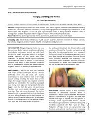
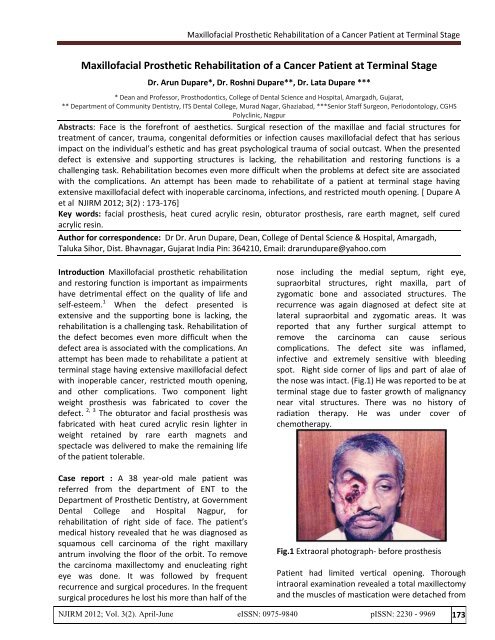
complications. The defect site was inflamed,<br />
infective and extremely sensitive with bleeding<br />
spot. Right side corner <strong>of</strong> lips and part <strong>of</strong> alae <strong>of</strong><br />
the nose was intact. (Fig.1) He was reported to be at<br />
terminal stage due to faster growth <strong>of</strong> malignancy<br />
near vital structures. There was no history <strong>of</strong><br />
radiation therapy. He was under cover <strong>of</strong><br />
chemotherapy.<br />
Fig.1 Extraoral photograph- before prosthesis<br />
<strong>Patient</strong> had limited vertical opening. Thorough<br />
intraoral examination revealed a total maxillectomy<br />
and the muscles <strong>of</strong> mastication were detached from<br />
NJIRM 2012; Vol. 3(2). April-June eISSN: 0975-9840 pISSN: 2230 - 9969 173
<strong>Maxill<strong>of</strong>acial</strong> <strong>Prosthetic</strong> <strong>Rehabilitation</strong> <strong>of</strong> a <strong>Cancer</strong> <strong>Patient</strong> at Terminal Stage<br />
maxilla <strong>of</strong> the right side. The presented defect<br />
situation corresponded to a class I situation<br />
(resected performed along the palatal midline)<br />
according to the Aramany classification <strong>of</strong> defects. 4<br />
He had difficulty in speech, mastication, swallowing<br />
and to maintain oral hygiene. A left side maxilla and<br />
the mandibular arch were completely dentulous<br />
with healthy teeth and normal occlusion. Tongue<br />
size and function was normal, but speech was<br />
altered.<br />
Treatment plan: A patient was at terminal stage <strong>of</strong><br />
life. The defect area was extensive with restricted<br />
mouth opening. Moreover, intimate contact <strong>of</strong> the<br />
prosthesis to tissue for support and retention<br />
cannot be utilized due to presence <strong>of</strong> malignancy<br />
and infected sensitive areas. The bone or teeth on<br />
right side to support the large and bulky prosthesis<br />
was lacking. Hence prosthetic rehabilitation <strong>of</strong> a<br />
case was challenging. He was very much distressed<br />
because <strong>of</strong> the extensive facial disfigurement and<br />
open surgical cavity with bleeding spots. He wanted<br />
us to give him some option to cover the defect till<br />
his survival.<br />
A case was considered to fabricate two -piece<br />
interim prosthesis separately to cover intraoral<br />
defects by obturator prosthesis and extraoral<br />
defects by facial prosthesis. This two-piece<br />
construction makes the insertion <strong>of</strong> the prosthesis<br />
easy and is done piece by piece making it less a<br />
struggle for the patient with limited mouth opening.<br />
The fabrication <strong>of</strong> intraoral obturator prosthesis<br />
was essential to prevent communication <strong>of</strong> food<br />
between the oral and nasal cavities. Moreover, the<br />
most important function this obturator served in<br />
the case was to support the facial prosthesis<br />
through the healthy teeth and bone present on left<br />
side. The extraoral facial prosthesis to fabricate was<br />
consisting <strong>of</strong> cheek, part <strong>of</strong> nose and eye. The heat<br />
cured acrylic resin was used to fabricate these two<br />
prostheses. Rare earth magnets were used to link<br />
the two portions. The case option <strong>of</strong> prosthesis was<br />
given to the patient that he readily accepted. He<br />
was ready to wear spectacle to hold the facial<br />
prosthesis.<br />
Clinical procedure: Fabrication <strong>of</strong> obturator<br />
prosthesis: A sterilized wet cotton gauze was<br />
placed in the operated cavity to prevent flow <strong>of</strong> the<br />
impression material in throat. An impression <strong>of</strong> the<br />
dentulous and supporting area to prepare obturator<br />
was then made with an alginate impression<br />
material. Working cast was prepared from this<br />
impression. The undercuts present in the defect<br />
were blocked out. To reduce weight simple plate<br />
type obturator prosthesis was fabricated and was<br />
extended on defect side so as to reach to the facial<br />
prosthesis. Healthy teeth and bone present on left<br />
side were used for retention and support to<br />
obturator and also to the facial prosthesis. The<br />
extension <strong>of</strong> the flange <strong>of</strong> obturator was done on<br />
left side in buccal sulcus area and on buccal surface<br />
<strong>of</strong> the teeth.<br />
Fabrication <strong>of</strong> facial prosthesis: Facial moulage was<br />
done to obtain a working cast to orient the<br />
prosthesis properly to the rest <strong>of</strong> the face. The<br />
obturator prosthesis was inserted in the mouth and<br />
the nasal opening was blocked with gauze. Plastic<br />
tube was placed in the mouth for air intake. The<br />
operated cavity was lined with sterilized cotton<br />
gauge. The face was coated with petroleum jelly. An<br />
impression for facial defect was made with alginate<br />
impression material. Alginate was mixed and<br />
applied to skin surface. Gauze squares were placed<br />
all over the alginate surface to provide mechanical<br />
retention for rigid plaster backing. Dental plaster<br />
was then spread over the entire surface to a<br />
sufficient thickness. After setting <strong>of</strong> plaster set<br />
impression was removed, grasping it on the sides<br />
and lifting it gently. The impression was inspected<br />
for any void or distortion. The impression was<br />
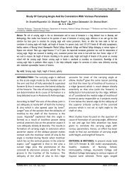
poured in type-III dental stone <strong>of</strong> about 5 millimeter<br />
thickness and after setting it was retrieved (Fig.2).<br />
Fig.2 Facial moulage<br />
All the undercuts on affected area <strong>of</strong> the cast was<br />
blocked. A wax pattern for facial prosthesis was<br />
NJIRM 2012; Vol. 3(2). April-June eISSN: 0975-9840 pISSN: 2230 - 9969 174
<strong>Maxill<strong>of</strong>acial</strong> <strong>Prosthetic</strong> <strong>Rehabilitation</strong> <strong>of</strong> a <strong>Cancer</strong> <strong>Patient</strong> at Terminal Stage<br />
sculpted with required thickness. Over extension on<br />
lower side <strong>of</strong> mandible was given to avoid visibility<br />
<strong>of</strong> the defect while opening <strong>of</strong> mouth. A<br />
prefabricated acrylic eye shell was selected <strong>of</strong><br />
shape, size and color after matching with left eye.<br />
The position <strong>of</strong> the pupil and the sclera was<br />
adjusted to mimic the left eye position while the<br />
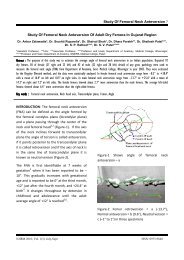
patient gazes forward. A wax pattern was carved<br />
around the acrylic eye shell on the model. Upper<br />
and lower eyelids were carved such that the area<br />
covered by them on sclera mimicked the left eye<br />
(Fig.3).<br />
means <strong>of</strong> securing a facial prosthesis. Hooks were<br />
placed in the frame <strong>of</strong> spectacle and the loops in<br />
the facial prosthesis for stability and in position.<br />
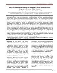
Fig.4 Tissue surface <strong>of</strong> obturator & facial prosthesis<br />
with magnet<br />
Fig.3 Waxed-up facial prosthesis<br />
For orientation <strong>of</strong> the eye, a wax pattern was<br />
checked on patient face. This pattern without the<br />
eye shell was then invested. Clear heat cured acrylic<br />
resin was used for facial prosthesis. Skin matched<br />
colored stains were added in heat cured acrylic<br />
resin to fabricate facial surface <strong>of</strong> the prosthesis.<br />
Self cured acrylic resin was used to stabilize the eye<br />
shell section. The inner surface <strong>of</strong> the prosthesis<br />
after curing was further hollowed and carved to<br />
reduce thickness and weight and also to eliminate<br />
any contact with infected area. The eye shell was<br />
then attached to its position with self cured acrylic<br />
resin. Stains were mixed and painted in<br />
predetermined sequence and quantity to achieve<br />
staining to create lifelike appearance.<br />
Attachment <strong>of</strong> retentive devises: A projection on<br />
inner surface <strong>of</strong> facial prosthesis and to cover the<br />
obturator prosthesis was made with self cured<br />
acrylic resin. For attaching the rare earth magnets<br />
to this final prosthesis the obturator was first placed<br />
in patient’s mouth and second magnet was<br />
attached to the first magnet on the obturator using<br />
self cured acrylic resin (Fig.4). A spectacle with<br />
broad frame was used that provided an excellent<br />
Delivery <strong>of</strong> prosthesis and instructions: Both the<br />
parts <strong>of</strong> the prosthesis were decided to wear<br />
separately for convenience. <strong>Patient</strong> was trained to<br />
insert, to stabilize in position (Fig.5) and also to<br />
remove the facial and obturator prosthesis for<br />
cleaning and during sleep.<br />
Fig.5 Extraoral photograph with prosthesis<br />
Discussion : The treatment <strong>of</strong> maxillary defects is<br />
always limited by difficulties with retention,<br />
movable tissue beds and lack <strong>of</strong> sufficient bone<br />
support. 5 When the defect is large then the solid<br />
bulky prosthesis is heavy in weight and to maintain<br />
its position is difficult. In case trismus is present,<br />
making execution <strong>of</strong> the procedures extremely<br />
cumbersome and for the patient, manipulation <strong>of</strong><br />
the prosthesis into and out <strong>of</strong> the mouth becomes a<br />
Herculean task, as it requires multiple paths <strong>of</strong><br />
insertion and removal. To overcome these problems<br />
<strong>of</strong> retention skin adhesives, spectacles,<br />
engagements <strong>of</strong> undercuts using flexible materials<br />
and implants have been advocated. 6<br />
NJIRM 2012; Vol. 3(2). April-June eISSN: 0975-9840 pISSN: 2230 - 9969 175
<strong>Maxill<strong>of</strong>acial</strong> <strong>Prosthetic</strong> <strong>Rehabilitation</strong> <strong>of</strong> a <strong>Cancer</strong> <strong>Patient</strong> at Terminal Stage<br />
The use <strong>of</strong> frames by attaching eye prosthesis has<br />
advantageous when the patients has undergone<br />
maxillectomy in combination with an orbital<br />
exenterations as reported by Beumer J et al. 2<br />
The rare earth magnets have been used in dentistry<br />
3<br />
since 1960 and Federick. in the year 1976<br />
presented a technique for fabrication <strong>of</strong> a sectional<br />
interim maxillary obturator with retention<br />
augmented by magnet. The use <strong>of</strong> rare earth<br />
magnet achieves a more life like appearance and<br />
keeps facial prosthesis independent to external<br />
support.<br />
The fabrication <strong>of</strong> flexible obturator for patients<br />
with severely limited mouth opening using either<br />
silicone or even a flexible vinyl resin mouth guard<br />
material can be the choice according to Lauciello` et<br />
al. 7 However, these materials are far from ideal and<br />
in an average maxillectomy case it would be<br />
inadequate.<br />
<strong>Prosthetic</strong> rehabilitation for facial defects has<br />
several advantages over surgical reconstruction as it<br />
is quite inexpensive, allows for periodic examination<br />
and cleaning and is also an alternative to surgery in<br />
unsuitable candidates. Acrylic resin was introduced<br />
to dental pr<strong>of</strong>ession in 1937 for both intra and extra<br />
oral prosthesis. The fabrication process <strong>of</strong> acrylic<br />
resin is relatively short and the clinician has a lot <strong>of</strong><br />
control over the color, shape and size <strong>of</strong> prostheses.<br />
Heat cured acrylic resins are routinely used for<br />
maxill<strong>of</strong>acial prosthesis.<br />
References :<br />
1. Kreissl ME. Zygoma implant supported<br />
prosthetic rehabilitation after partial<br />
maxillectomy using surgical navigation. A<br />
clinical report. J Prosthet Dent 2007;97:121-28<br />
2. Beumer J III, Curtis TA, Marunick MT.<br />
<strong>Maxill<strong>of</strong>acial</strong> rehabilitation: Prosthodontic and<br />
surgical consideration. 2 nd edition St. Louis<br />
Ishiyaku euro America Inc. 1996 ;408-16.<br />
3. Federick DR. A magnetically retained interim<br />
maxillary obturator. J Prosthet Dent<br />
1976;36:671-75.<br />
4. Aramany MA. Basic principles <strong>of</strong> obturator<br />
design for partially edentulous patients Part I:<br />
Classification. J Prosthet Dent 1978; 40:554-57.<br />
5. Taylor TD, Flyer A, La Ville WE. Alternate<br />
obturation for the maxillectomy patient with<br />
severely limited mandibular opening. J Prosthet<br />
Dent 1985;53:83-85.<br />
6. Chalian VA, DraneJB, Standish SM. <strong>Maxill<strong>of</strong>acial</strong><br />
prosthetics- A Multidisciplinary Practice,4 th<br />
edition Williams & Wilkin Co. page: 1972;142-46<br />
7. Chen MS, Udagama A, Drane JB. Evaluation <strong>of</strong><br />
facial prostheses for head and neck cancer<br />
patients. J Prosthet Dent 1981; 46:538-44.<br />
In the case heat cured acrylic resin was used to<br />
fabricate facial and simple obturator prosthesis.<br />
Polymeric coated rare earth magnets (Neodeniumiron-boron)<br />
and broad frame spectacle was used for<br />
retention and support <strong>of</strong> this maxill<strong>of</strong>acial<br />
prosthesis.<br />
Conclusion: An attempt has been made to<br />
rehabilitate a patient at terminal stage associated<br />
with extensive maxill<strong>of</strong>acial defect and<br />
complications. A two components, obturator and<br />
facial prosthesis fabricated with heat cured acrylic<br />
resin lighter in weight retained by rare earth<br />
magnets and spectacle was delivered to make the<br />
remaining life <strong>of</strong> the patient tolerable.<br />
NJIRM 2012; Vol. 3(2). April-June eISSN: 0975-9840 pISSN: 2230 - 9969 176