Morphometric Study Of Pterion In Dry Skull Of Gujarat Region
Morphometric Study Of Pterion In Dry Skull Of Gujarat Region
Morphometric Study Of Pterion In Dry Skull Of Gujarat Region

You also want an ePaper? Increase the reach of your titles
YUMPU automatically turns print PDFs into web optimized ePapers that Google loves.
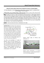
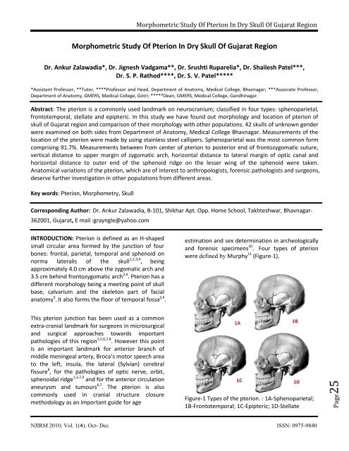
Page25<strong>Morphometric</strong> <strong>Study</strong> <strong>Of</strong> <strong>Pterion</strong> <strong>In</strong> <strong>Dry</strong> <strong>Skull</strong> <strong>Of</strong> <strong>Gujarat</strong> <strong>Region</strong><strong>Morphometric</strong> <strong>Study</strong> <strong>Of</strong> <strong>Pterion</strong> <strong>In</strong> <strong>Dry</strong> <strong>Skull</strong> <strong>Of</strong> <strong>Gujarat</strong> <strong>Region</strong>Dr. Ankur Zalawadia*, Dr. Jignesh Vadgama**, Dr. Srushti Ruparelia*, Dr. Shailesh Patel***,Dr. S. P. Rathod****, Dr. S. V. Patel******Assistant Professor, **Tutor, ****Professor and Head, Department of Anatomy, Medical College, Bhavnagar; ***Associate Professor,Department of Anatomy, GMERS, Medical College, Gotri; *****Dean, GMERS, Medical College, Gandhinagar.Abstract: The pterion is a commonly used landmark on neurocranium; classified in four types: sphenoparietal,frontotemporal, stellate and epipteric. <strong>In</strong> this study we have found out morphology and location of pterion ofskull of <strong>Gujarat</strong> region and comparison of their morphology with other populations. 42 skulls of unknown genderwere examined on both sides from Department of Anatomy, Medical College Bhavnagar. Measurements of thelocation of the pterion were made by using stainless steel callipers. Sphenoparietal was the most common formcomprising 91.7%. Measurements between from center of pterion to posterior end of frontozygomatic suture,vertical distance to upper margin of zygomatic arch, horizontal distance to lateral margin of optic canal andhorizontal distance to outer end of the sphenoid ridge on the lesser wing of the sphenoid were taken.Anatomical variations of the pterion, which are of interest to anthropologists, forensic pathologists and surgeons,deserve further investigation in other populations from different areas.Key words: <strong>Pterion</strong>, Morphometry, <strong>Skull</strong>Corresponding Author: Dr. Ankur Zalawadia, B-101, Shikhar Apt. Opp. Home School, Takhteshwar, Bhavnagar-362001, <strong>Gujarat</strong>, E mail :grayngle@yahoo.comINTRODUCTION: <strong>Pterion</strong> is defined as an H-shapedsmall circular area formed by the junction of fourbones: frontal, parietal, temporal and sphenoid onnorma lateralis of the skull 1,2,3,4 , beingapproximately 4.0 cm above the zygomatic arch and3.5 cm behind frontozygomatic arch 2,4 . <strong>Pterion</strong> has adifferent morphology being a meeting point of skullbase, calvarium and the skeleton part of facialanatomy 5 . It also forms the floor of temporal fossa 2,4 .estimation and sex determination in archeologicallyand forensic specimens 10 . Four types of pterionwere defined by Murphy 11 (Figure-1).This pterion junction has been used as a commonextra-cranial landmark for surgeons in microsurgicaland surgical approaches towards importantpathologies of this region 1,5,6,7,8 . However this pointis an important landmark for anterior branch ofmiddle meningeal artery, Broca’s motor speech areato the left, insula, the lateral (Sylvian) cerebralfissure 8 , for the pathologies of optic nerve, orbit,sphenoidal ridge 1,3,7,9 and for the anterior circulationaneurysm and tumours 6,7 . The pterion is alsocommonly used in cranial structure closuremethodology as an important guide for ageFigure-1 Types of the pterion. : 1A-Sphenoparietal;1B-Frontotemporal; 1C-Epipteric; 1D-StellateNJIRM 2010; Vol. 1(4). Oct- Dec. ISSN: 0975-9840
Page26<strong>Morphometric</strong> <strong>Study</strong> <strong>Of</strong> <strong>Pterion</strong> <strong>In</strong> <strong>Dry</strong> <strong>Skull</strong> <strong>Of</strong> <strong>Gujarat</strong> <strong>Region</strong>The sphenoparietal type is defined as a suturalpattern in which the sphenoid and parietal bonesare in direct contact. Conversely, thefrontotemporal type is a sutural pattern in whichthe frontal and temporal bones are in direct contact.The stellate type is characterized by articulation offour bones (frontal, parietal, temporal and sphenoid)at a point.The epipteric type is defined by presence of a smallsutural bone between the parietal bone and thegreater wing of the sphenoid bone. Present studywas conducted to determine and map the locationof the pterion, and to define its type, in skulls ofwestern <strong>In</strong>dia.Figure-2 Distance measurements on lateral aspectof skull– P-FZ: the distance from the center of the pterionto the posterolateral aspect of the frontozygomaticfissure,– P-ZA: the vertical distance from the center of thepterion to the zygomatic arch,– P-LWS: the horizontal distance from the internalaspect of the center of the pterion to the outer endof the sphenoid ridge on the lesser wing of thesphenoid,– P-OC: the horizontal distance from the internalaspect of the center of the pterion to the lateralmargin of the optic canal. The data obtained wereanalyzed using standard statistical software.MATERIAL AND METHOD: Total 42 dry human adultaged skull of unknown sex without any grosspathology or abnormality were studied.On both the left and right sides of each skull, thesutural pattern of the pterion was determinedbased on descriptions (sphenoparietal,frontotemporal, stellate and epipteric types). Acircle of smallest radius was drawn connecting thefour bones involved in the formation of the pterion,the center of which was taken as the center of thepterion. Following measurements taken twice thenaveraged so as to minimize bias errors, usingstandard stainless steel callipers with an accuracy of0.1 mm, of the distances between the pterion andspecific identifiable bony landmarks (Figure-2,3):Figure-3 Distance measurements in interior of skullRESULTS: All types of the pterion were found inskull of <strong>Gujarat</strong> region of western <strong>In</strong>dia (Table-1).The sphenoparietal pterion was the predominanttype in both sides. A single stellate type wasobserved in left side of skull while on the oppositeside there was sphenoparietal type; this was theonly asymmetry of pterion seen.Table-1 Frequency of pterion types observed onthe right and left sides of the skullRight Left Both side<strong>Pterion</strong> type side siden=42 n=42 n=82Sphenoparietal 92.9% 90.5% 91.7%Frontotemporal 2.4% 2.4% 2.4%Stellate 0 2.4% 1.2%Epipteric 4.8% 4.8% 4.8%NJIRM 2010; Vol. 1(4). Oct- Dec. ISSN: 0975-9840
Page27<strong>Morphometric</strong> <strong>Study</strong> <strong>Of</strong> <strong>Pterion</strong> <strong>In</strong> <strong>Dry</strong> <strong>Skull</strong> <strong>Of</strong> <strong>Gujarat</strong> <strong>Region</strong>The means and associated standard deviations ofthe various linear measurements taken from thecenter of the pterion are shown in (Table 2). Noobvious differences between the right and left sideswere observed for any of the measurements(p>0.05).Table-2 Mean and associated standard deviationsof the linear distance (cm) from pterion to specificidentifiable bony landmarksRight side (n=42) Left side (n=42)DistanceMean±SD cmMean±SD cmP-FZ 3.73±0.51 3.55±0.42P-ZA 3.12±0.44 2.97±0.33P-LWS 1.36±0.35 1.33±0.22P-OC 4.52±0.32 4.37±0.23DISCUSSION: Knowledge and understanding of thetype and location of the pterion and its relation tosurrounding bony landmarks is important, especiallywith respect to neurosurgery. Detailed informationcan only readily be obtained from an examination ofdry skulls to determine more precise relationshipsbetween bony landmarks and the underlying softtissues.<strong>In</strong> the present study sphenoparietal,Frontotemporal, stellate and epipteric types ofpterion were observed (Table-1). Sphenoparietaltype of pterion is most common seen in Asiatic<strong>In</strong>dians (95.1%) 12 , Northern <strong>In</strong>dians (87.72%) 3 , South<strong>In</strong>dians (93.55%) 14 and Nigerians (87.79% and82.1%) 12,15 same as in this study (91.7%); while itwas significantly lower in Korean (76.5%) 16 andKenyan (66%) 17 populations as compared to thisstudy (Table-3).Table-3 Comparison of the percentage of pterion types in different populationsType of pterion<strong>Study</strong>/Population n (skull), sexSphenoparietal Frontotemporal Stellate Epipteric% % % %Saxena et al 12 ., 1988, Nigerian, n=40,87.79 10.11 5.06 3.79unknown sexSaxena et al 12 ., 1988, <strong>In</strong>dian, n=72,95.3 3.46 1.38 11.79unknown sexManjunath et al. 14 , 1993, South <strong>In</strong>dian,93.55 3.52 2.93 17.3n=172, unknown sexAsala et al. 15 , 1996, Nigerian, n=212,82.1 23.6 - 5.7unknown sexLee et al. 16 , 2001, Korean, n=149, unknown 76.5 - - 40.3sexSaxena et al. 3 , 2003, North <strong>In</strong>dian, n=203, 87.72 10.01 5.17 0both sexOguz et al. 7 , 2004, Turkish, n=26, male 88 10 0 2Mwachaka et al. 17 , Kenyan, n=50, both sex 66 15 7 12Present study, 2009, Western <strong>In</strong>dian, n=42,both sex91.7 2.4 1.2 4.8The incidence of the frontotemporal type of pterionhas also been observed to vary in different groups,being reported of 10.11–23.6% in Nigerians 12,15 ,15% of Kenyans 17 , 41.1% of Papuan skulls 18 , whichare significantly higher than present study. <strong>In</strong> thepresent study the frequency of a frontotemporaltype pterion was 2.4% being closest to thatreported in other populations of <strong>In</strong>dia (Table-3) 12,14 .According to previous studies, the sphenoparietalNJIRM 2010; Vol. 1(4). Oct- Dec. ISSN: 0975-9840
Page28<strong>Morphometric</strong> <strong>Study</strong> <strong>Of</strong> <strong>Pterion</strong> <strong>In</strong> <strong>Dry</strong> <strong>Skull</strong> <strong>Of</strong> <strong>Gujarat</strong> <strong>Region</strong>type of pterion is the dominant form in humanswhereas the frontotemporal type is dominant innonhuman primates 3,7,12,16,19,20 . <strong>In</strong> primate evolution,the anterosuperior segment of the squamous partof the temporal bone of lower primates becamedetached from its parent and incorporated into theposterosuperior angle of the greater wing of thesphenoid bone of humans, thereby changing thepterion pattern from the frontotemporal type ofnonhuman primates to the sphenoparietal type ofhumans 3,19 .An epipteric type pterion was observed in a smallnumber of skulls (4.8%) in the present study, beingsignificantly less than that reported in Nigerians(23.6%) 15 , Australian Aborigines (18.5%) 11 andNorthern <strong>In</strong>dians (10.01%) 3 but similar to that inSouth <strong>In</strong>dians (3.52%) 14 .The pterion has been reported to lie 4.0 cm abovethe zygomatic arch and 3.0–3.5 cm behind thefrontozygomaticsuture 2 . <strong>In</strong> the present study thepterion was 3.73±0.51 cm and 3.55±0.42 cm behindthe frontozygomatic suture, and 3.12±0.44 cm and2.97±0.33 cm above the zygomatic arch on the rightand left sides respectively (Table 2).The lesser wing of the sphenoid is a common sitefor meningiomas. The pterional approach may beused to reach the tumor, particularly if it is locatedlaterally, in which case the distance between theinternal aspect of the pterion and the lateral end ofthe sphenoid ridge is an important measurement.<strong>In</strong> the present study this distance was found to be1.40±0.33 cm and 1.48±0.32 cm on the right andleft sides respectively (Table 2). The pterionalapproach may also be used to access the optic canal,in which case the distance between the internalaspect of the pterion and the medial margin of theoptic canal is an important measurement. Thisdistance was found to be 4.39±0.40 cm and4.36±0.40 cm on the right and left sides respectively(Table 2).CONCLUSION: Knowledge of the location andrelations of the pterion is important in relation tosurgical intervention, particularly with respect tothe course of the branches of the middle meningealartery and Broca’s motor speech area on the leftside. The distances between the pterion and thelesser wing of the sphenoid and optic canal are ofpractical importance in surgical approaches to theseregions via the pterion.ACKNOWLEDGEMENTS: We are thankful to, Dean,Govt. Medical College, Bhavnagar, <strong>Gujarat</strong> (<strong>In</strong>dia)for providing facility to conduct research project.REFERENCES:1. Ersoy M, Evliyaoglu C, Bozkurt MC, Konuksan B,Tekdemir I, Keskil IS. Epipteric bones in the pterionmay be surgical pitfall. Minim <strong>In</strong>vasive Neurosurg2003;46:364–365.2. Moore KL, Dalley AF. Clinically oriented anatomy,4th edn. Lippincott Williams & Wilkins, Baltimore,1999;836–842.3. Saxena RC, Bilodi AKS, Mane SS, Kumar A. <strong>Study</strong>of pterion in skulls of awadh area-in and aroundLucknow. Kathmandu Univ Med J 2003;1:32–33.4. Standring S, Ellis H, Healy JC, Johnson D. Gray’sanatomy, 39th edn. Elsevier Churchill Livingstone,London, 2005;442–471.5. Feng WF, Qi ST, Huang SP, Huang LJ. Surgicaltreatment of anterior circulation aneurysm viapterion keyhole approach. Di Yi Jun Yi Da Xue XueBao 2005;25,546-8.6. Cheng WY, Lee HT, Sun MH, Shen CC. A pterionkeyhole approach for the treatment of anteriorcirculation aneurysms. Minim <strong>In</strong>vasive Neurosurg2006;9:257–262.7. Oguz O, Sanli SG, Bozkir MG, Soames RW. Thepterion in Turkish male skulls. Surg Radiol Anat2004;26:220–4.8. Urzi F, Ianello A, Torrisi A, Foti P, Mortellaro NF,Cavallaro M. Morphological variability of pterion inthe human skull. Ital J Anat Embryol 2003;108:83–117.9. Lang J. The pterion region and its clinicallyimportant distance to the optic nevre, dimensionsand shape of the recess or the temporal pole.Neurochirurgia (Stuttg) 1984;27:31–35.10. Lovejoy CO. Meindl RS. Mensforth RP, Barton TJ.Multifactorial determination of skeletal age atdeath: a method a blind tests of its accuracy. Am JPhys Anthropol 1985;68:1-14.NJIRM 2010; Vol. 1(4). Oct- Dec. ISSN: 0975-9840
Page29<strong>Morphometric</strong> <strong>Study</strong> <strong>Of</strong> <strong>Pterion</strong> <strong>In</strong> <strong>Dry</strong> <strong>Skull</strong> <strong>Of</strong> <strong>Gujarat</strong> <strong>Region</strong>11. Murphy T. The pterion in the Australianaborigine. Am J Phys Anthropol 1956;14:225-44.12. Saxena SK, Jain SP, Chowdhary DS. Acomparative study of pterion formation and itsvariations in the skulls of Nigerians and <strong>In</strong>dians.Anthropol Anz 1988;46:75-82.13. Wang Q, Opperman LA, Havil LM, Carlson DS,Dechow PC. <strong>In</strong>heritance of sutural pattern at thepterion in Rhesus Monkey skulls. Anat Rec DiscovMol Cell Evol Biol 2006;288:1042-9.14. Manjunath KY, Thomas IM. <strong>Pterion</strong> variants andepipteric ossicles in South <strong>In</strong>dian skulls. J Anat Soc<strong>In</strong>dia 1993;42:85-94.15. Asala SA, Mbajiorgu FE. Epigenetic variation inthe Nigerian skull: sutural pattern at the pterion.East Afr Med J 1996;73:484-6.16. Lee UY, Park DK, Kwon SO, Paik DJ, Han SH.Morphological analysis of the pterion in Korean.Korean J Phys Anthropol 2001;14:281-9.17. Mwachaka PM, Hassanali J, Odula P. Suturalmorphology of the pterion and asterion amongadult Kenyans. Braz J Morphol Sci 2009;26:4-7.18. Williams LP, Bannister LH, Berry MM, Collins P,Dyson M, Dussek JE, Ferguson MWJ. Gray’sanatomy, 38th Edn. Churchill Livingstone, London,1998;568,59519. Ashley-Montagu FM. The anthropologicalsignificance of the pterion in the Primates. Am JPhys Anthropol 1933;18:159-336.20. Matsumara G, Kida K, Ichikawa R, Kodama G.<strong>Pterion</strong> and epipteric bones in Japanese adults andfetuses with special reference to their formationand variations. Acta Anatomica Nipponica1991;66:462-71.NJIRM 2010; Vol. 1(4). Oct- Dec. ISSN: 0975-9840