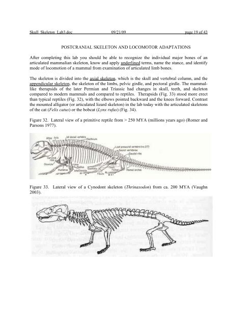
POSTCRANIAL SKELETON AND LOCOMOTOR ADAPTATIONS ...
POSTCRANIAL SKELETON AND LOCOMOTOR ADAPTATIONS ...
POSTCRANIAL SKELETON AND LOCOMOTOR ADAPTATIONS ...

Create successful ePaper yourself
Turn your PDF publications into a flip-book with our unique Google optimized e-Paper software.
Skull_Skeleton_Lab3.doc 09/21/09 page 19 of 43<br />
<strong>POSTCRANIAL</strong> <strong>SKELETON</strong> <strong>AND</strong> <strong>LOCOMOTOR</strong> <strong>ADAPTATIONS</strong><br />
After completing this lab you should be able to recognize the individual major bones of an<br />
articulated mammalian skeleton, know and apply underlined terms, name the stance, and identify<br />
mode of locomotion of a mammal from examination of articulated limb bones.<br />
The skeleton is divided into the axial skeleton, which is the skull and vertebral column, and the<br />
appendicular skeleton, the skeleton of the limbs, pelvic girdle, and pectoral girdle. The mammallike<br />
therapsids of the later Permian and Triassic had changes in skull, teeth, and skeleton<br />
compared to modern mammals and compared to reptiles. Therapsids (Fig. 33) stood more erect<br />
than typical reptiles (Fig. 32), with the elbows pointed backward and the knees forward. Contrast<br />
the mounted alligator (or articulated lizard skeleton) in the lab today with the articulated skeletons<br />
of the cat (Felis catus) or the bobcat (Lynx rufus) (Fig. 34).<br />
Figure 32. Lateral view of a primitive reptile from > 250 MYA (millions years ago) (Romer and<br />
Parsons 1977).<br />
Figure 33. Lateral view of a Cynodont skeleton (Thrinaxodon) from ca. 200 MYA (Vaughn<br />
2003).
Skull_Skeleton_Lab3.doc 09/21/09 page 20 of 43<br />
Figure 34. Lateral view of cat (Felis catus) skeleton (Hickman et al. 1997) and of a bobcat (Lynx<br />
rufus) skeleton (photograph of UMD collection specimen).<br />
Figure 35. Lateral view of a tree shrew (order Scandentia) skeleton (Vaughn 2003).<br />
The broad adaptive radiation of mammals means that many mammal skeletons do not look like<br />
the tree shrew (Fig. 35). Early mammals were four-footed (quadrupedal) and each foot had five<br />
toes (pentadactyl). Humans retain portions of the ancestral arrangement, with modifications for<br />
the bipedal gait.<br />
Stance describes the way an animal stands. Locomotion describes how an animal moves. Humans<br />
stand on the soles of the feet in a plantigrade stance. While walking and jogging, the stance is still<br />
plantigrade. When humans sprint, the stance can become digitigrade (on toes or digits). Look at<br />
the different articulated skeletons that are available and Figs. 32-35, as well as the figures and<br />
pictures below and determine if they have a digitigrade or plantigrade stance.
Skull_Skeleton_Lab3.doc 09/21/09 page 21 of 43<br />
Forelimb. Identify the bones of the forelimb on skeletons of several species. A detailed view of<br />
the forelimb of some of the species in the laboratory is given in Fig. 36. The pectoral girdle of<br />
most mammals is composed of two elements. The scapula is a large, typically flat bone dorsal to<br />
the ribs. It is embedded in back muscles and does not articulate directly to the axial skeleton. A<br />
clavicle, or collar bone, is usually present. This rod-like bone extends from the scapula to the<br />
sternum, providing a firm support for the front limbs. The clavicle is often reduced or absent in<br />
mammals that run on hard ground, absorbing the shock of the feet hitting the ground with soft<br />
tissues around the scapula.<br />
Figure 36. Forelimbs of some of the specimens available in the lab today. Included are the<br />
Norway rat (Rattus norvegicus), echidna or spiny anteater (Tachyglossa sp.), and the rabbit<br />
(Oryctolagus cuniculus).<br />
See Color Handout<br />
The pectoral limb consists of three major parts. The proximal humerus has a large head which is<br />
articulated to the scapula by a ball-and-socket joint. Distally (away from the base or point of<br />
attachment) the humerus meets the ulna and the radius. The ulna has a proximal extension, the<br />
olecranon process, which serves as the short arm of the lever for the muscles extending the<br />
forearm. The ulna and radius are usually able to rotate around each other, enhancing mobility of<br />
the forefoot. The forefoot or hand (manus) includes three different sets of bones. The first,<br />
proximal group forms the carpus, or wrist. The individual elements are called the carpals. They<br />
are followed by the metacarpals, one for every digit. The most distal series of bones is called the<br />
phalanges (singular, phalanx). In the primitive pattern, the first digit of the forelimb, or pollex,<br />
has two phalanges. The rest of the digits contain three. The ancestral mammalian phalangeal<br />
formula is 2-3-3-3-3, a reduction from the general reptilian formula of 2-3-4-5-3.<br />
Hindlimb. Identify the bones of the hindlimb on skeletons of several species. A detailed view of<br />
the hindlimb of some of the species in the laboratory is given in Fig. 37. The hip or pelvic girdle<br />
is composed of two symmetrical halves, the innominate bones, each of which was formed by the<br />
fusion of three bones. The ilia (singular, ilium) extend anterodorsally and articulate with the<br />
lower vertebral column. The ischia (singular, ischium) extend posteriorly and form the bony part<br />
of the rump. The pubic bones project anteroventrally and are joined at their distal ends. These<br />
three pairs of bones, together with their articulated vertebrae, form a ring through which the<br />
reproductive, urinary, and digestive tracts leave the body.<br />
The pelvic limb is similar to the front limb. The proximal bone (corresponding to the humerus) is<br />
the femur. The middle segment of the hindlimb includes the larger tibia and narrower fibula.<br />
These two bones are often partly fused. Many mammals have a patella or knee-cap. This bone is<br />
formed independently from the other leg bones; it protects the knee joint. The hindfoot, or pes,<br />
consists of three series of bony elements. The most proximal are the tarsals. The largest of the<br />
tarsals, the calcaneum or heel bone, has a posterior process which serves as an attachment site for<br />
the tendon of the extensor muscle of the hind foot. The tarsals are followed by the metatarsals<br />
and, as in the manus, the distal phalanges. The number of the phalanges is the same as in the<br />
corresponding digits of the forefoot. The first digit (the "big toe") is the hallux. Carpals and<br />
tarsals are referred to collectively as podials, metacarpals and metatarsals together are called<br />
metapodials.
Skull_Skeleton_Lab3.doc 09/21/09 page 22 of 43<br />
Figure 37. Hindlimb of some of the specimens available in the lab today. Included are the<br />
Norway rat (Rattus norvegicus), echidna or spiny anteater (Tachyglossa sp.), and the rabbit<br />
(Oryctolagus cuniculus).<br />
Adaptations to Distinctive Habitats<br />
See Color Handout<br />
Terrestrial adaptations.--Early mammals were small, terrestrial, pentadactyl quadrupeds. They<br />
had the ancestral limb structure discussed above with a stance in which the sole of the foot<br />
contacts the ground as the animal walks. This plantigrade stance persists or has developed<br />
secondarily in many mammals. When running many plantigrade mammals increase the effective<br />
length of the leg by lifting the heel off the ground for cursorial (running) locomotion. Some<br />
carnivores that rely on running speed and endurance to catch their prey are always in a digitigrade<br />
stance. Compare the lengths and relative positions of the various limb bones for digitigrade and<br />
plantigrade mammals.<br />
Some prey species stand only on the longest, medial digits. The side toes were reduced, and the<br />
remaining digits strengthened and elongated. The claws increased in size to support the toes and<br />
eventually surrounded the tips, creating hooves which are the only parts of the feet to touch the<br />
ground. Members of two living orders show this unguligrade stance--most artiodactyls and the<br />
horse. Most artiodactyls have two digits of equal size on each foot. Pairs of metacarpals and<br />
metatarsals are often fused to form a single cannon bone to reinforce stability and strength.<br />
Horses (Equidae) have only the medial digit in each foot. Many artiodactyls and Equids have also<br />
reduced and fused the ulna to the radius and the fibula to the tibia. This results in only a single<br />
bone in the middle segment of the limbs. The trend towards long limbs specialized for high speed<br />
is generally accompanied by reduced lateral mobility.<br />
Figure 38. Front and lateral views of a horse (Equus caballus) limb and front view of a cow (Bos<br />
taurus) limb in the laboratory. Also included are 2 front views of an Irish elk (Megaloceros<br />
giganteus) forelimb at the Smithsonian Museum of Natural History.<br />
See Color Handout<br />
A foot with an even number of toes is called paraxonic; the toes are situated beside (para) the<br />
axis of symmetry of the foot. A foot with an odd number of toes is mesaxonic; the axis of<br />
symmetry passes through the middle (meso) of the foot. Examine mounted limbs of unguligrade<br />
mammals (Fig. 38). Compare lengths and arrangement of elements with those of digitigrade<br />
mammals. What joint(s) restrict lateral movement in the unguligrade limb?<br />
When a running squirrel or rabbit jumps forward, the stronger and longer hind legs leave the<br />
ground last, and the front legs touch it first when the animal lands. The hind limbs provide the<br />
major part of the thrust. Several mammals, including lagomorphs (other than pikas) and many<br />
rodents, use this saltatorial locomotion. In contrast, kangaroo rats and kangaroos are completely<br />
bipedal. Their front feet are not used for locomotion. The hind feet are elongated and the usually<br />
strong, long tails provide support and counterbalance. Locomotion of animals like the kangaroo is<br />
termed ricochetal.
Skull_Skeleton_Lab3.doc 09/21/09 page 23 of 43<br />
Figure 39. Hindlimb of saltatory and ricochetal mammals, rabbit (Oryctolagus cuniculus) and the<br />
kangaroo (Macropodidae). Tail vertebrae are missing from the kangaroo. Note the relative lengths<br />
of elements in the fore- and hind limbs.<br />
See Color Handout<br />
Some large terrestrial mammals like the elephant have graviportal limbs. The leg bones are in a<br />
straight, vertical line, and the feet are large and broad, distributing the weight of the animal over a<br />
larger area. Elephants are partially digitigrade animals. The digits are arrayed in a semi-circular<br />
pattern, around an enormous heel pad. This thick cushion of dense connective tissue carries the<br />
bulk of the animal's weight.<br />
Figure 40. Mastodon (Mammut) skeleton in Smithsonian Museum of Natural History. Note the<br />
thick limbs and the position of the animals “toes”. Next to this figure is a close-up drawing of the<br />
foot of an elephant (mastodon and mammoth feet would be similar).<br />
Fossorial adaptations. Many mammals dig occasionally, e.g., coyotes (Canis latrans) digging up<br />
a cache or enlarging a burrow to shelter the young. Other mammals dig burrows and use them as<br />
a permanent home. Badgers (Taxidea taxus), armadillos (Edentata:Dasypodidae), prairie dogs<br />
(Rodentia:Sciuridae:Cynomys), or marmots (Rodentia:Sciuridae:Marmota spp.) spend large<br />
amounts of time excavating and maintaining burrows. Adaptations for this semifossorial way of<br />
life are increasingly small ears and stronger limbs and claws. These prey species also keep senses<br />
of sight and hearing and rapid mobility above ground. Compare the general body shape of a<br />
badger with that of a raccoon. What modifications for semifossorial life appear in the badger?<br />
Figure 41. Scanned images of raccoon (Procyon lotor) and badger (Taxidea taxus).
Skull_Skeleton_Lab3.doc 09/21/09 page 24 of 43<br />
The step from a semifossorial animal to one living and foraging permanently underground<br />
requires greater anatomical change. Fossorial mammals generally have sturdy, compact bodies,<br />
the length of neck and tail is reduced, tactile and olfactory senses are improved, the pinnae are<br />
small or absent, and the eyes are small and sometimes non-functional. Specialized digging limbs<br />
have evolved. Fossorial insectivores (the moles) and marsupials (the marsupial mole, Notoryctes)<br />
have front limbs with large hands, strong claws, and powerful muscles. The palms of the hands<br />
point backwards. Fossorial rodents (like pocket gophers (e.g., Geomys bursarius) and mole rats)<br />
tend to use powerful, procumbent incisors for digging. The limbs are robust, but generally are<br />
used only to move the loosened soil backwards.<br />
Figure 42. Skeleton of a mole (Condylura cristata). Compare the structures of the hind limbs and<br />
the forelimbs, and the structure of limbs in this skeleton compared to others.<br />
See Color Handout<br />
Examine the skin of a mole. Brush the hair in different directions. What is the advantage of this<br />
type of pelage to a fossorial mammal?<br />
Arboreal adaptations. Even mammals without obvious adaptations climb trees, either to reach<br />
food or to find places to rest or escape. Many smaller rodents and insectivores with unspecialized<br />
hands are excellent climbers and forage in trees and shrubs. Cats, martens, raccoons, bears, and<br />
even goats and some foxes spend time in trees.<br />
Tree squirrels have an essentially terrestrial structure. Their arboreal specializations are limited to<br />
very sharp claws and long bushy tails that aid in balance. They tend to live in the trees, where<br />
they have their nests, but they also forage on the ground. Tree squirrels are considered scansorial<br />
("scampering") animals. A similar body shape is found in the tree shrews (Order Scandentia)<br />
(Fig. 35). Do you see differences in the skull and dentition of a tree shrew compared to the<br />
squirrel skull next to the case? What are they?<br />
Mammals specialized for life in trees are called arboreal. They often have opposable digits or<br />
prehensile tails, or both. Monkeys and most apes are essentially arboreal animals. Animals like<br />
the orangutan that have a "hand" on every limb are termed quadrumanal. Gibbons employ one<br />
form of locomotion above all others: they hang by their hands and move forward by swinging<br />
from branch to branch. This brachiation is often compared to an up-side-down bipedal walk.<br />
Brachiating mammals have especially long fingers and short thumbs. Tree sloths hang from hooklike<br />
claws on manus and pes. Their arboreal locomotion resembles a slow up-side-down<br />
quadrupedal walk. Sloths are bradypodal ("slow-footed") animals. On the ground they walk on<br />
the sides of their feet.
Skull_Skeleton_Lab3.doc 09/21/09 page 25 of 43<br />
Figure 43. Skeleton of a two-toed sloth (Edentata: Megalonychidae). Note the claws gripping<br />
around the branch.<br />
See Color Handout<br />
Some arboreal mammals like the colugos ("flying lemurs" which are not lemurs and do not fly)<br />
and "flying" squirrels (which do not fly but are squirrels!) have increased surface areas with flaps<br />
of skin that extend between their hind and front legs. This patagium is spread out when the animal<br />
leaps from one tree to another, allowing the animal to glide. Such glissant animals can travel great<br />
distances in one leap and control the direction of glide and rate of descent. No glissant mammal<br />
really flies. Nonetheless gliding is a successful strategy and has developed independently in<br />
several orders.<br />
Figure 46. Flying squirrel (Glaucomys sabrinus) study skin with extended patagium.<br />
See Color Handout<br />
Examine the skulls of arboreal mammals. Do they have the orbits directed anteriorly to provide<br />
binocular vision from the overlapping visual ranges? What is the selective advantage of depth<br />
perception, which binocular vision makes possible, in these forms?<br />
Specimen list<br />
Order Family Species Common name<br />
Insectivora Talpidae Condylura cristata Star-nosed mole<br />
Carnivora Felidae Lynx rufus Bobcat<br />
Carnivora Felidae Felis catus Cat<br />
Scandentia Tupaiidae Tree shrew<br />
Rodentia Muridae Rattus norvegicus Rat<br />
Monotremata Tachyglossidae Tachyglossa sp. Spiny anteater<br />
Lagomorpha Leporidae Oryctolagus cuniculus Domestic rabbit<br />
Edentata Dasypodidae Dasypus novemcinctus armadillo<br />
Carnivora Otariidae Seal<br />
Perissodactyla Equidae Equus caballus Horse<br />
Artiodactyla Bovidae Bos taurus Cow<br />
Diprotodontia Macropodidae Kangaroo<br />
Rodentia Sciuridae Sciurus carolinensis Gray squirrel<br />
Rodentia Sciuridae Glaucomys sabrinus Flying squirrel<br />
Edentata Megalonychidae Two-toed sloth<br />
Chiroptera Pteropodidae Fruit bat<br />
Primates Galagonidae Bushbaby<br />
Class Reptilia<br />
Order Crocodylia Alligatoridae Alligator mississippiensis American alligator