Cervical Node Metastasis as the First Sign of Cancer of the Caecum
Cervical Node Metastasis as the First Sign of Cancer of the Caecum
Cervical Node Metastasis as the First Sign of Cancer of the Caecum

Create successful ePaper yourself
Turn your PDF publications into a flip-book with our unique Google optimized e-Paper software.
several lymph nodes <strong>of</strong> which two were enlarged, with fixation,<br />
firmness and a white-grey colour. Histological examination<br />
showed proliferation <strong>of</strong> cohesive neopl<strong>as</strong>tic cells, with a sharp<br />
interface towards <strong>the</strong> residual nodal tissue and multiple are<strong>as</strong><br />
<strong>of</strong> necrosis. Immunohistochemistry w<strong>as</strong> positive for cytokeratin<br />
20, and negative for cytokeratin 7 and leukocyte common<br />
antigen, reflecting <strong>the</strong> same pr<strong>of</strong>ile <strong>of</strong> <strong>the</strong> caecal tumour, thus<br />
<strong>as</strong>sociating both neopl<strong>as</strong>ms (3). Comparative microscopic<br />
slides <strong>of</strong> both tumours with immunohistochemistry are shown<br />
in Figure 3. The tumour w<strong>as</strong> pathologically staged <strong>as</strong> pT3 pN1<br />
pM1 (stage IV), poorly-differentiated (G3) according to <strong>the</strong><br />
International Union Against <strong>Cancer</strong> (4). The postoperative<br />
course w<strong>as</strong> uneventful and <strong>the</strong> patient w<strong>as</strong> discharged on <strong>the</strong><br />
13th postoperative day. However, he died nine months after<br />
surgery and chemo<strong>the</strong>rapy.<br />
Discussion<br />
Met<strong>as</strong>t<strong>as</strong>es to <strong>the</strong> cervical lymph nodes from an unknown<br />
primary tumour are rare, representing about 2% <strong>of</strong> all new<br />
head and neck cancers (5), and <strong>the</strong> primary lesion, in <strong>the</strong>se<br />
3590<br />
ANTICANCER RESEARCH 27: 3589-3592 (2007)<br />
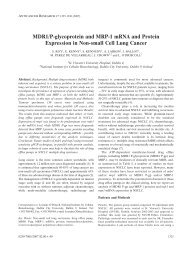
Figure 1. Computed tomography <strong>of</strong> <strong>the</strong> neck with intravenous contr<strong>as</strong>t showing an enlarged cervical lymph node (arrow).<br />
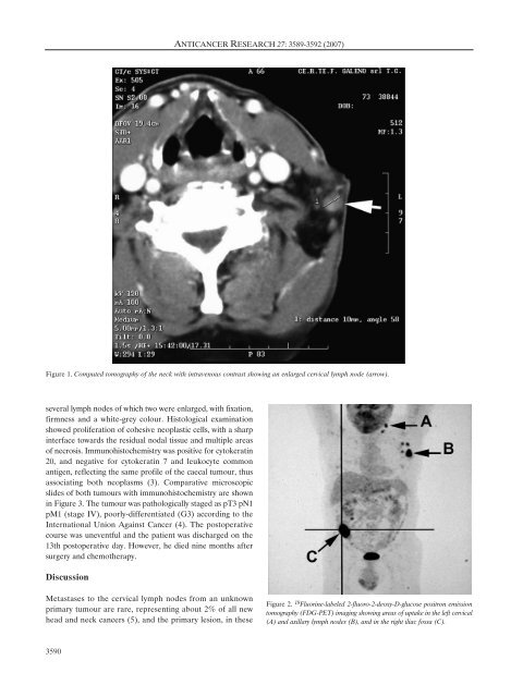
Figure 2. 18 Fluorine-labeled 2-fluoro-2-deoxy-D-glucose positron emission<br />
tomography (FDG-PET) imaging showing are<strong>as</strong> <strong>of</strong> uptake in <strong>the</strong> left cervical<br />
(A) and axillary lymph nodes (B), and in <strong>the</strong> right iliac fossa (C).