Pleomorphic Lipoma - Anticancer Research
Pleomorphic Lipoma - Anticancer Research
Pleomorphic Lipoma - Anticancer Research
You also want an ePaper? Increase the reach of your titles
YUMPU automatically turns print PDFs into web optimized ePapers that Google loves.
ANTICANCER RESEARCH 24: 3157-3160 (2004)<br />
<strong>Pleomorphic</strong> <strong>Lipoma</strong>: A Definite Histopathological Entity<br />
Abstract. <strong>Pleomorphic</strong> lipomas are rare benign tumours<br />
that can resemble a variety of malignant tissue tumour on<br />
histological examination. We describe a case of pleomorphic<br />
lipoma arising on the posterior aspect of the neck of a 70year-old<br />
man, successfully treated by surgical excision. A<br />
review of the literature is presented, summarizing the<br />
principal clinical and morphological characteristics of this<br />
rare tumor.<br />
<strong>Lipoma</strong>s are considered the most common benign soft<br />
tissue neoplasm of the head and neck (1,2). There are many<br />
variants of lipomas, which are differentiated based on the<br />
amount and type of mesenchymal elements present. One<br />
such variant is pleomorphic lipoma. This is a rare, benign,<br />
pseudosarcomatous, soft tissue neoplasm, which typically<br />
occurs in males (4:1) between the ages of 50 and 70 years<br />
(3,4). Its typical location is in the sub-cutis of the neck and<br />
of the shoulder. Currently there are fewer than 150 cases<br />
reported in the world literature (5). We report on a rare<br />
case of pleomorphic lipoma.<br />
Materials and Methods<br />
A 70-year-old man presented in our Department with a 6-year<br />
history of a mass on the posterior aspect of the neck, gradually<br />
increasing in size. Clinically the lesion appeared as an isolated,<br />
Correspondence to: Dott. Alfonso Baldi, Via G. Orsi 25, 80128<br />
Naples, Italy. Fax: +390815569693, e-mail: alfonsobaldi@tiscali.it<br />
Key Words: <strong>Pleomorphic</strong> lipoma.<br />
PAOLO PERSICHETTI 1 , FILIPPO DI LELLA 1 , GIOVANNI F. MARANGI 1 ,<br />
BARBARA CAGLI 1 , PIERFRANCO SIMONE 1 , STEFANIA TENNA 1 ,<br />
CARLA RABITTI 2 , ROBERTO CASSANDRO 3 , VINCENZO ESPOSITO 4,5 ,<br />
ANGELA M. GROEGER 5,6,7 and ALFONSO BALDI 8<br />
1 Department of Plastic and Reconstructive Surgery and<br />
2 Department of Pathology, Campus BioMedico University, Rome;<br />
3 Institute of Cardio-Thoracic <strong>Research</strong>, Respiratory Section. V. Monaldi Hospital, Naples;<br />
4 Third Division of Infective Diseases, D. Cotugno Hospital, Naples, Italy;<br />
5 International Society for the Study of Comparative Oncology (ISSCO), Silver Spring, MD, U.S.A.;<br />
6 Division of Vascular Surgery, Cardarelli Hospital, Naples, Italy;<br />
7 Department of Cardio-Thoracic Surgery, University of Vienna, Vienna, Austria;<br />
8 Department of Biochemistry and Biophysic "F. Cedrangolo", Section of Pathology, Second University of Naples, Italy<br />
0250-7005/2004 $2.00+.40<br />
mobile, non-tender subcutaneous mass measuring 5 x 3 cm<br />
transversally oriented situated at C7 level. There was no sign of<br />
cervical adenopathy. Moreover, there was no history of a previous<br />
trauma at this site.<br />
The excisional biopsy specimen was fixed in 10% bufferedformalin<br />
and paraffin-embedded. Sections of 5-Ì were stained with<br />
haematoxylin-eosin, haematoxylin-van Gieson and PAS-haematoxylin.<br />
Other sections were stained with immunohistochemical<br />
procedure, using avidin-biotin peroxidase complex (ABC) and<br />
antibodies specific for CD34 and S100 (all the reagents were from<br />
Dako, Carpinteria, CA, USA).<br />
The patient was treated with surgery and is disease-free at 36<br />
months of follow-up.<br />
Results<br />
Intra-operatively the mass appeared capsulated and well<br />
circumscribed. Light microscopic examination revealed a<br />
completely excised pleomorphic lipoma based on the<br />
presence of mature fat cells, collagen fibers and "floret-like"<br />
multinucleated giant cells (Figure 1). The immunohistochemical<br />
analysis revealed that more than 80% of<br />
neoplastic cells were positive for CD34 and S-100, while<br />
cytokeratin was completely negative (data not shown).<br />
The light microscopy and immunohistochemical findings<br />
were diagnostic for a pleomorphic lipoma.<br />
Discussion<br />
We describe, here, a rare case of pleomorphic lipoma of the<br />
neck in an adult male (70-year-old). The diagnosis of this<br />
rare lesion is difficult and should be considered in every<br />
growing mass of the head and neck region (5). In our case it<br />
3157
was based on the pathological examination of the tissue<br />
sample guided by immunohistological methods and on the<br />
clinical history of the patient.<br />
<strong>Pleomorphic</strong> lipoma was first described in the early 80s<br />
and, in recent years, it has been shown that spindle cell<br />
lipoma and pleomorphic lipoma are regarded as a single<br />
clinical, histological, immunohistochemical and<br />
cytogenetic entity in the spectrum of benign lipogenic<br />
neoplasms (1-4). Differential diagnosis between spindle<br />
cell lipoma/pleomorphic lipoma, well-differentiated<br />
liposarcoma and atypical lipomas relies on clinical and<br />
histopathological examination. <strong>Pleomorphic</strong> lipoma<br />
typically arises in the sub-cutis of the neck and the<br />
shoulder as a progressively enlarging mass. The average<br />
period necessary for diagnosis is 3.3 years. There are still<br />
ANTICANCER RESEARCH 24: 3157-3160 (2004)<br />
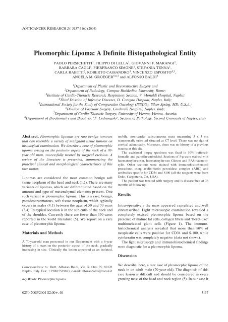
Figure 1. Section through the tumour showing loosely-textured fibrous stroma containing pleomorphic hyperchromatic cells, chronic inflammatory cells<br />
incuding occasional mast cells, and characteristic multinucleated floret-like giant cells with multiple peripherally placed nuclei. Haematoxylin and eosin;<br />
original magnification X 40.<br />
3158<br />
less than 150 cases of pleomorphic lipoma reported in the<br />
literature (5).<br />
Fine-needle aspiration has been reported as being<br />
effective in evaluating subcutaneous lesion especially in the<br />
head and neck region (5). However, pleomorphic lipoma<br />
can masquerade as a malignancy on fine-needle aspiration,<br />
therefore histological confirmation should be obtained prior<br />
to definitive therapy (6,7). In our experience, we do not<br />
usually perform FNA because of the possibility of falsenegative<br />
results. We routinely evaluate subcutaneous masses<br />
by US. Atypical results or difficult interpretation of US are<br />
subsequently evaluated by MRI and eventually by excisional<br />
biopsy. In the case presented, we decided to directly<br />
perform histological examination of the mass, due to clinical<br />
presentation.
Surgical treatment of pleomorphic lipoma involves<br />
complete surgical excision with clear margins, as simple<br />
enucleation is inadequate and is associated with a high<br />
recurrence rate. In the case presented, the patient is<br />
disease-free at 36 months of follow-up, thus confirming the<br />
reported excellent cure rates for this tumor (8).<br />
In conclusion, pleomorphic lipoma is a rare benign,<br />
psueodsarcomatous soft tissue neoplasm typically occurring<br />
in the sub-cutis of the neck and shoulder, that can resemble<br />
a variety of malignant soft tissue tumors. Therefore, careful<br />
examination of the clinical setting, as well as of the<br />
histopathological characteristics of this kind of tumors is<br />
essential for a correct diagnosis and to avoid unnecessary<br />
and often disfiguring surgery.<br />
Acknowledgements<br />
This work was supported by FUTURA-Onlus and I.S.S.C.O.<br />
References<br />
1 Enzinger FM and Weiss SW: Soft Tissue Tumors, 3rd ed. St.<br />
Louis: CV Mosby, 351-466: 1995.<br />
Persichetti et al: <strong>Pleomorphic</strong> <strong>Lipoma</strong><br />
2 Krandsorf FM: Benign soft-tissue tumors in a large referral<br />
population: distribution of specific diagnoses by age, sex, and<br />
location. Am J Roentgenol 164: 395-402, 1995.<br />
3 Enzinger FM: Benign lipomatous tumors simulating a<br />
sarcoma. In: Martin RG, Ayala AG, eds. Management of<br />
Primary Bone and Soft Tissue Tumors. Chicago: Year Book<br />
Publishing, 11-24, 1977.<br />
4 Shmookler BM and Enzinger FM: <strong>Pleomorphic</strong> lipoma: a<br />
benign tumor simulating liposarcoma. Cancer 47: 126-133,<br />
1981.<br />
5 Yencha MW and Hodge JJ: <strong>Pleomorphic</strong> lipoma: case report<br />
and literature review. Dermatol Surg 26: 375-380, 2000.<br />
6 Rigby HS, Wilson YG, Cawthorn SJ and Ibrahim NB: Fineneedle<br />
aspiration of pleomorphic lipoma: a potential pitfall of<br />
cytodiagnosis. Cytopathology 4: 55-58, 1993.<br />
7 Dundas KE, Wong MP and Suen KC: Two unusual benign<br />
lesions of the neck masquerading as malignancy on fine-needle<br />
aspiration cytology. Diagn Cytopathol 12: 272-279, 1995.<br />
8 Bryant J: <strong>Pleomorphic</strong> lipoma of the scalp. J Dermatol Surg<br />
Oncol 7: 323-325, 1981.<br />
Received May 18, 2004<br />
Accepted August 2, 2004<br />
3159


