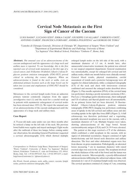
Cervical Node Metastasis as the First Sign of Cancer of the Caecum
Cervical Node Metastasis as the First Sign of Cancer of the Caecum
Cervical Node Metastasis as the First Sign of Cancer of the Caecum

Create successful ePaper yourself
Turn your PDF publications into a flip-book with our unique Google optimized e-Paper software.
ANTICANCER RESEARCH 27: 3589-3592 (2007)<br />
Abstract. The unusual c<strong>as</strong>e <strong>of</strong> an adenocarcinoma <strong>of</strong> <strong>the</strong><br />
caecum undiagnosed until <strong>the</strong> appearance <strong>of</strong> a large neck and<br />
axillary m<strong>as</strong>s is reported. To our knowledge, this is <strong>the</strong> first<br />
reported c<strong>as</strong>e <strong>of</strong> cervical node met<strong>as</strong>t<strong>as</strong>is <strong>as</strong> <strong>the</strong> first sign <strong>of</strong> a<br />
caecal cancer, and 18 fluorine-18-labeled 2-fluoro-2-deoxy-Dglucose<br />
positron emission tomography (FDG-PET) proved<br />
critical in achieving <strong>the</strong> correct diagnosis. When an<br />
adenocarcinoma is found in <strong>the</strong> neck or axilla, even an<br />
abdominal primary location such <strong>as</strong> <strong>the</strong> large bowel can be<br />
taken into account and employment <strong>of</strong> FDG-PET should be<br />
considered.<br />
Met<strong>as</strong>t<strong>as</strong>es to <strong>the</strong> cervical lymph nodes from an unknown<br />
primary tumour commonly originate from <strong>the</strong> upper<br />
aerodigestive tract (1) and <strong>the</strong> need for a careful work-up<br />
in patients with <strong>as</strong>ymmetric enlargement <strong>of</strong> cervical nodes<br />
h<strong>as</strong> been stressed since 1952 (2). We report <strong>the</strong> unusual c<strong>as</strong>e<br />
<strong>of</strong> an adenocarcinoma <strong>of</strong> <strong>the</strong> caecum undiagnosed until <strong>the</strong><br />
appearance <strong>of</strong> a large neck and axillary m<strong>as</strong>s.<br />
C<strong>as</strong>e Report<br />
<strong>Cervical</strong> <strong>Node</strong> <strong>Met<strong>as</strong>t<strong>as</strong>is</strong> <strong>as</strong> <strong>the</strong> <strong>First</strong><br />
<strong>Sign</strong> <strong>of</strong> <strong>Cancer</strong> <strong>of</strong> <strong>the</strong> <strong>Caecum</strong><br />
LUIGI BASSO 1 , LUCIANO IZZO 1 , ERIKA CALISI 1 , GIUSEPPE CAVALLARO 1 , UMBERTO COSTI 1 ,<br />
ANTONIO CIARDI 2 , FRANCESCA FORNARI 1 , ANDREA POLISTENA 1 and GIORGIO DE TOMA 1<br />
1 Cattedra di Chirurgia Generale, Divisione di Chirurgia "B", Department <strong>of</strong> Surgery "Pietro Valdoni" and<br />
2 Department <strong>of</strong> Experimental Medicine and Pathology, University <strong>of</strong> Rome<br />
"La Sapienza" <strong>First</strong> Medical School, Policlinico "Umberto I", Rome, Italy<br />
A 73-year-old male came under our care three months after<br />
he noticed a lump on <strong>the</strong> left side <strong>of</strong> his neck. His previous<br />
bowel habits were regular and stayed unchanged. Eight weeks<br />
after <strong>the</strong> outbreak <strong>of</strong> <strong>the</strong>se two lumps, before coming under<br />
our observation, his attending General Practitioner requested<br />
a cervical ultr<strong>as</strong>onography (US) which showed multiple<br />
Correspondence to: Pr<strong>of</strong>essor Luigi B<strong>as</strong>so, Department <strong>of</strong> Surgery<br />
"Pietro Valdoni", University <strong>of</strong> Rome "La Sapienza" Medical<br />
School, Policlinico "Umberto I", viale del Policlinico 155, 00161<br />
Rome, Italy. Tel: +39 06 49972167, Fax: +39 06 49972197, e-mail:<br />
luigi.b<strong>as</strong>so@uniroma1.it / lgb<strong>as</strong>so@yahoo.it<br />
Key Words: Occult, primary, neck, axilla, cancer, caecum, lymph<br />
nodes, positron-emission tomography, PET.<br />
0250-7005/2007 $2.00+.40<br />
enlarged lymph nodes on <strong>the</strong> left side <strong>of</strong> <strong>the</strong> neck, with a<br />
maximum diameter <strong>of</strong> 1.5 cm. A month later, after<br />
unsuccessful conservative treatment, <strong>the</strong> patient w<strong>as</strong> referred<br />
to our surgical outpatient department. General examination<br />
w<strong>as</strong> unremarkable, except for enlarged left cervical and left<br />
axillary nodes, which one month before were clinically normal.<br />
General blood results, physical examination, careful<br />
<strong>as</strong>sessment <strong>of</strong> tonsils and a posterior laryngoscopy were all<br />
negative for clinical indications, while a computed tomography<br />
(CT) <strong>of</strong> <strong>the</strong> neck and chest with intravenous contr<strong>as</strong>t<br />
confirmed and <strong>as</strong>sessed <strong>the</strong> enlarged nodes described above<br />
(Figure 1). Fine-needle <strong>as</strong>piration (FNA) <strong>of</strong> <strong>the</strong> cervical lump<br />
w<strong>as</strong> performed, showing a poorly met<strong>as</strong>tatic carcinoma (CK+,<br />
Pan Leu–). Oesophago-g<strong>as</strong>tro-duodenoscopy, chest radiograph<br />
and US <strong>of</strong> salivary and thyroid glands were all unremarkable.<br />
As no primary lesion had yet been detected, 18 fluorinelabeled<br />
2-fluoro-2-deoxy-D-glucose positron emission<br />
tomography (FDG-PET) imaging w<strong>as</strong> performed, confirming<br />
are<strong>as</strong> <strong>of</strong> uptake in <strong>the</strong> left cervical and axillary are<strong>as</strong> and also<br />
revealing uptake in <strong>the</strong> right iliac fossa (RIF) (Figure 2). A full<br />
colonoscopy w<strong>as</strong> <strong>the</strong>refore performed and a vegetating,<br />
partially ulcerated neopl<strong>as</strong>m w<strong>as</strong> seen in <strong>the</strong> caecum, with a<br />
maximum diameter <strong>of</strong> 5 cm. Multiple biopsies were taken,<br />
showing adenocarcinoma <strong>of</strong> <strong>the</strong> large bowel. Serum<br />
carcinoembryonic antigen (CEA) and CA 19-9 were elevated.<br />
The patient at this stage received a CT <strong>of</strong> <strong>the</strong> abdomen,<br />
which confirmed a solid neopl<strong>as</strong>m in <strong>the</strong> caecum, with no<br />
sign(s) <strong>of</strong> abdominal secondaries. Surgery consisted <strong>of</strong> right<br />
hemicolectomy and left lateral neck dissection and axillary<br />
dissection. The liver and o<strong>the</strong>r abdominal organs were free<br />
from dise<strong>as</strong>e on gross examination. Pathologically, an ulcerated<br />
lesion me<strong>as</strong>uring 7 cm in maximum diameter with raised<br />
everted edges w<strong>as</strong> seen in <strong>the</strong> caecum. The neopl<strong>as</strong>m involved<br />
<strong>the</strong> whole thickness <strong>of</strong> <strong>the</strong> wall <strong>of</strong> <strong>the</strong> bowel and <strong>the</strong> lumen w<strong>as</strong><br />
moderately stenotic. Microscopically, a poorly-differentiated<br />
adenocarcinoma infiltrating <strong>the</strong> subserosal adipose tissue, with<br />
met<strong>as</strong>t<strong>as</strong>is in 1 out <strong>of</strong> 23 examined lymph nodes w<strong>as</strong> observed.<br />
The cervical specimen consisted <strong>of</strong> a lump <strong>of</strong> 3 cm, containing<br />
3589
several lymph nodes <strong>of</strong> which two were enlarged, with fixation,<br />
firmness and a white-grey colour. Histological examination<br />
showed proliferation <strong>of</strong> cohesive neopl<strong>as</strong>tic cells, with a sharp<br />
interface towards <strong>the</strong> residual nodal tissue and multiple are<strong>as</strong><br />
<strong>of</strong> necrosis. Immunohistochemistry w<strong>as</strong> positive for cytokeratin<br />
20, and negative for cytokeratin 7 and leukocyte common<br />
antigen, reflecting <strong>the</strong> same pr<strong>of</strong>ile <strong>of</strong> <strong>the</strong> caecal tumour, thus<br />
<strong>as</strong>sociating both neopl<strong>as</strong>ms (3). Comparative microscopic<br />
slides <strong>of</strong> both tumours with immunohistochemistry are shown<br />
in Figure 3. The tumour w<strong>as</strong> pathologically staged <strong>as</strong> pT3 pN1<br />
pM1 (stage IV), poorly-differentiated (G3) according to <strong>the</strong><br />
International Union Against <strong>Cancer</strong> (4). The postoperative<br />
course w<strong>as</strong> uneventful and <strong>the</strong> patient w<strong>as</strong> discharged on <strong>the</strong><br />
13th postoperative day. However, he died nine months after<br />
surgery and chemo<strong>the</strong>rapy.<br />
Discussion<br />
Met<strong>as</strong>t<strong>as</strong>es to <strong>the</strong> cervical lymph nodes from an unknown<br />
primary tumour are rare, representing about 2% <strong>of</strong> all new<br />
head and neck cancers (5), and <strong>the</strong> primary lesion, in <strong>the</strong>se<br />
3590<br />
ANTICANCER RESEARCH 27: 3589-3592 (2007)<br />
Figure 1. Computed tomography <strong>of</strong> <strong>the</strong> neck with intravenous contr<strong>as</strong>t showing an enlarged cervical lymph node (arrow).<br />
Figure 2. 18 Fluorine-labeled 2-fluoro-2-deoxy-D-glucose positron emission<br />
tomography (FDG-PET) imaging showing are<strong>as</strong> <strong>of</strong> uptake in <strong>the</strong> left cervical<br />
(A) and axillary lymph nodes (B), and in <strong>the</strong> right iliac fossa (C).
instances, is later to be commonly found in <strong>the</strong> upper<br />
aerodigestive tract. In a historical series <strong>of</strong> 157 patients <strong>of</strong><br />
<strong>the</strong> Memorial Sloan-Kettering Center <strong>of</strong> New York, U.S.A.<br />
with unexplained cervical node met<strong>as</strong>t<strong>as</strong>is, 79 patients were<br />
reported to have epidermoid carcinoma, 29 adenocarcinoma,<br />
13 anapl<strong>as</strong>tic carcinoma and 11 melanoma (1). Evaluation <strong>of</strong><br />
patients with cervical node met<strong>as</strong>t<strong>as</strong>is <strong>of</strong> occult origin should,<br />
<strong>the</strong>refore, include: thorough medical history, detailed<br />
physical examination <strong>of</strong> <strong>the</strong> skin, evaluation <strong>of</strong> <strong>the</strong> upper<br />
respiratory and digestive systems complemented by<br />
B<strong>as</strong>so et al: <strong>Cervical</strong> <strong>Met<strong>as</strong>t<strong>as</strong>is</strong> <strong>as</strong> <strong>Sign</strong> <strong>of</strong> Caecal <strong>Cancer</strong><br />
Figure 3. Comparative slides, showing positive immunohistochemistry (cytokeratin 20) <strong>of</strong> poorly-differentiated adenocarcinoma in both <strong>the</strong> specimen <strong>of</strong><br />
<strong>the</strong> large bowel (A) and <strong>of</strong> <strong>the</strong> cervical lymph nodes (B) (PAP-DAB, x250).<br />
endoscopy, careful <strong>as</strong>sessment <strong>of</strong> tonsils, salivary and thyroid<br />
glands, FNA <strong>of</strong> <strong>the</strong> m<strong>as</strong>s(es), chest radiographs and imaging<br />
studies (US, CT scan, magnetic resonance imaging,<br />
scintigraphy). Recently, FDG-PET h<strong>as</strong> shown itself to be a<br />
useful diagnostic imaging tool in <strong>the</strong>se patients. Thoracic and<br />
abdominal primaries (especially from <strong>the</strong> lungs, oesophagus,<br />
stomach, ovary, biliary tract, or pancre<strong>as</strong>) should be sought<br />
in c<strong>as</strong>e <strong>of</strong> an adenocarcinoma discovered in a cervical lump.<br />
A recent review showed that <strong>of</strong> 52 patients with head and<br />
neck met<strong>as</strong>t<strong>as</strong>es from an occult primary site reported in <strong>the</strong><br />
3591
literature, 33 (63.5%) had met<strong>as</strong>t<strong>as</strong>es to <strong>the</strong> cervical lymph<br />
nodes, 3 (5.8%) to <strong>the</strong> thyroid gland, 6 (11.5%) to <strong>the</strong><br />
paran<strong>as</strong>al sinus, 3 (5.8%) to <strong>the</strong> tonsils, 3 (5.8%) to <strong>the</strong><br />
parotid gland, 2 (3.8%) to <strong>the</strong> ear, and 1 each (1.9%) to <strong>the</strong><br />
larynx and to <strong>the</strong> mandibula, respectively. Of <strong>the</strong> 33 patients<br />
with met<strong>as</strong>t<strong>as</strong>es to <strong>the</strong> cervical lymph nodes, 17 (51.5%) had<br />
secondaries from a primary in <strong>the</strong> g<strong>as</strong>trointestinal tract, but<br />
only 2 out <strong>of</strong> 33 (6.1%) had primary abdominal digestive<br />
neopl<strong>as</strong>ms, one arising from <strong>the</strong> liver and one being a<br />
carcinoid tumour <strong>of</strong> <strong>the</strong> terminal ileum (6, 7). In any c<strong>as</strong>e,<br />
all <strong>of</strong> <strong>the</strong>se advanced stage IV malignancies imply ominous<br />
outcomes.<br />
To our knowledge, this is <strong>the</strong> first reported c<strong>as</strong>e <strong>of</strong><br />
cervical node met<strong>as</strong>t<strong>as</strong>is <strong>as</strong> <strong>the</strong> first sign <strong>of</strong> a caecal cancer.<br />
It is not known how cancer cells could have reached <strong>the</strong><br />
neck from <strong>the</strong> large bowel, without grossly involving <strong>the</strong><br />
hepatic filter. Lymphatic spread through retrograde or<br />
unusual flow should be considered, while haematogenous<br />
dissemination can be ruled out due to <strong>the</strong> existence <strong>of</strong><br />
hepatic and pulmonary filters. Indeed, somewhat bizarre<br />
secondaries can be <strong>the</strong> first sign <strong>of</strong> abdominal malignancies,<br />
such <strong>as</strong> Sister Mary Joseph’s umbilical nodule, which,<br />
however, is located at an abdominal level (8).<br />
FDG-PET allows detection <strong>of</strong> <strong>the</strong> primary tumour in about<br />
21% <strong>of</strong> c<strong>as</strong>es located above <strong>the</strong> diaphragm (9), while it proves<br />
more sensitive than CT and monitoring <strong>of</strong> CEA and CA 19-9<br />
levels in detecting abdominal secondaries and/or recurrences<br />
<strong>of</strong> colorectal cancer (10, 11). In <strong>the</strong> c<strong>as</strong>e we report, FDG-PET<br />
proved critical in identifying <strong>the</strong> primary lesion, while CT had<br />
initially been aimed only to <strong>the</strong> chest and neck, <strong>as</strong> <strong>the</strong>y are <strong>the</strong><br />
common site <strong>of</strong> origin <strong>of</strong> <strong>the</strong>se tumours.<br />
In conclusion, distant met<strong>as</strong>t<strong>as</strong>es to <strong>the</strong> cervical, or<br />
axillary lymph nodes from an unknown primary neopl<strong>as</strong>m<br />
are rare and have a poor prognosis. When an<br />
adenocarcinoma is found in <strong>the</strong>se are<strong>as</strong>, even an<br />
abdominal primary location such <strong>as</strong> <strong>the</strong> large bowel should<br />
be taken into account and employment <strong>of</strong> FDG-PET<br />
should be considered.<br />
3592<br />
ANTICANCER RESEARCH 27: 3589-3592 (2007)<br />
References<br />
1 Spiro RH, DeRose G and Strong EW: <strong>Cervical</strong> node met<strong>as</strong>t<strong>as</strong>is<br />
<strong>of</strong> occult origin. Am J Surg 46: 441-446, 1983.<br />
2 Martin H and Romieu C: The diagnostic significance <strong>of</strong> a "lump<br />
in <strong>the</strong> neck". Postgrad Med 11: 491-500, 1952.<br />
3 Chu P, Wu E and Weiss LM: Cytokeratin 7 and cytokeratin 20<br />
expression in epi<strong>the</strong>lial neopl<strong>as</strong>ms: a survey <strong>of</strong> 435 c<strong>as</strong>es. Mod<br />
Pathol 13: 962-972, 2000.<br />
4 International Union Against <strong>Cancer</strong>. TNM Cl<strong>as</strong>sification <strong>of</strong><br />
Malignant Tumours. Sobin LH and Wittekind Ch (eds.). Sixth<br />
Edition. Hoboken, NJ, USA, Jossey-B<strong>as</strong>s, 2002.<br />
5 Grau C, Johansen LV, Jakobsen J, Geertsen P, Andersen E and<br />
Jensen BB: <strong>Cervical</strong> lymph node met<strong>as</strong>t<strong>as</strong>es from unknown<br />
primary tumours. Results from a national survey by <strong>the</strong> Danish<br />
Society for Head and Neck Oncology. Radio<strong>the</strong>r Oncol 55: 121-<br />
129, 2000.<br />
6 Imamura S and Suzuki H: Head and neck met<strong>as</strong>t<strong>as</strong>es from<br />
occult abdominal primary site: a c<strong>as</strong>e report and literature<br />
review. Acta Otolaryngol 124: 107-112, 2004.<br />
7 Welling RE and Taggart JP: Carcinoid tumour met<strong>as</strong>tatic to<br />
neck. Arch Surg 110: 111-113, 1975.<br />
8 Gabriele R, Borghese M, Conte M and B<strong>as</strong>so L: Sister Mary<br />
Joseph's nodule from adenocarcinoma <strong>of</strong> <strong>the</strong> cecum. Report <strong>of</strong><br />
a c<strong>as</strong>e. Dis Colon Rectum 47: 115-117, 2004.<br />
9 Safa AA, Tran LM, Rege S, Brown CV, Mandelkern MA,<br />
Wang MB, Sadeghi A and Juillard G: The role <strong>of</strong> positron<br />
emission tomography in occult primary head and neck cancers.<br />
<strong>Cancer</strong> J Sci Am 5: 214-218, 1999.<br />
10 Johnson K, Bakhsh A, Young D, Martin TE Jr and Arnold M:<br />
Correlating computed tomography and positron emission<br />
tomography scan with operative findings in met<strong>as</strong>tatic<br />
colorectal cancer. Dis Colon Rectum 44: 354-357, 2001.<br />
11 Liu FY, Chen JS, Changchien CR, Yeh CY, Liu SH, Ho KC<br />
and Yen TC: Utility <strong>of</strong> 2-fluoro-2-deoxy-D-glucose positron<br />
emission tomography in managing patients <strong>of</strong> colorectal cancer<br />
with unexplained carcinoembryonic antigen elevation at<br />
different levels. Dis Colon Rectum 48: 1900-1912, 2005.<br />
Received April 16, 2007<br />
Revised July 12, 2007<br />
Accepted July 24, 2007