An overview of sexually transmitted diseases. Part III ... - Dermatology
An overview of sexually transmitted diseases. Part III ... - Dermatology
An overview of sexually transmitted diseases. Part III ... - Dermatology

You also want an ePaper? Increase the reach of your titles
YUMPU automatically turns print PDFs into web optimized ePapers that Google loves.
The HIV epidemic has drastically altered the medical<br />
community’s approach to <strong>sexually</strong> <strong>transmitted</strong><br />
<strong>diseases</strong> (STDs). For generations, multiple<br />
STDs occurred in individual patients. However, not<br />
until HIV did these infections interact so significantly<br />
with each other. Clinically, HIV can alter the presentation<br />
or treatments <strong>of</strong> other STDs as in, for example, a<br />
case <strong>of</strong> coexistent HIV and scabies. Conversely, an<br />
increasing recognition <strong>of</strong> the ways in which STDs influence<br />
HIV transmission has underscored the importance<br />
<strong>of</strong> early detection and treatment <strong>of</strong> all STDs. In 1998,<br />
the Centers for Disease Control and Prevention (CDC)<br />
published Guidelines for the Treatment <strong>of</strong> Sexually<br />
Transmitted Diseases, 1 and in addition the CDC published<br />
recommendations for the prevention <strong>of</strong> HIV<br />
through early detection and treatment <strong>of</strong> other STDs. 2<br />
From the Department <strong>of</strong> <strong>Dermatology</strong> at the University <strong>of</strong> Texas—<br />
Houston Health Science Center and St Joseph Hospital,<br />
Houston a ; the Department <strong>of</strong> <strong>Dermatology</strong> at the University <strong>of</strong><br />
Texas—Southwestern Medical School and Veterans Affairs<br />
Hospital, Dallas b ; and the Departments <strong>of</strong> <strong>Dermatology</strong>,<br />
Microbiology & Immunology and Internal Medicine at the<br />
University <strong>of</strong> Texas Medical Branch, Galveston. c<br />
Reprint requests: Stephen K. Tyring, MD, PhD, Department <strong>of</strong><br />
<strong>Dermatology</strong>, Route 1070, University <strong>of</strong> Texas Medical Branch,<br />
Galveston, TX 77555-1070. E-mail: styring@utmb.edu.<br />
16/2/105158<br />
doi:10.1067/mjd.2000.105158<br />
CONTINUING MEDICAL EDUCATION<br />
<strong>An</strong> <strong>overview</strong> <strong>of</strong> <strong>sexually</strong> <strong>transmitted</strong> <strong>diseases</strong>.<br />
<strong>Part</strong> <strong>III</strong>. Sexually <strong>transmitted</strong> <strong>diseases</strong> in<br />
HIV-infected patients<br />
Adam Czelusta, MD, a <strong>An</strong>gela Yen-Moore, MD, b Melody Van der Straten, MD, c<br />
Daniel Carrasco, MD, c and Stephen K. Tyring MD, PhD c Houston, Dallas, and Galveston, Texas<br />
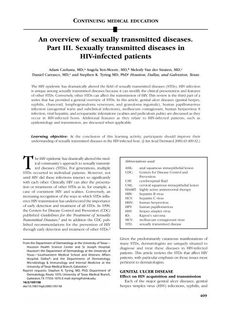
The HIV epidemic has dramatically altered the field <strong>of</strong> <strong>sexually</strong> <strong>transmitted</strong> <strong>diseases</strong> (STDs). HIV infection<br />
is unique among <strong>sexually</strong> <strong>transmitted</strong> <strong>diseases</strong> because it can modify the clinical presentation and features<br />
<strong>of</strong> other STDs. Conversely, other STDs can affect the transmission <strong>of</strong> HIV. This review is the third part <strong>of</strong> a<br />
series that has provided a general <strong>overview</strong> <strong>of</strong> STDs. In this article, genital ulcer <strong>diseases</strong> (genital herpes,<br />
syphilis, chancroid, lymphogranuloma venereum, and granuloma inguinale), human papillomavirus<br />
infection (anogenital warts and subclinical infections), molluscum contagiosum, human herpesvirus 8<br />
infection, viral hepatitis, and ectoparasitic infestations (scabies and pediculosis pubis) are discussed as they<br />
occur in HIV-infected hosts. Additional features as they relate to HIV-infected patients, such as<br />
epidemiology and transmission, are discussed when applicable.<br />
Learning objective: At the conclusion <strong>of</strong> this learning activity, participants should improve their<br />
understanding <strong>of</strong> <strong>sexually</strong> <strong>transmitted</strong> <strong>diseases</strong> in the HIV-infected host. (J Am Acad Dermatol 2000;43:409-32.)<br />
Abbreviations used:<br />
ASIL: anal squamous intraepithelial lesion<br />
CDC: Centers for Disease Control and<br />
Prevention<br />
CSF: cerebrospinal fluid<br />
CSIL: cervical squamous intraepithelial lesion<br />
HAART: highly active antiretroviral therapy<br />
HBV: hepatitis B virus<br />
HCV: hepatitis C virus<br />
HHV: human herpesvirus<br />
HPV: human papillomavirus<br />
HSV: herpes simplex virus<br />
KS: Kaposi’s sarcoma<br />
MCV: molluscum contagiosum virus<br />
STD: <strong>sexually</strong> <strong>transmitted</strong> disease<br />
Given the predominantly cutaneous manifestations <strong>of</strong><br />
many STDs, dermatologists are uniquely situated to<br />
diagnose and treat these <strong>diseases</strong> in HIV-infected<br />
patients. This article reviews the STDs that affect HIV<br />
patients, with particular emphasis on those issues most<br />
pertinent to dermatologists.<br />
GENITAL ULCER DISEASE<br />
Effect on HIV acquisition and transmission<br />
Each <strong>of</strong> the major genital ulcer <strong>diseases</strong>, genital<br />
herpes simplex virus (HSV) infections, syphilis, and<br />
409
410 Czelusta, Yen-Moore, and Tyring J AM ACAD DERMATOL<br />
SEPTEMBER 2000<br />
chancroid, have been associated with an increased<br />
risk <strong>of</strong> acquiring and transmitting HIV. One estimation<br />
suggests that STDs increase the overall risk <strong>of</strong><br />
acquiring HIV about 3 to 5 times. 3 Cross-sectional<br />
studies performed in Nairobi, Kenya have consistently<br />
found that HIV seropositivity was more<br />
common in persons with either a history or clinical<br />
evidence <strong>of</strong> genital ulcer disease, 4-7 and one<br />
prospective study from this same region showed an<br />
increased risk <strong>of</strong> seroconversion in patients with<br />
genital ulcer disease. 8 Similarly, Telzak et al 9 found<br />
that 2.9% <strong>of</strong> men with genital ulcer disease turned<br />
HIV-positive, whereas only 1% <strong>of</strong> men without genital<br />
ulcer disease seroconverted. Clearly, a relationship<br />
between genital ulcer disease and HIV transmission<br />
exists; subsequently, prevention <strong>of</strong> genital ulcer<br />
disease should decrease transmission <strong>of</strong> HIV. A<br />
Tanzanian study showed that communities that<br />
improved their recognition and treatment <strong>of</strong> STDs<br />
saw a decrease in the incidence <strong>of</strong> HIV infection in<br />
their population. 10 This is the first documented<br />
intervention involving treatment <strong>of</strong> STDs that was<br />
associated with a decrease in HIV incidence in a<br />
defined population. Thus, through the early recognition<br />
and treatment <strong>of</strong> all STDs, including genital<br />
ulcer disease, HIV transmission can be reduced.<br />
Numerous studies have isolated HIV from genital<br />
ulcer exudates. 11-14 Mechanisms by which genital<br />
ulcer disease appear to facilitate HIV transmission<br />
have been suggested. Disruption <strong>of</strong> the genital<br />
mucosa is associated with the recruitment <strong>of</strong> inflammatory<br />
cells such as CD4 + T lymphocytes and<br />
macrophages. The presence <strong>of</strong> these cells can facilitate<br />
transmission <strong>of</strong> HIV virions from HIV-infected<br />
persons to uninfected persons or provide additional<br />
targets for HIV entry in HIV-negative persons who<br />
are being exposed to the virus. 14-17<br />
Studies implicating the specific agents <strong>of</strong> genital<br />
ulcer disease have focused mostly on genital herpes,<br />
syphilis, and chancroid. Several reports have shown an<br />
association between genital HSV lesions and the acquisition<br />
<strong>of</strong> HIV infection in the affected population.<br />
4,9,18-23 Keet et al 23 showed in a prospective<br />
cohort that pre-existing herpes simplex virus type 2<br />
(HSV-2) seropositivity was a predictor <strong>of</strong> HIV seroconversion.<br />
HIV virions have been shown in HSV ulcers, 24<br />
and in one study HIV RNA was isolated from herpetic<br />
lesions on 67% <strong>of</strong> the days that lesions were present. 13<br />
Similarly, arguments implicating syphilis and chancroid<br />
in the acquisition <strong>of</strong> HIV can be made. Two studies<br />
from Baltimore document strong associations<br />
between HIV seroconversion and either positive<br />
syphilis serology or a history <strong>of</strong> syphilis. 25,26 One<br />
cross-sectional study from Kenya found a similar association<br />
5 ; however, the difficulty in making this associ-<br />
ation statistically significant may reflect the difficulty<br />
in identifying cases <strong>of</strong> syphilis with active genital<br />
ulcers during the study. 8 <strong>An</strong> outbreak <strong>of</strong> chancroid in<br />
Jackson, Mississippi showed a strong association<br />
between this infection and HIV seropositivity, 27 and<br />
most <strong>of</strong> the ulcers in an aforementioned Kenyan<br />
study were due to chancroid and were associated<br />
with increased HIV seropositivity. 6 Lymphogranuloma<br />
venereum and granuloma inguinale are rare <strong>diseases</strong>,<br />
and, as such, they have not been extensively studied<br />
in relation to HIV transmission. Granuloma inguinale<br />
has been associated with a significant number <strong>of</strong> HIV<br />
infections, 28 and its eradication (in an effort to prevent<br />
HIV transmission) has been suggested. 29<br />
Clinical features and treatment<br />
Genital herpes. Genital herpes is the most common<br />
cause <strong>of</strong> genital ulceration worldwide, 30 and its<br />
association with HIV was noticed as early as 1981. 31<br />
Although genital herpes is most commonly caused<br />
by HSV-2, an increasing number <strong>of</strong> cases are suspected<br />
to be caused by HSV-1. 1,32 In HIV-infected<br />
patients, genital herpes can result in severe and atypical<br />
clinical presentations, and treatment may<br />
require increased doses <strong>of</strong> antiviral medications.<br />
Suppressive therapy for HSV appears to significantly<br />
improve survival in HIV-positive patients.<br />
Clinically, HIV-infected patients may experience<br />
an increased number and size <strong>of</strong> lesions in both primary<br />
and reactivated HSV infections as compared<br />
with HIV-uninfected patients. 31,33,34 The vesicles and<br />
ulcers are more painful and heal slower than those<br />
experienced by an immunocompetent host. 35 As<br />
CD4 cell counts drop and immunosuppression worsens,<br />
recurrent outbreaks increase in frequency and<br />
severity. 36,37 Chronic HSV-2 ulcers <strong>of</strong> more than 1<br />
month in duration are an AIDS-defining illness in<br />
HIV-infected patients. 38,39<br />
Atypical HSV presentations occur relatively <strong>of</strong>ten<br />
in HIV-infected patients. <strong>Part</strong>icularly severe lesions<br />
have been reported on patients’ lower backs, buttocks,<br />
or perianal regions, and these lesions may<br />
expand to 20 cm in diameter (Fig 1). 31,40 Such ulcers<br />
commonly become impetiginized and require intensive<br />
long-term therapy. 31 Several case reports<br />
describe HSV-2 presenting as hyperkeratotic verrucous<br />
lesions resembling condyloma in severely<br />
immunocompromised patients. 41-44 These masses<br />
can become large 41,43 and may represent concurrent<br />
HSV and cytomegalovirus infection. 44 A recent<br />
report describes a patient with a pseudotumor <strong>of</strong> the<br />
tongue that was discovered to be an atypical recurrence<br />
<strong>of</strong> HSV-2. 45 HSV may also cause esophagitis,<br />
hepatitis, pneumonitis, or life-threatening disseminated<br />
infections in AIDS patients. 46
J AM ACAD DERMATOL<br />
VOLUME 43, NUMBER 3<br />
Acyclovir, valacyclovir, and famciclovir are recognized<br />
by the CDC as appropriate therapies for primary<br />
or recurrent genital herpes in the HIV-infected<br />
patient. 1 As in the immunocompetent patient, therapy<br />
should start as soon as possible, preferably during<br />
the prodromal period. Therapy is then continued<br />
until clinical resolution is obtained. 1 For acyclovir<br />
and famciclovir, increased dosages above those recommended<br />
for immunocompetent persons may be<br />
required. For example, whereas the recommended<br />
oral dose <strong>of</strong> acyclovir is either 400 mg 3 times per<br />
day or 200 mg 5 times per day, regimens <strong>of</strong> 400 mg<br />
given 5 times per day have been useful in immunocompromised<br />
patients. 1 Likewise, famciclovir 250<br />
mg twice daily is recommended for suppression <strong>of</strong><br />
genital herpes in the immunocompetent person, but<br />
500 mg twice daily has been effective in decreasing<br />
both the rate <strong>of</strong> recurrences and the rate <strong>of</strong> subclinical<br />
shedding among HIV-seropositive persons. 47<br />
When given at doses <strong>of</strong> 8 g per day to markedly<br />
immunocompromised persons for suppression <strong>of</strong><br />
cytomegalovirus infections, valacyclovir was associated<br />
with either thrombotic thrombocytopenic purpura<br />
or the hemolytic uremic syndrome. 1,48,49 However, no<br />
cause-and-effect relationship was ascertained. When<br />
taken as a dose <strong>of</strong> 500 mg twice daily, valacyclovir<br />
appeared safe and effective for the suppression <strong>of</strong><br />
genital HSV in HIV-seropositive persons and was<br />
superior to acyclovir 400 mg twice daily. 50 Therefore<br />
acyclovir, valacyclovir, and famciclovir all appear useful<br />
for treatment and suppression <strong>of</strong> HSV in immunocompromised<br />
persons. 47-55<br />
Interestingly, acyclovir treatment <strong>of</strong> HSV infections<br />
in HIV-positive patients may <strong>of</strong>fer a significant<br />
survival benefit. Eight randomized controlled trials,<br />
combined in a meta-analysis, showed that patients<br />
treated with acyclovir had a significant survival<br />
advantage compared with those who went untreated.<br />
51 Two multicenter clinical trials noted that<br />
patients given acyclovir and zidovudine lived longer<br />
than those given only zidovudine. 56,57 These studies<br />
suggest that long-term suppressive acyclovir therapy<br />
prolongs survival in AIDS patients with extensive histories<br />
<strong>of</strong> HSV infections.<br />
The mechanism by which this occurs is unclear.<br />
Studies demonstrate that acute or reactivated HSV<br />
infection may stimulate HIV replication. 58-60<br />
Furthermore, Mole et al 61 documented increased<br />
plasma HIV viral loads in HIV patients experiencing<br />
an outbreak <strong>of</strong> HSV. By reducing or attenuating<br />
the occurrences <strong>of</strong> HSV outbreaks, acyclovir therapy<br />
may help reduce these deleterious effects <strong>of</strong><br />
the infections. Clearly, further investigation<br />
regarding this issue is required, as evidenced by<br />
the study <strong>of</strong> Gallant et al, 62 which found no asso-<br />
Czelusta, Yen-Moore, and Tyring 411<br />
Fig 1. HIV-positive patient. Large suprapubic ulcer due to<br />
HSV type 2.<br />
ciation between survival and acyclovir use in HIVinfected<br />
patients.<br />
A preliminary report from the CDC noted that<br />
6.4% <strong>of</strong> HSV isolates from 140 HIV-positive patients<br />
were resistant to acyclovir compared with less than<br />
1% <strong>of</strong> isolates from immunocompetent persons. 63<br />
Typically, resistance to acyclovir is also associated<br />
with resistance to the other thymidine kinase<br />
inhibitors. Resistance rates before the advent <strong>of</strong><br />
these medicines appear similar to those currently<br />
seen in the population. Resistance does not appear<br />
to be increased or induced by long-term suppressive<br />
therapy with these medications. 64 In contrast, the<br />
increased HSV acyclovir-resistance rates seen in HIVpositive<br />
patients may reflect the increased replication<br />
<strong>of</strong> HSV in these patients; therefore resistance<br />
may actually be reduced by long-term suppressive<br />
thymidine kinase inhibitor therapy. 65<br />
Resistance to acyclovir may be mediated by mutations<br />
in either the HSV thymidine kinase or HSV<br />
DNA polymerase genes with decreased substrate<br />
affinity or by decreased or absent production <strong>of</strong> the
412 Czelusta, Yen-Moore, and Tyring J AM ACAD DERMATOL<br />
SEPTEMBER 2000<br />
Fig 2. HIV-positive patient. Primary syphilis: perianal<br />
chancres.<br />
HSV thymidine kinase. 66 If resistance is suspected,<br />
HSV cultures and susceptibility testing are indicated.<br />
As such, the virus remains susceptible to foscarnet<br />
(which competitively blocks the pyrophosphate<br />
binding site <strong>of</strong> viral DNA polymerase) and cid<strong>of</strong>ovir<br />
(which is a nucleotide analogue not dependent on<br />
viral thymidine kinase). 67 In cases <strong>of</strong> acyclovir resistance,<br />
intravenous foscarnet, 40 mg/kg <strong>of</strong> body<br />
weight every 8 hours, or topical cid<strong>of</strong>ovir 1% applied<br />
to the lesions daily is suggested therapy from the<br />
CDC. 1<br />
Foscarnet resistance has been reported, and this<br />
may make cid<strong>of</strong>ovir the most viable option. 68,69<br />
Regardless, these two medicines have important<br />
potential side effects. Foscarnet is nephrotoxic and<br />
can produce severe hypocalcemia, and in a phase I/II<br />
trial, topical cid<strong>of</strong>ovir gel in concentrations <strong>of</strong> 3%<br />
and 5% was associated with local toxicity and delayed<br />
healing. 70<br />
Syphilis. The interactions between syphilis and<br />
HIV gained widespread attention after a 1987 case<br />
series described HIV-infected patients whose syphilis<br />
infection was either refractory to appropriate thera-<br />
pies or displayed a rapid progression from primary<br />
syphilis to neurosyphilis. 71 Since this report, many<br />
investigators have studied the atypical features <strong>of</strong><br />
syphilis in the setting <strong>of</strong> HIV disease, but the significance<br />
<strong>of</strong> these features remains unclear. Syphilis in<br />
the HIV patient appears to progress more rapidly<br />
through the clinical stages <strong>of</strong> syphilis, <strong>of</strong>ten will have<br />
an atypical clinical presentation, may have a refractory<br />
course after appropriate intramuscular penicillin,<br />
and may lead to unusual serologic test results (Fig 2).<br />
Numerous reports describe an accelerated progression<br />
through the syphilitic stages in HIV patients. 71-74<br />
This progression may be related to level <strong>of</strong> immunosuppression;<br />
one study described HIV-infected<br />
patients initially presenting to physicians with undiagnosed<br />
secondary syphilis when their CD4 cell counts<br />
were less than 500 cells/mm 3 . 75 Others reported an<br />
association between accelerated ulcerating syphilis<br />
(malignant syphilis) and advancing HIV disease. 76,77<br />
<strong>An</strong> association between advancing HIV disease and<br />
progression to neurosyphilis has also been noted. 76 In<br />
contradiction to these findings, Dowell et al 78 found<br />
no association between HIV stage and syphilitic progression<br />
or relapse after treatment.<br />
Although the clinical presentation is commonly the<br />
same in the HIV-infected host as it is in the otherwise<br />
healthy host, unusual features may be seen. The primary<br />
stage <strong>of</strong> syphilis may consist <strong>of</strong> multiple or more<br />
extensive chancres in the HIV patient. 79 Secondary<br />
syphilis affecting the immunocompetent host can present<br />
in a multitude <strong>of</strong> fashions, and likewise, the HIV<br />
patient can exhibit a wide range <strong>of</strong> cutaneous manifestations<br />
in this stage. Several reports document that<br />
patients with HIV are more likely to progress to neurosyphilis<br />
in the first 2 years after diagnosis, <strong>of</strong>ten<br />
despite appropriate therapy. 71-73,80-85 Unusual manifestations<br />
<strong>of</strong> neurosyphilis 86 have been documented<br />
in HIV-infected patients, as have unusual gummatous<br />
lesions. 87 Furthermore, rapidly developing cases <strong>of</strong><br />
ocular syphilis 82,88,89 and syphilitic aortitis 90 have been<br />
reported in the HIV-infected population. Encephalitis<br />
and arteritis are other potential atypical presentations<br />
<strong>of</strong> syphilis in the HIV-infected host.<br />
For most people with coinfections <strong>of</strong> HIV and<br />
syphilis, laboratory tests can be interpreted as they<br />
would in an immunocompetent person. 1 However,<br />
atypical serologic responses in HIV-infected patients<br />
with syphilis do occur. Most reports involve falsepositive<br />
responses to the two nontreponemal<br />
screening tests: the rapid plasma reagin card test and<br />
the Venereal Disease Research Laboratory (VDRL)<br />
test. This sort <strong>of</strong> false-positive test is termed a “biologic<br />
false positive.” Several studies document an<br />
increased rate <strong>of</strong> biologic false positives in HIVinfected<br />
patients when compared with controls. 91-93
J AM ACAD DERMATOL<br />
VOLUME 43, NUMBER 3<br />
In addition, one study noted an increased rate <strong>of</strong> biologic<br />
false-positive findings among those HIV<br />
patients who acquired their disease through intravenous<br />
drug abuse and in those who were coinfected<br />
with the hepatitis B virus (HBV), as compared<br />
with homosexual HIV-positive controls. 94 Other<br />
Czelusta, Yen-Moore, and Tyring 413<br />
Fig 3. HIV-positive patient. Biopsy-confirmed, seronegative secondary syphilis.<br />
Fig 4. Photomicrograph <strong>of</strong> biopsy specimen <strong>of</strong> secondary syphilis shows dense lichenoid infiltrate<br />
with abundant plasma cells and histiocytes.<br />
abnormalities reported in serologic tests involve<br />
delayed titer responses after treatment <strong>of</strong> syphilis in<br />
HIV-infected patients. Typically, a patient’s biologic<br />
titers are expected to drop 4-fold after treatment. In<br />
HIV-infected patients, studies have documented<br />
both a normal serologic titer response after syphilis
414 Czelusta, Yen-Moore, and Tyring J AM ACAD DERMATOL<br />
SEPTEMBER 2000<br />
Table I. Recommended management for syphilis in HIV-infected patients 1<br />
Stage Treatment Follow-up<br />
Primary, secondary, or early latent 2.4 × 10 6 U intramuscular benzathine Clinical and serologic exams at 3, 6, 9, 12,<br />
syphilis penicillin G once and 24 mo<br />
Late latent syphilis or syphilis <strong>of</strong> 2.4 × 10 6 U <strong>of</strong> intramuscular Clinical and serologic exams at 6, 12, 18,<br />
unknown duration benzathine penicillin G weekly for and 24 mo<br />
3 wk<br />
Neurosyphilis or ocular syphilis* 3-4 × 10 6 U <strong>of</strong> intravenous aqueous If CSF pleocytosis was initially present,<br />
crystalline penicillin G every 4 h for CSF exams every 6 mo (for up to 2 yr)<br />
10-14 days (or) intramuscular until this parameter returns to normal<br />
procaine penicillin 2.4 × 10 6 U daily<br />
and oral probenicid 500 mg 4 times<br />
per day for 10-14 days<br />
*Some experts recommend the addition <strong>of</strong> a single intramuscular dose <strong>of</strong> 2.4 × 10 6 U <strong>of</strong> benzathine penicillin G to the conclusion <strong>of</strong> therapy<br />
in cases <strong>of</strong> neurosyphilis.<br />
treatment 75,95,96 and a delayed serologic titer<br />
response after syphilis treatment. 79,97-99 Finally,<br />
other reports describe cases <strong>of</strong> seronegative syphilis,<br />
accelerated loss <strong>of</strong> antibody reactivity after treatment,<br />
antibody production to decreased numbers <strong>of</strong><br />
treponemal antigens, and the return <strong>of</strong> titers to nonreactive<br />
as immunosuppression advances (Fig<br />
3). 100-105 When serologic tests are inconsistent with<br />
clinical suspicion, other tests are indicated, such as<br />
lesional biopsy (Fig 4), dark-field microscopy, or<br />
direct fluorescent antibody staining <strong>of</strong> lesion material.<br />
1 Still, the histologic features <strong>of</strong> a biopsied lesion<br />
may be altered in HIV-infected patients. 106<br />
Progression to neurosyphilis despite appropriate<br />
therapy 71-73,80-85 has prompted some experts to recommend<br />
regular cerebrospinal fluid (CSF) examinations<br />
in HIV-infected patients when they are first<br />
diagnosed with either primary or secondary syphilis 1<br />
in addition to current recommendations for CSF<br />
examinations in HIV-infected patients with late latent<br />
syphilis. * These experts recommend modification <strong>of</strong><br />
treatment based on the CSF laboratory results.<br />
However, the CSF results in this population may be<br />
difficult to interpret, given the fact that HIV-positive<br />
patients have high rates <strong>of</strong> CSF abnormalities without<br />
syphilis infection. 1<br />
General treatment guidelines as recommended<br />
by the CDC are outlined in Table I. In the management<br />
<strong>of</strong> patients with primary, secondary, or early<br />
latent syphilis, some specialists recommend supple-<br />
*Early latent syphilis is defined as those asymptomatic patients<br />
with titers suggestive <strong>of</strong> a current syphilis infection who, within<br />
the past year, acquired syphilis as reflected either by a documented<br />
seroconversion, indisputable symptoms <strong>of</strong> primary or<br />
secondary syphilis, or a sex partner who had primary, secondary,<br />
or early latent syphilis in the past year. Almost all other patients<br />
have either late latent syphilis or syphilis <strong>of</strong> unknown duration.<br />
mental antibiotic therapy in addition to that listed,<br />
and some experts recommend a CSF examination at<br />
6 months after therapy in this group. 1 Those patients<br />
in this group who do not respond to treatment but<br />
whose CSF findings remain normal are treated with<br />
intramuscular benzathine penicillin G 2.4 × 10 6 U<br />
weekly for 3 weeks. 1<br />
If during the follow-up <strong>of</strong> any patient with late<br />
latent syphilis or syphilis <strong>of</strong> unknown duration, he or<br />
she develops symptoms or a rise in antibody titers,<br />
then CSF should be examined and retreatment<br />
administered appropriately. 1 In any HIV-infected<br />
patient with syphilis <strong>of</strong> any stage, penicillin desensitization<br />
is recommended if the patient is allergic to<br />
penicillin. 1 The management <strong>of</strong> syphilis in the HIVinfected<br />
host is a complex issue in which further<br />
study is clearly necessary.<br />
Chancroid. Chancroid is the most common cause<br />
<strong>of</strong> genital ulceration in Africa, 107 and outbreaks have<br />
been reported in Europe, Canada, and the United<br />
States. 108,109 Men are most commonly affected, and<br />
the typical clinical presentation is single or multiple<br />
s<strong>of</strong>t-edged ulcers that are accompanied by inguinal<br />
adenopathy in 40% to 70% <strong>of</strong> cases. 110 Although relatively<br />
few studies have evaluated chancroid in HIVpositive<br />
patients, the clinical presentation appears to<br />
have only minor differences from HIV-negative<br />
patients, and the rate <strong>of</strong> treatment failures may be<br />
slightly increased in HIV-positive patients. 111-115<br />
Ulcer size was consistently unaffected by HIV<br />
serostatus in all studies surveyed. 111-114 The only differences<br />
reported include a longer ulcer duration<br />
113,114 and a greater number <strong>of</strong> ulcers at initial<br />
presentation 113 in HIV-positive patients. Nonetheless,<br />
atypical presentations can occur as evidenced<br />
by an HIV-positive patient from New York who had<br />
chancroid that presented as a chronic penile ulcer
J AM ACAD DERMATOL<br />
VOLUME 43, NUMBER 3<br />
Fig 5. HIV-positive patient. Chancroid with typical “kissing” ulcers.<br />
accompanied by the development <strong>of</strong> ulcers on his<br />
legs and digits 115 (Fig 5).<br />
Two studies that evaluated histologic features <strong>of</strong><br />
punch biopsy specimens from chancroid ulcers<br />
found that specimens from HIV-negative and -positive<br />
patients had no notable differences. 114,116 As in<br />
the immunocompetent host, diagnosis <strong>of</strong> chancroid<br />
in the HIV-positive patient is ideally made by culture<br />
<strong>of</strong> the causative organism, Haemophilus ducreyi, or,<br />
if this is not possible, by the presence <strong>of</strong> a clinical<br />
presentation typical <strong>of</strong> chancroid (ie, genital ulcers,<br />
usually painful, <strong>of</strong>ten multiple, with ragged edges<br />
and a base covered by a necrotic, yellow-gray exudate),<br />
and negative laboratory evaluations for both<br />
Treponema pallidum and HSV. 1<br />
Several clinical trials report the efficacy <strong>of</strong> antibiotic<br />
therapy for chancroid on the basis <strong>of</strong> their subjects’<br />
HIV serostatus. 112-114,117-120 Studies <strong>of</strong> a single<br />
quinolone (fleroxacin) dose demonstrated that HIVpositive<br />
patients are cured <strong>of</strong> chancroid less frequently<br />
with these treatments than are HIV-negative<br />
patients. 117,118 However, despite the apparent consistent<br />
difference in response rates, none <strong>of</strong> these studies<br />
achieved statistical significance in this comparison.<br />
One study actually extended the quinolone therapy <strong>of</strong><br />
HIV-negative patients to 5 days and compared it with<br />
single-dose therapy in HIV-positive patients. 112 Again,<br />
while the HIV-positive patients were cured at a lower<br />
rate than the HIV-negative patients, the difference was<br />
not statistically significant. Single-dose ceftriaxone and<br />
single-dose azithromycin demonstrate statistically sig-<br />
Czelusta, Yen-Moore, and Tyring 415<br />
nificant failure rates in treating HIV-positive patients<br />
with chancroid when compared with HIV-negative<br />
patients. 119,120 Similarly, 7-day erythromycin therapy<br />
has shown significantly lower rates <strong>of</strong> curing chancroid<br />
infections in HIV-infected patients at 1 and 2 weeks<br />
after starting therapy, 113,114,120 but when these<br />
patients are observed to 3 weeks, the cure rates are<br />
not significantly different between HIV-positive and<br />
-negative patients. 114 Current CDC recommendations<br />
for treatment <strong>of</strong> chancroid in immunocompetent and<br />
HIV-infected patients are the same: oral azithromycin<br />
1 g in a single dose, intramuscular ceftriaxone 250 mg<br />
in a single dose, oral cipr<strong>of</strong>loxacin 500 mg twice daily<br />
for 3 days, or oral erythromycin base 500 mg 4 times<br />
daily for 7 days. 1 In addition, the CDC notes that HIVpositive<br />
patients may require longer courses <strong>of</strong> therapy<br />
than those listed and that any <strong>of</strong> the regimens may<br />
fail. For this reason, close follow-up <strong>of</strong> HIV-positive<br />
chancroid patients with chancroid is essential.<br />
Granuloma inguinale. Granuloma inguinale,<br />
caused by Calymmatobacterium granulomatis, is<br />
endemic to the Caribbean, South America, South<br />
Africa, Southeast India, Papua New Guinea, and<br />
among the Aborigines in central Australia, 121 but it is<br />
extremely rare in North America. 122 It typically<br />
involves the genitalia with a chronic, destructive,<br />
beefy red, nontender granulomatous ulcer, and diagnosis<br />
is made in both HIV-positive and HIV-negative<br />
patients by visualizing characteristic intracytoplasmic<br />
Donovan bodies on microscopic evaluation <strong>of</strong> either<br />
tissue smears or biopsy specimens. 123 Few data are
416 Czelusta, Yen-Moore, and Tyring J AM ACAD DERMATOL<br />
SEPTEMBER 2000<br />
Table II. Recommended therapy for granuloma inguinale and lymphogranuloma venereum 1<br />
Infection Recommended therapy Alternative therapy<br />
Granuloma inguinale* Oral trimethoprim-sulfamethoxazole Oral cipr<strong>of</strong>loxacin 750 mg twice daily (or)<br />
double-strength tablet twice daily oral erythromycin base 500 mg 4 times<br />
(or) oral doxycycline 100 mg twice daily until lesions heal or for at least 3 wk<br />
daily until lesions heal or for at least<br />
3 wk<br />
Lymphogranuloma venereum Aspiration or incision and drainage <strong>of</strong> Aspiration or incision and drainage <strong>of</strong><br />
buboes if necessary and oral buboes if necessary and oral erythromycin<br />
doxycycline 100 mg twice daily for 3 wk base 500 mg 4 times daily for 3 wk<br />
*Gentamicin (1 mg/kg intravenously every 8 h) should be strongly considered in HIV-positive patients.<br />
available about the effects <strong>of</strong> HIV infection on this<br />
disease, but it appears that HIV-positive patients<br />
have ulcers that persist for a longer duration and<br />
may require more intensive antibiotic therapy as<br />
compared with HIV-negative patients. 124 A prospective<br />
case-control study <strong>of</strong> 50 patients (21 HIV-positive<br />
and 29 HIV-negative) in India demonstrated<br />
that, while the ulcer size and clinical presentations <strong>of</strong><br />
HIV-positive and HIV-negative patients with granuloma<br />
inguinale were not significantly different, the<br />
mean duration to complete ulcer healing was significantly<br />
longer in HIV-positive patients (25.7 vs 16.8<br />
days) and associated with greater tissue destruction.<br />
124 A retrospective study <strong>of</strong> pregnant women<br />
with granuloma inguinale demonstrated no difference<br />
in pregnancy outcome between HIV-negative<br />
and HIV-positive patients. 125 Although extragenital<br />
dissemination <strong>of</strong> granuloma inguinale has been<br />
reported in association with HIV infection, 126 most<br />
case reports note the failure <strong>of</strong> traditional antibiotic<br />
treatments. 127-129 In one case, a patient who did not<br />
respond to therapy or relapsed after treatment with<br />
doxycycline, tetracycline, erythromycin, cephalexin,<br />
ceftriaxone, and trimethoprim-sulfamethoxazole<br />
finally responded to a combination <strong>of</strong> trimethoprimsulfamethoxazole<br />
and <strong>of</strong>loxacin. 129<br />
Currently the CDC recommends the same treatments<br />
for granuloma inguinale in HIV-negative and<br />
HIV-positive patients (Table II) with the note that<br />
gentamicin (1 mg/kg administered intravenously<br />
every 8 hours) should be strongly considered in HIVpositive<br />
patients. 1 Although granuloma inguinale is a<br />
relatively rare infection, O’Farell, Windsor, and<br />
Becker 28 have recommended a unified public health<br />
effort for its eradication based on its ease <strong>of</strong> treatment<br />
and strong association with HIV transmission.<br />
LYMPHOGRANULOMA VENEREUM<br />
Lymphogranuloma venereum, caused by the L 1 ,<br />
L 2 , and L 3 immunotypes <strong>of</strong> Chlamydia trachomatis,<br />
may present clinically as primary (papule), secondary<br />
(inguinal), and tertiary (rectal) lesions. 130,131<br />
Lymphogranuloma venereum occurs most frequently<br />
in tropical countries and is rare in the United<br />
States. 132 In both HIV-positive and HIV-negative<br />
patients, diagnosis is made by a combination <strong>of</strong> clinical<br />
presentation and high chlamydial complement<br />
fixation antibody titers (≥ 1:64). No studies have<br />
been performed on patients with concomitant HIV<br />
infection and lymphogranuloma venereum, and<br />
remarkably little anecdotal evidence regarding clinical<br />
features and treatment has been published.<br />
Retrospective analysis <strong>of</strong> 27 cases <strong>of</strong> lymphogranuloma<br />
venereum seen in a Paris hospital found 6<br />
cases <strong>of</strong> concomitant HIV and lymphogranuloma<br />
venereum infection. HIV appeared to have no effect<br />
on the clinical presentation in each <strong>of</strong> these 6<br />
cases. 133 Similarly, Heaton et al 134 reported a case in<br />
which an HIV-positive pregnant woman had uncomplicated<br />
lymphogranuloma venereum.<br />
Nonetheless, an atypical presentation <strong>of</strong><br />
Parinaud’s oculoglandular syndrome (unilateral follicular<br />
conjunctivitis followed by enlargement <strong>of</strong><br />
preauricular lymph nodes) with associated inguinal<br />
lymphadenopathy due to Chlamydia trachomatis<br />
immunotype L 2 has been reported. This patient<br />
responded well to surgical management, topical<br />
(ocular) cefazolin and gentamicin, and oral tetracycline.<br />
135 The CDC recommends the same treatments<br />
for lymphogranuloma venereum in HIV-negative and<br />
HIV-positive patients (see Table II), with the note<br />
that disease duration may be prolonged in HIV-positive<br />
patients. 1<br />
HUMAN PAPILLOMAVIRUS<br />
Subclinical genital human papillomavirus<br />
infections<br />
Subclinical genital human papillomavirus (HPV)<br />
can affect the cervix, vagina, vulva, penis, anus, or<br />
any other genital skin. 136 The same types can also
J AM ACAD DERMATOL<br />
VOLUME 43, NUMBER 3<br />
Fig 6. HIV-positive patient. Oral condyloma acuminatum (ie, HPV type 6).<br />
infect the oral epithelium (Fig 6). The association<br />
between certain HPV types (eg, HPV types 16, 18, 31,<br />
and 45) and the development <strong>of</strong> dysplastic lesions in<br />
the cervix is well known. 137 The dysplastic lesion,<br />
termed a cervical squamous intraepithelial lesion<br />
(CSIL), occurs in the transformation zone along the<br />
squamocolumnar junction near the cervical os. CSIL<br />
is a precursor to cervical cancer. Similarly, the anal<br />
canal has a squamocolumnar junction and transformation<br />
zone that is affected by HPV infections.<br />
Specifically, the anal squamous intraepithelial lesion<br />
(ASIL) and invasive anal cancer appear to be associated<br />
with HPV infections, most notably HPV type 16<br />
infections. 138-141<br />
Genital HPV infections occur more commonly in<br />
HIV-infected men and women when compared with<br />
age-matched healthy control populations. 140,142-145<br />
The lesions are more frequently diffuse, dysplastic,<br />
and subclinical in HIV patients, whereas control populations<br />
more commonly have condylomatous<br />
lesions and less commonly have subclinical and dysplastic<br />
lesions. 146 In addition, HIV-positive patients<br />
tend to be infected with more HPV types than control<br />
populations. 144,147-149 As CD4 cell count decreases,<br />
shedding <strong>of</strong> HPV and extent <strong>of</strong> disease appear to<br />
increase. 150,151<br />
Given these clinical features, HPV replication and<br />
disease progression appear to be potentiated by HIV<br />
infections. 150,151 The mechanism behind this phenomena<br />
is unclear. HIV appears to influence gene<br />
transcription in HPV. 152,153 This may lead to a defect<br />
in the host’s local immune defenses and, when<br />
Czelusta, Yen-Moore, and Tyring 417<br />
accompanied by the systemic immunosuppression<br />
<strong>of</strong> HIV infection, may explain the increased severity<br />
<strong>of</strong> HPV infections observed in this setting.<br />
Regardless <strong>of</strong> the exact mechanism, it is clear that<br />
HIV-infected persons have higher rates <strong>of</strong> cervical,<br />
anal, and other genital cancers (Figs 7 and 8). Those<br />
persons most at risk are women with a history <strong>of</strong><br />
abnormal Papanicolaou (pap) smears and men or<br />
women who participate in receptive anal intercourse<br />
or have a history <strong>of</strong> anal condyloma. Not surprisingly,<br />
anal cancer is currently the fourth most common<br />
reportable cancer among HIV-positive men 154 and is<br />
about 7 times more common in homosexual men<br />
with HIV than those who are HIV seronegative.<br />
Likewise, cervical cancer in an HIV patient is an<br />
AIDS-defining illness. 38<br />
Clinically, full genital examinations in HIV-positive<br />
patients are extremely important. In women, the<br />
CDC recommends two pap smears and pelvic examinations<br />
during the first year after a diagnosis <strong>of</strong> HIV. 1<br />
If the results are normal, yearly pap smears and<br />
pelvic examinations are indicated thereafter. 1<br />
Abnormal results should be managed in consultation<br />
with a gynecologist, and current CDC recommendations<br />
regarding this issue are listed in the “Interim<br />
Guidelines for Management <strong>of</strong> Abnormal Cervical<br />
Cytology.” 155 Some research suggests that pap<br />
smears are an insensitive method to screen for cervical<br />
cancer in AIDS patients. 156 Subsequently, some<br />
experts support colposcopy as a regular screening<br />
tool in patients when they are initially diagnosed<br />
with HIV. 156 Nonetheless, a more recent study con-
418 Czelusta, Yen-Moore, and Tyring J AM ACAD DERMATOL<br />
SEPTEMBER 2000<br />
Fig 7. HIV-positive patient. Adenocarcinoma <strong>of</strong> the anus.<br />
(Courtesy <strong>of</strong> Axel Hoke, MD, Novato, Calif.)<br />
tradicts these arguments by finding that pap smears<br />
are adequately sensitive in HIV-infected patients. 157<br />
The CDC specifically states that colposcopy is not<br />
indicated as a regular screening tool for women with<br />
HIV. 1<br />
There is no established method <strong>of</strong> screening for<br />
anal disease in HIV patients. Certainly inspection <strong>of</strong><br />
the anal region is an important part <strong>of</strong> the evaluation.<br />
Often, dysplastic lesions are highlighted in<br />
white after the application <strong>of</strong> acetic acid. Moreover,<br />
ASIL may present as Bowen’s disease <strong>of</strong> the perianal<br />
region. 151 This usually presents as pigmented<br />
papules or plaques. Condylomata acuminata may<br />
harbor dysplastic lesions, and anal cancer frequently<br />
originates from epithelium external to the anal<br />
verge. <strong>An</strong>al cancer may present with perirectal bleeding,<br />
constipation, or tenesmus, and occasionally<br />
perirectal induration or erythema. For these reasons,<br />
external inspection is an important tool in the<br />
screening for anal disease in HIV-infected patients.<br />
Palefsky 151 supports an ASIL screening process<br />
that would test only the persons at highest risk for<br />
Fig 8. HIV-positive patient. Verrucous carcinoma <strong>of</strong> the<br />
penis.<br />
ASIL in whom treatment would be most appropriate.<br />
Specifically, the group would include HIV-positive<br />
men with a history <strong>of</strong> receptive anal intercourse who<br />
might benefit from aggressive treatment <strong>of</strong> premalignant<br />
lesions. Excluded from this group would be<br />
HIV-infected patients with a poor prognosis, as<br />
determined by either CD4 cell counts or HIV plasma<br />
RNA levels. In addition, HIV-negative men who participate<br />
in receptive anal intercourse or have a history<br />
<strong>of</strong> perianal or intra-anal condylomas may represent<br />
an appropriate group for screening. Palefsky<br />
recognizes that similar risk groups in women might<br />
be appropriate for screening but admits that too few<br />
studies have evaluated ASIL in women for sound scientific<br />
recommendations regarding this population.<br />
<strong>An</strong>ogenital warts<br />
In the immunocompetent host anogenital warts<br />
are most commonly caused by HPV types 6 and 11.<br />
Studies have demonstrated that when compared<br />
with normal hosts, HIV-infected patients’ anogenital<br />
warts more frequently represent infections with mul
J AM ACAD DERMATOL<br />
VOLUME 43, NUMBER 3<br />
tiple HPV types, including oncogenic types. 146,158-60<br />
Clinically, although HIV patients may have the same<br />
condylomas as normal persons, they may also have<br />
more extensive or even disseminated condylomas<br />
that may be relatively refractory to treatment.<br />
Furthermore, HIV-infected patients’ condylomas are<br />
associated with a significant risk <strong>of</strong> transformation<br />
into squamous cell carcinoma.<br />
Proper diagnosis is essential in these cases.<br />
Usually, anogenital warts are diagnosed by clinical<br />
acumen, but in HIV-infected patients biopsy should<br />
be considered before therapy so that appropriate<br />
diagnosis <strong>of</strong> dysplastic changes or squamous cell<br />
cancer can be ascertained early in the disease management<br />
process. 1,158 Treatment options available to<br />
the HIV-infected host do not differ from those available<br />
to the immunocompetent host and are discussed<br />
by Brown, Tyring, and Yen-Moore 161 in part II<br />
<strong>of</strong> this series on <strong>sexually</strong> <strong>transmitted</strong> <strong>diseases</strong>. Some<br />
clinicians advocate treatment by excision and electrodesiccation<br />
because <strong>of</strong> the poor response and frequent<br />
recurrences after topical treatments 162 and<br />
the association between HPV and cancer in this population.<br />
158,163<br />
Notwithstanding, some studies have evaluated<br />
nonsurgical treatment modalities for genital warts<br />
in the immunocompromised host. Podophyllotoxin<br />
has been studied for genital warts in HIV-positive<br />
Tanzanian patients, 164 but given the association <strong>of</strong><br />
HPV in HIV-infected patients with squamous cell<br />
cancer, this therapy may be inappropriate for this<br />
population. 165 Interferon has been studied in this<br />
population, 166,167 as has imiquimod. 168 Although<br />
both have some efficacy in treating HPV infection in<br />
HIV patients, neither appears to be effective as<br />
monotherapy in completely clearing clinical lesions<br />
from the most severely immunocompromised<br />
patients. Use <strong>of</strong> imiquimod as adjunctive therapy<br />
after surgical or cytodestructive treatment <strong>of</strong><br />
condyloma acuminatum does appear effective in<br />
HIV-seropositive and in other immunocompromised<br />
patients in terms <strong>of</strong> significant delays or prevention<br />
<strong>of</strong> recurrences. Cid<strong>of</strong>ovir gel has been<br />
studied in a phase I/II trial <strong>of</strong> HIV-positive patients<br />
with condylomata acuminata and appears safe and<br />
potentially effective in this population. 169 Finally,<br />
Orlando et al 170 recently reported that relapse<br />
rates <strong>of</strong> condyloma in HIV-infected patients<br />
decreased with improved treatment <strong>of</strong> their underlying<br />
HIV infection with antiretroviral medication.<br />
Successful treatment <strong>of</strong> condylomas thus appears<br />
easier when a person’s underlying HIV infection is<br />
better controlled. Clearly, treatment <strong>of</strong> HPV infections<br />
in HIV-infected patients is an issue that<br />
deserves further study.<br />
Czelusta, Yen-Moore, and Tyring 419<br />
MOLLUSCUM CONTAGIOSUM<br />
The association between HIV infection and molluscum<br />
contagiosum was first noticed in 1983<br />
through an autopsy study <strong>of</strong> 10 patients with<br />
AIDS. 171 Many reports <strong>of</strong> severe and atypical infections<br />
have surfaced, and in AIDS patients, the prevalence<br />
<strong>of</strong> molluscum contagiosum lesions ranges<br />
from 5% to 18%. 172-176 Dann and Tabibian 177 document<br />
molluscum contagiosum as one <strong>of</strong> the 3 most<br />
common reasons nondermatologists referred HIVinfected<br />
patients to a university-based immunosuppression<br />
skin clinic.<br />
In HIV-infected patients, molluscum contagiosum<br />
manifests itself most commonly when immune function<br />
has been dramatically reduced. Several studies<br />
document that molluscum contagiosum infection is<br />
a clinical sign <strong>of</strong> marked HIV progression and very<br />
low CD4 cell counts. 176,178-181 Specifically, when the<br />
CD4 cell count drops below 200/mm 3 , the incidence<br />
<strong>of</strong> molluscum contagiosum appears to increase dramatically.<br />
182 The unfortunate clinical correlate with<br />
this finding is that AIDS patients in whom molluscum<br />
contagiosum occurs have a poor prognosis,<br />
with a median survival time <strong>of</strong> 12 months in one<br />
study. 176 The presence <strong>of</strong> mollusca, however, does<br />
not appear to be an independent prognostic indicator<br />
after accounting for immunosuppression.<br />
Considerable debate remains as to whether the<br />
disease is caused by the reactivation <strong>of</strong> latent virus or<br />
whether it represents a recently acquired infection<br />
complicating patients’ progressive immunosuppression.<br />
The molluscum contagiosum virus commonly<br />
infects the general population. In an Australian study<br />
incorporating both HIV-positive and HIV-negative<br />
patients, 23% <strong>of</strong> the studied population had antibodies<br />
consistent with either a current or previous<br />
infection. 183 As the age <strong>of</strong> the studied population<br />
increased, so did the frequency <strong>of</strong> molluscum contagiosum<br />
antibodies. 183 These findings were believed<br />
to support the theory that mollusca in AIDS patients<br />
reflect the reactivation <strong>of</strong> a latent infection. 184<br />
However, other studies contradict this supposition.<br />
Molecular research demonstrates that molluscum<br />
contagiosum viruses can be divided into two<br />
major types (designated MCV-1 and MCV-2) based<br />
upon restriction fragment cleavage patterns <strong>of</strong> the<br />
viruses’ genome. 185 Although it is not yet clear what<br />
clinical implications these types may have, the ratio<br />
<strong>of</strong> MCV-1 to MCV-2 in one Japanese population was<br />
found to be 13:1. 186 MCV-1 occurred in highest frequency<br />
in children and adult women, whereas MCV-<br />
2 occurred more frequently in adult men and<br />
patients with HIV. 186 This study was consistent with<br />
an earlier Australian study that showed HIV-positive<br />
patients were significantly more likely to be infected
420 Czelusta, Yen-Moore, and Tyring J AM ACAD DERMATOL<br />
SEPTEMBER 2000<br />
Fig 9. HIV-positive patient. Molluscum contagiosum.<br />
by the MCV-2 virus than control patients. 187 The<br />
presence <strong>of</strong> MCV-2 in these patients may suggest<br />
that HIV-positive patients are manifesting an adult<br />
acquired infection because young and healthy<br />
patients are more commonly infected with MCV-1. 187<br />
Clinically, molluscum contagiosum in HIV-positive<br />
persons appears to be <strong>transmitted</strong> in both sexual and<br />
nonsexual patterns. Lesions in healthy, <strong>sexually</strong><br />
active adults commonly occur on the lower<br />
abdomen, inner thighs, and genitalia. HIV-infected<br />
patients may have lesions with this distribution, but<br />
lesions on the face and neck are more common (Fig<br />
9). In one study <strong>of</strong> mostly homosexual HIV-positive<br />
men, 14 <strong>of</strong> 27 patients had lesions on the face and<br />
neck, whereas only 7 patients had lesions in locations<br />
associated with sexual transmission. 188<br />
Ophthalmologists have described many cases <strong>of</strong><br />
complicated eyelid mollusca in HIV-infected<br />
patients. 189,190 The lesions can become quite large<br />
(up to 2 cm) 191 and numerous (numbering up to the<br />
hundreds). 192 They are prone to autoinoculation,<br />
and in male patients, shaving the beard area has<br />
been reported to cause particularly severe infec-<br />
tions, with lesions encompassing their entire<br />
face. 184,193,194 Certainly, numerous lesions on a<br />
patient who is not yet diagnosed with HIV disease<br />
should prompt discussion <strong>of</strong> an HIV test. 195<br />
Atypical molluscum contagiosum is common in<br />
HIV patients. Lesions may resemble comedones,<br />
abscesses, furuncles, condylomas, syringomas, keratoacanthomas,<br />
basal cell carcinomas, ecthyma, a<br />
sebaceous nevus <strong>of</strong> Jadassohn, and a cutaneous<br />
horn. 196-201 Importantly, disseminated fungal infections,<br />
specifically cryptococcosis, 202,203 Penicillium<br />
marneffei infection, and histoplasmosis, 204 are<br />
reported to clinically mimic molluscum contagiosum<br />
and should be included in the differential diagnosis<br />
for these patients.<br />
Because <strong>of</strong> the atypical nature <strong>of</strong> mollusca in the<br />
HIV-positive patient, diagnosis is in large part dependent<br />
on biopsy. Studies evaluating the microscopic<br />
and ultrastructural (electron microscopic) features<br />
<strong>of</strong> mollusca identified no major differences between<br />
samples taken from healthy patients as compared<br />
with patients with AIDS. 184,205 One possible exception<br />
to this might be that AIDS patients are less likely<br />
to have the inflammation and lymphocytic infiltrates<br />
associated with mollusca regression in their<br />
tissue. Interestingly, Smith et al 206 noted ultrastructural<br />
evidence for the presence <strong>of</strong> viral particles in<br />
“immunocompetent” skin adjacent to mollusca in<br />
HIV-infected patients. This was thought to help<br />
explain the high recurrence rates <strong>of</strong> lesions after<br />
treatment. 206,207<br />
Molluscum contagiosum in HIV-positive patients<br />
is notoriously difficult to treat, 208 and unlike otherwise<br />
healthy hosts, there is no evidence that lesions<br />
spontaneously resolve. Most available evidence<br />
regarding the management <strong>of</strong> molluscum contagiosum<br />
in HIV-infected patients is anecdotal. Some<br />
potential treatment modalities are listed in Table <strong>III</strong>.<br />
Perhaps the most widely used methods are curettage<br />
209 and cryosurgery. Tretinoin may serve as a<br />
helpful adjunct to any locally destructive therapy<br />
through daily applications. 192 Although this medicine<br />
does appear to diminish the appearance <strong>of</strong> new<br />
lesions and help eliminate old lesions, its use is<br />
limited by local irritation. Nonetheless, some<br />
researchers have reported success in AIDS patients<br />
by using nightly tretinoin as an adjunct to cantharidin<br />
(applied for up to 24 hours) on body and<br />
facial lesions followed by curettage for recalcitrant<br />
lesions. 192 One study using trichloroacetic acid peels<br />
yielded an average reduction in molluscum contagiosum<br />
lesion counts <strong>of</strong> 40.5% in 7 HIV-seropositive<br />
patients. 210 Intralesional 211 and systemic interferon<br />
212 have been used with minimal to moderate success<br />
in treating AIDS patients with mollusca.
J AM ACAD DERMATOL<br />
VOLUME 43, NUMBER 3<br />
Imiquimod has been studied in AIDS patients and<br />
certainly deserves consideration as a potential therapy.<br />
213 Crude podophyllin extract might be a poor<br />
choice in an HIV-infected patient, given the predisposition<br />
for the development <strong>of</strong> cancer in these<br />
patients and podophyllin’s link to the mutagens<br />
quercetin and kaempherol. 165 Cid<strong>of</strong>ovir is a<br />
nucleotide analog that is used most commonly for<br />
the treatment <strong>of</strong> cytomegalovirus retinitis in AIDS<br />
patients. 214 Because <strong>of</strong> its activity against DNA viruses,<br />
it may be effective as either an intravenous or topical<br />
therapy for recalcitrant mollusca. A recent case<br />
report describes resolution <strong>of</strong> extensive facial lesions<br />
in 3 AIDS patients after beginning either intravenous<br />
or topical cid<strong>of</strong>ovir therapy. 215 The authors <strong>of</strong> this<br />
report point out that the patients’ improvement<br />
most closely correlates with the use <strong>of</strong> cid<strong>of</strong>ovir, but<br />
that it is impossible to exclude the possible immune<br />
system–enhancing effects <strong>of</strong> antiretroviral therapy.<br />
At least 3 case reports describe the reduction in<br />
number <strong>of</strong> patients’ extensive mollusca after beginning<br />
potent combination antiretroviral treatment.<br />
190,216,217 In one early case, a patient improved<br />
after initiation <strong>of</strong> zidovudine, 190 and in a more recent<br />
case, a patient improved after starting ritonavir. 216<br />
This highlights an important point: every attempt<br />
should be made to optimize treatment <strong>of</strong> the HIV<br />
infection in patients afflicted with molluscum contagiosum<br />
because this will make treatment <strong>of</strong> the molluscum<br />
contagiosum infection more feasible. 218<br />
HUMAN HERPESVIRUS 8<br />
Human herpesvirus 8 (HHV-8), formerly known<br />
as Kaposi’s sarcoma–associated herpesvirus, was<br />
originally identified in Kaposi’s sarcoma (KS) from<br />
AIDS patients. 221 It has been linked with all other<br />
forms <strong>of</strong> KS as well. 222-226 HHV-8 is also associated<br />
with a rare type <strong>of</strong> non-Hodgkin’s lymphoma,<br />
termed primary effusion lymphoma, 227,228 and with<br />
the plasma cell variant <strong>of</strong> Castleman’s disease. 229,230<br />
Furthermore, patients with HIV-associated KS are at<br />
a significantly greater risk for the development <strong>of</strong><br />
non-Hodgkin’s lymphoma than their unaffected<br />
counterparts. 231-234 Schwartz 235 discussed the major<br />
issues relating to KS in a comprehensive review, and<br />
a similar discussion would be inappropriate here.<br />
Although the biology <strong>of</strong> HHV-8 is not entirely understood,<br />
it appears to be a <strong>sexually</strong> <strong>transmitted</strong> infection<br />
in the United States and Western Europe.<br />
Most cases <strong>of</strong> AIDS-associated KS have appeared<br />
in men who participated in promiscuous homosexual<br />
activities or had a history <strong>of</strong> STDs. 236-244<br />
Furthermore, homosexual men whose partners live<br />
in areas <strong>of</strong> high HHV-8 prevalence, such as San<br />
Francisco or New York City, appear to be at an<br />
Czelusta, Yen-Moore, and Tyring 421<br />
Table <strong>III</strong>. Treatment modalities for molluscum<br />
contagiosum<br />
Surgical<br />
Curettage 192,209<br />
Electrodesiccation 192,218<br />
Cryotherapy 219<br />
Laser surgery 220<br />
Cytodestructive<br />
Cantharadin 192<br />
Iodine 192,218<br />
Lactic acid 207<br />
Phenol 192,207<br />
Salicylic acid 207<br />
Silver nitrate 192<br />
Tretinoin 192<br />
Trichloroacetic acid 210<br />
Chemotherapeutic/antiviral<br />
Cid<strong>of</strong>ovir 215<br />
Interferon 211,212<br />
Imiquimod 213<br />
increased risk <strong>of</strong> acquiring the virus. Those persons<br />
who acquired HIV non<strong>sexually</strong> (eg, hemophiliacs or<br />
intravenous drug abusers) have much lower rates <strong>of</strong><br />
KS than those people who contracted HIV from<br />
homosexual or bisexual contacts.<br />
Many studies have focused on shedding <strong>of</strong> HHV-8<br />
into semen with variable results. Most studies were<br />
unable to detect HHV-8 in either the semen <strong>of</strong><br />
healthy patients or HIV patients, 245-249 and detection<br />
<strong>of</strong> HHV-8 in the semen <strong>of</strong> patients with KS proved<br />
similarly difficult. However, two controversial studies<br />
reported high rates <strong>of</strong> HHV-8 in the semen <strong>of</strong><br />
healthy patients. 250,251 Oral secretions in patients<br />
with KS appear to consistently shed HHV-8 virions.<br />
252,253 Other suggested, but less investigated<br />
methods <strong>of</strong> transmission, include oral-anal contacts<br />
or exposure to feces. 254-256 Further investigation<br />
regarding this virus, its transmission, and its relationship<br />
to HIV is indicated.<br />
KS was originally described in HIV-seronegative<br />
persons (ie, classic KS). This form usually presents as<br />
purple plaques or papules on the lower extremities<br />
<strong>of</strong> elderly men <strong>of</strong> Mediterranean and/or Jewish<br />
decent. Patients with classic KS may die with this sarcoma<br />
but rarely die from it. In HIV-positive persons,<br />
however, a much wider variety <strong>of</strong> lesions (eg, patches,<br />
plaques, papules, nodules, ulcers) may appear<br />
anywhere on the skin or mucous membranes (Fig<br />
10). Visceral involvement <strong>of</strong> KS is common in HIVseropositive<br />
persons and is associated with significant<br />
morbidity and mortality. KS has been much less<br />
common in the past 3 years since the availability <strong>of</strong><br />
highly active antiretroviral therapy (HAART), which
422 Czelusta, Yen-Moore, and Tyring J AM ACAD DERMATOL<br />
SEPTEMBER 2000<br />
suggests that the development or resolution <strong>of</strong> KS is<br />
tightly linked to immune system control <strong>of</strong> HHV-8.<br />
HEPATITIS B VIRUS AND HEPATITIS C<br />
VIRUS<br />
Both hepatitis B virus and hepatitis C virus (HBV<br />
and HCV, respectively) commonly coinfect HIVseropositive<br />
persons. Sexual transmission <strong>of</strong> HBV,<br />
however, appears to be more frequent than with HCV.<br />
Discussion <strong>of</strong> the cutaneous manifestations <strong>of</strong> those<br />
viruses as well as their treatment and prophylaxis can<br />
be found in part II <strong>of</strong> this 3-part STD review. 161<br />
The relationship between hepatitis C infection<br />
and sporadic porphyria cutanea tarda in the<br />
immunocompetent host is well documented. 257,258<br />
Similarly, sporadic porphyria cutanea tarda can occur<br />
in the HIV-positive patient in association with HCV<br />
infection. 259-262 Some reports evaluate the potential<br />
<strong>of</strong> HIV infection as an independent c<strong>of</strong>actor in the<br />
development <strong>of</strong> porphyria cutanea tarda. 262-265 One<br />
recent report describes a case in which an HCV- and<br />
HIV-infected man whose porphyria cutanea tarda<br />
subsided 18 months after starting HAART for HIV<br />
infection. 259 Regardless, any patients presenting with<br />
porphria cutanea tarda should be evaluated for both<br />
HCV and HIV infection.<br />
ECTOPARASITIC INFESTATIONS<br />
Scabies<br />
Scabies occurs commonly in young adults who<br />
acquire it through sexual contact. In 1848, Danielssen<br />
Fig 10. HIV-positive patient. Kaposi’s sarcoma.<br />
and Boeck first described a particularly contagious<br />
and fulminant form <strong>of</strong> scabies in Norwegian patients<br />
immunosuppressed as a consequence <strong>of</strong> Hansen’s<br />
disease. These patients’ infestations were characterized<br />
by thick, friable plaques. This form <strong>of</strong> scabies,<br />
Norwegian or crusted scabies, has emerged as yet<br />
another harbinger <strong>of</strong> HIV infection. Published reports<br />
<strong>of</strong> atypical and crusted scabies infestations in association<br />
with HIV infection have become increasingly frequent<br />
since the first such report in 1986. 266-298<br />
The defining clinical features <strong>of</strong> scabies in the<br />
HIV-positive patient are <strong>of</strong>ten determined by the<br />
degree <strong>of</strong> immunocompromise. 299 The typical presentation<br />
<strong>of</strong> a person infested with scabies (papules<br />
and burrows in the axillae, groin, or digital web<br />
spaces associated with complaints <strong>of</strong> nocturnal pruritus)<br />
occurs in HIV-infected patients with relatively<br />
normal immune function. However, as patients<br />
become progressively more immunosuppressed, the<br />
more contagious and fulminant forms <strong>of</strong> scabies<br />
become apparent. 267-269,271,272,282 This conversion<br />
from ordinary scabies to more severe and unusual<br />
infestations has been documented in individual<br />
patients who experience declines in their CD4 cell<br />
counts to below 200/mm 3 . 266,272,274,285 Still, as noted<br />
by Portu et al, 295 although low CD4 cell counts may<br />
be more commonly associated with the severe and<br />
unusual forms <strong>of</strong> scabies, these fulminant infections<br />
can occur in early stages <strong>of</strong> HIV disease.<br />
These severe and unusual forms <strong>of</strong> scabies can be<br />
divided into two overlapping and broad categories:
J AM ACAD DERMATOL<br />
VOLUME 43, NUMBER 3<br />
papular (also known as atypical or exaggerated) scabies<br />
and crusted (also known as Norwegian or<br />
hyperkeratotic) scabies. 285,299,300 The papular forms<br />
are characterized by generalized papules, each <strong>of</strong><br />
which is topped by a scabietic burrow, which may be<br />
scaly. Patients complain <strong>of</strong> severe pruritus with this<br />
form. 266,273,277,285 The crusted forms are characterized<br />
by thick, friable, white-gray plaques, which may<br />
also be diffuse, but are commonly localized to individual<br />
body regions including the scalp, face, back,<br />
buttocks, nails, and feet (Fig 11). The plaques are<br />
<strong>of</strong>ten associated with fissuring that may be mild to<br />
severe. 267-272 Furthermore, as a patient’s lesions<br />
become crusted, they tend to become less pruritic.<br />
The distinction between papular and crusted scabies<br />
is not mutually exclusive, and some reports document<br />
patients with lesions characteristic <strong>of</strong> both<br />
forms. 270,284<br />
Infestations may be mistaken for eczema, psoriasis,<br />
contact dermatitis, drug reactions, seborrheic<br />
dermatitis, Darier’s disease, or dermatophytosis.<br />
Scabies must be suspected in any HIV-infected person<br />
with an atypical or pruritic rash. Similarly, young<br />
patients with HIV risk factors in whom papular or<br />
crusted scabies develops without an apparent underlying<br />
cause should be suspected <strong>of</strong> having HIV infection.<br />
Skin scrapings are <strong>of</strong>ten diagnostic in crusted<br />
or papular scabies and may be taken from any nonexcoriated<br />
region or from underneath the nails; however,<br />
if scrapings are negative and clinical suspicion<br />
remains, a skin biopsy can be very helpful. 285,299,300<br />
It must be noted that a key feature <strong>of</strong> crusted scabies<br />
infestations in the HIV-infected patient is an<br />
exceptionally high mite burden. Whereas an immunocompetent<br />
host is estimated to have 10 to 15 live<br />
female mites during an infestation, 301 individual<br />
crusts in crusted scabies may harbor thousands <strong>of</strong><br />
mites. Furthermore, although the scabies mite has a<br />
limited life span (
424 Czelusta, Yen-Moore, and Tyring J AM ACAD DERMATOL<br />
SEPTEMBER 2000<br />
reinfected from other untreated personal contacts or<br />
from improper cleaning <strong>of</strong> bedding and clothing.<br />
The CDC recommendation for HIV-infected<br />
patients with crusted or papular scabies is for “consultation<br />
with an expert.” 1 Five percent permethrin<br />
cream is the best topical agent currently available for<br />
treatment <strong>of</strong> crusted or papular scabies. 300 Although<br />
topical lindane preparations have been used frequently<br />
in published reports, this medicine has the<br />
potential for serious neurotoxicity. In crusted scabies,<br />
deep fissures <strong>of</strong>ten enhance the systemic absorption<br />
<strong>of</strong> lindane resulting in potentially high serum concentrations<br />
<strong>of</strong> the neurotoxin. Furthermore, many HIVpositive<br />
patients have AIDS-related neurologic<br />
changes, which may predispose them to any neurotoxic<br />
effects. At least one death in an HIV-infected<br />
patient with scabies has been attributed to lindane. 290<br />
For these reasons, permethrin is the preferred treatment<br />
in HIV-positive patients. 300<br />
Taplin and Meinking, 300 among others, have recommended<br />
ivermectin as an oral alternative to topical<br />
therapies for the treatment <strong>of</strong> scabies. Several<br />
studies 302-305 document the safety and efficacy <strong>of</strong><br />
ivermectin in the treatment <strong>of</strong> scabies, including one<br />
such study that evaluated the medicine’s effects on<br />
HIV-positive patients who were taking multiple medications<br />
including antiretrovirals and antifungals. 305<br />
However, no reports have directly compared the efficacy<br />
<strong>of</strong> ivermectin with the currently recommended<br />
regimens (permethrin or lindane), and it is not yet<br />
approved by the FDA for this purpose.<br />
In the treatment <strong>of</strong> crusted scabies, it is also clear<br />
that patients benefit from keratolytic agents (eg, 6%<br />
salicylic acid) or, if possible, manual debridement.<br />
This serves to decrease the patient’s mite load and to<br />
facilitate the penetration <strong>of</strong> topical medications.<br />
Some researchers support combined topical permethrin,<br />
keratolytics, and oral ivermectin as a potential<br />
regimen in the management <strong>of</strong> these patients. 298,300<br />
It is believed that the combination <strong>of</strong> topical therapy<br />
and oral therapy maximizes the penetration <strong>of</strong> antiscabietic<br />
medications into the crusts. Therapy for<br />
the infestation in crusted or papular scabies needs to<br />
be persistent because the high mite burdens in these<br />
patients are difficult to eradicate. Special attention<br />
should be paid to the head and neck as well as<br />
underneath the fingernail. Skin scrapings should be<br />
collected <strong>of</strong>ten, and treatment continued until<br />
repeated scrapings yield no mites. Permethrin treatments<br />
may be needed 2 to 3 times per week for up<br />
to 6 weeks to eradicate the menace.<br />
A unique complication that accompanies crusted<br />
scabies is bacteremia. This is seen when the<br />
patients have severe fissuring associated with their<br />
crusts and has led to the patients’ death on several<br />
occasions. Streptococcal, 268 staphylococcal, 271 and<br />
Pseudomonas spp 274,284 have caused bacteremia in<br />
patients with crusted scabies, and antibiotic prophylaxis<br />
appropriate to the bacterial flora pr<strong>of</strong>ile <strong>of</strong><br />
the hospital is highly recommended in these cases<br />
to avoid this complication. 283,288,299,300<br />
Pediculosis pubis<br />
Very little has been written about the effect that<br />
HIV infection has on infestation with Phthirus pubis.<br />
It appears that the ectoparasite behaves the same<br />
clinically regardless <strong>of</strong> HIV serostatus. The CDC recommends<br />
the same treatment for HIV-positive<br />
patients with pediculosis pubis as it does HIV-negative<br />
patients. 1 Permethrin 1% cream rinse applied for<br />
10 minutes, lindane 1% shampoo applied for 4 minutes,<br />
or pyrethrins with piperonyl butoxide applied<br />
for 10 minutes are all appropriate therapies when<br />
accompanied by appropriate decontamination <strong>of</strong><br />
bedding and clothing. Pediculosis <strong>of</strong> the eyes should<br />
be treated with an occlusive ophthalmic ointment<br />
twice daily for 10 days.<br />
In summary, STDs may enhance both the acquisition<br />
and the transmission <strong>of</strong> HIV. This relationship is<br />
especially true <strong>of</strong> STDs causing genital ulcers. STDs<br />
in HIV-seropositive persons may differ qualitatively<br />
or quantitatively, or both, in both clinical and laboratory<br />
parameters from STDs in immuncompetent<br />
persons. Such measures as education, which may<br />
lead to early recognition <strong>of</strong> the signs and symptoms<br />
<strong>of</strong> STDs, have the potential to positively affect the<br />
HIV epidemic. Therefore enhanced efforts should be<br />
devoted to the prevention and treatment <strong>of</strong> STDs.<br />
REFERENCES<br />
1. Centers for Disease Control and Prevention. 1998 Guidelines<br />
for the treatment <strong>of</strong> <strong>sexually</strong> <strong>transmitted</strong> <strong>diseases</strong>. MMWR<br />
1998;47(RR-1):1-116.<br />
2. Centers for Disease Control and Prevention. HIV prevention<br />
through early detection and treatment <strong>of</strong> other <strong>sexually</strong><br />
<strong>transmitted</strong> <strong>diseases</strong>—United States. MMWR 1998;47(RR-<br />
12):1-24.<br />
3. Wasserheit JN. Epidemiological synergy: interrelationships<br />
between human immunodeficiency virus infection and other<br />
<strong>sexually</strong> <strong>transmitted</strong> <strong>diseases</strong>. Sex Transm Dis 1992;29:61-77.<br />
4. Greenblatt RM, Lukehart SA, Plummer FA, Quinn TC, Critchlow<br />
CW, Ashley RL, et al. Genital ulceration as a risk factor for<br />
human immunodeficiency virus infection. AIDS 1988;2:47-50.<br />
5. Kreiss JK, Koech D, Plummer FA, Holmes KK, Lightfoote M, Piot<br />
P, et al. AIDS virus infection in Nairobi prostitutes. N Engl J Med<br />
1986;314:414-8.<br />
6. Plourde PS, Plummer FA, Pepin J, Agoki E, Moss G, Ombette J,<br />
et al. Human immunodeficiency virus type 1 infection in<br />
women attending a <strong>sexually</strong> <strong>transmitted</strong> disease clinic in<br />
Kenya. J Infect Dis 1992;166:86-92.<br />
7. Simonsen JN, Cameron DW, Gakinya MN, Ndinya-Achola JO,<br />
D’Costa LJ, Karasira P, et al. Human immunodeficiency virus<br />
infection among men with <strong>sexually</strong> <strong>transmitted</strong> <strong>diseases</strong>. N<br />
Engl J Med 1988;319:274-8.
J AM ACAD DERMATOL<br />
VOLUME 43, NUMBER 3<br />
8. Plummer FA, Simonsen JN, Cameron DW, Ndinya-Achola JO,<br />
Kreiss JK, Gakinya MN, et al. C<strong>of</strong>actors in male-female sexual<br />
transmission <strong>of</strong> human immunodeficiency virus type 1. J<br />
Infect Dis 1991;163:233-9.<br />
9. Telzak EE, Chaisson MA, Bevier PJ, Stoneburner RL, Castro KG,<br />
Jaffe HN. HIV-1 seroconversion in patients with and without<br />
genital ulcer disease. <strong>An</strong>n Intern Med 1993;119:1181-6.<br />
10. Grosskurth H, Mosha F, Todd J, Mwijarubi E, Klokke A, Senkoro<br />
K, et al. Impact <strong>of</strong> improved treatment <strong>of</strong> <strong>sexually</strong> <strong>transmitted</strong><br />
<strong>diseases</strong> on HIV infection in rural Tanzania: randomized controlled<br />
trial. Lancet 1995;346:530-6.<br />
11. Plummer FA, Wainberg MA, Plourde P, Jessamine P, D’Costa LJ,<br />
Wamola IA, et al. Detection <strong>of</strong> human immunodeficiency virus<br />
type 1 (HIV-1) in genital ulcer exudate <strong>of</strong> HIV-1-infected men<br />
by culture and gene amplification. J Infect Dis 1990;161:810-1.<br />
12. Kreiss JK, Coombs R, Plummer F, Holmes KK, Nikora B, Cameron<br />
W, et al. Isolation <strong>of</strong> human immunodeficiency virus from genital<br />
ulcers in Nairobi prostitutes. J Infect Dis 1989;160:380-4.<br />
13. Schacker T, Ryncarz AJ, Goddard J, Diem K, Shaughnessy M,<br />
Corey L. Frequent recovery <strong>of</strong> HIV-1 from genital herpes simplex<br />
virus lesions in HIV-1-infected men. JAMA 1998;280:61-6.<br />
14. Cunningham AL, Turner RR, Miller AC, Para MF, Merigan TC.<br />
Evolution <strong>of</strong> recurrent herpes simplex lesions: an immunohistologic<br />
study. J Clin Invest 1985;75:226-33.<br />
15. Koelle DM, Abbo H, Peck A, Ziegweid K, Corey L. Direct recovery<br />
<strong>of</strong> HSV-specific T lymphocyte clones from recurrent genital<br />
HSV-2 lesions. J Infect Dis 1994;169:956-61.<br />
16. Heng MCY, Heng SY. Coinfection and synergy <strong>of</strong> human<br />
immunodeficiency virus-1 and herpes simplex virus-1. Lancet<br />
1994;343:255-8.<br />
17. De Vincenzi I. A longitudinal study <strong>of</strong> human immunodeficiency<br />
virus transmission by heterosexual partners. N Engl J<br />
Med 1994;331:341-6.<br />
18. Spinola SM, Orazi A, Arno JN, Fortney K, Kotylo P, Chen CY, et al.<br />
Haemophilus ducreyi elicits a cutaneous infiltrate <strong>of</strong> CD4 cells<br />
during experimental human infection. J Infect Dis 1996;173:<br />
394-402.<br />
19. Hook ED, Cannon RD, Nahmias AJ, Lee FF, Campbell CH, Glasser<br />
D, et al. Herpes simplex virus infection as a risk factor for<br />
human immunodeficiency virus infection in heterosexuals. J<br />
Infect Dis 1992;165:251-5.<br />
20. Boulos R, Ruff AJ, Nahmias A, Holt E, Harrison L, Magder L, et al.<br />
Herpes simplex virus type 2 infection, syphilis, and hepatitis B<br />
virus infection in Haitian women with human immunodeficiency<br />
virus type 1 and human T lymphotrophic virus type 1<br />
infections. J Infect Dis 1992;166:418-512.<br />
21. Stamm W, Handsfield H, Rompalo A, Ashley R, Roberts P, Corey<br />
L. The association between genital ulcer disease and acquisition<br />
<strong>of</strong> HIV infection in homosexual men. JAMA 1988;260:<br />
1429-33.<br />
22. Holmberg SD, Stewart JA, Gerber AR, Byers RH, Lee FK,<br />
O’Malley PM, et al. Prior herpes simplex virus type 2 infection<br />
as a risk factor for HIV infection. JAMA 1988;259:1048-50.<br />
23. Keet IP, Lee FK, van Griensven GJ, Lange JM, Nahmias A,<br />
Coutinho RA. HSV type-2 and other genital ulceration infections<br />
as a risk factor for HIV-1 acquisition. Genitourin Med<br />
1990;66:330-3.<br />
24. Gadkari DA, Quinn TC, Gangakhedkar RR, Mehendale SM,<br />
Divekar AD, Risbud AR, et al. HIV-1 DNA shedding in genital<br />
ulcers and its associated risk factors in Pune, India. J Acquir<br />
Immune Defic Syndr Hum Retrovirol 1998;18:277-81.<br />
25. Quinn TC, Cannon RO, Glasser D, Groseclose SL, Brathwaite WS,<br />
Fauci AS, et al. The association <strong>of</strong> syphilis with risk <strong>of</strong> human<br />
immunodeficiency virus infection in patients attending <strong>sexually</strong><br />
<strong>transmitted</strong> <strong>diseases</strong> clinics. Arch Intern Med 1990;150:<br />
1297-302.<br />
Czelusta, Yen-Moore, and Tyring 425<br />
26. Quinn TC, Glasser D, Cannon RO, Matuszak DL, Dunning RW,<br />
Kline RL, et al. Human immunodeficiency virus infection<br />
among patients attending clinics for <strong>sexually</strong> <strong>transmitted</strong> <strong>diseases</strong>.<br />
N Engl J Med 1988;318:197-203.<br />
27. Mertz KJ, Weiss JB, Webb RM, Levine WC, Lewis JS, Orle KA, et<br />
al. <strong>An</strong> investigation <strong>of</strong> genital ulcers in Jackson, Mississippi,<br />
with use <strong>of</strong> a multiplex polymerase chain reaction assay: high<br />
prevalence <strong>of</strong> chancroid and human immunodeficiency virus<br />
infection. J Infect Dis 1998;178:1060-6.<br />
28. O’Farrell N, Windsor I, Becker P. HIV infection among heterosexual<br />
attenders at a <strong>sexually</strong> <strong>transmitted</strong> <strong>diseases</strong> clinic in<br />
Durban. S Afr Med J 1991;80:17-20.<br />
29. O’Farrell N. Global eradication <strong>of</strong> donovanosis: an opportunity<br />
for limiting the spread <strong>of</strong> HIV-1 infection. Genitourin Med<br />
1995;71:27-31.<br />
30. Kinghorn GR. Genital herpes: natural history and treatment <strong>of</strong><br />
acute episodes. J Med Virol 1993;Suppl 1:33-8.<br />
31. Siegal FP, Lopez C, Hammer GS, Brown AE, Kornfeld SJ, Gold J,<br />
et al. Severe acquired immunodeficiency in male homosexuals,<br />
manifested by chronic perianal ulcerative herpes simplex<br />
lesions. N Engl J Med 1981;305:1439-44.<br />
32. Schomogyi M, Wald A, Corey L. Herpes simplex virus-2 infection:<br />
an emerging disease? Infect Dis Clin North Am 1998;12:<br />
47-61.<br />
33. Quinnan G, Masur H, Rook AH, Armstrong G, Fredrick WR,<br />
Epstein J, et al. Herpesvirus infections in the acquired immunodeficiency<br />
syndrome. JAMA 1984;252:72-7.<br />
34. Maier J, Bergman A, Ross M. Acquired immunodeficiency syndrome<br />
manifested by chronic primary genital herpes. Am J<br />
Obstet Gynecol 1986;155:756-8.<br />
35. Skinhoj P. Herpesvirus infections in the immunocompromised<br />
patient. Scand J Infect Dis 1985;47(Suppl):121-7.<br />
36. Bagdades E, Pillay D, Squire S, O’Neil C, Johnson MA, Griffiths<br />
PD. Relationship between herpes simplex virus ulceration and<br />
CD4+ cell counts in patients with HIV infection. AIDS 1992;<br />
6:1317-20.<br />
37. Augenbraun M, Feldman J, Chirgwin K, Zenilman J, Clarke L,<br />
DeHovitz J, et al. Increased genital shedding <strong>of</strong> herpes simplex<br />
virus type 2 in HIV-seropositive women. <strong>An</strong>n Intern Med 1995;<br />
123:845-7.<br />
38. Centers for Disease Control. 1993 Revised classification system<br />
for HIV infection and expanded surveillance case definition<br />
for AIDS among adolescents and adults. MMWR 1992;41(RR-<br />
17):1-19.<br />
39. Tayal SC, Pattman RS, McLelland J, Sviland L, Snow MH. <strong>An</strong><br />
indolent penile herpetic ulcer in a patient with previously<br />
undiagnosed human immunodeficiency virus infection. Br J<br />
Dermatol 1998;138:334-6.<br />
40. Horner P, Harris J. A herpes simplex skin ulcer in a patient with<br />
AIDS: an unusual presentation. Int J STD AIDS 1990;1:288-9.<br />
41. Gretzula J, Penneys N. Complex viral and fungal skin lesions <strong>of</strong><br />
patients with acquired immunodeficiency syndrome. J Am<br />
Acad Dermatol 1987;16:1151-4.<br />
42. Smith K, Skelton H, Frissman D, <strong>An</strong>gritt P.Verrucous lesions secondary<br />
to DNA viruses in patients infected with the human<br />
immunodeficiency virus in association with increased factor<br />
X<strong>III</strong>a–positive dermal dendritic cells. J Am Acad Dermatol<br />
1992;27:943-50.<br />
43. Tong P, Mutasim D. Herpes simplex virus infection masquerading<br />
as condyloma acuminata in a patient with HIV disease. Br<br />
J Dermatol 1996;134:797-800.<br />
44. Smith K, Skelton H, James W, <strong>An</strong>gritt P. Concurrent epidermal<br />
involvement <strong>of</strong> cytomegalovirus and herpes simplex virus in<br />
two HIV-infected patients. J Am Acad Dermatol 1991;25:500-6.<br />
45. Husak R, Tebbe B, Goerdt S, Wolfer LU, Zeichardt H, St<strong>of</strong>fler-<br />
Meilicke M, et al. Pseudotumor <strong>of</strong> the tongue caused by her-
426 Czelusta, Yen-Moore, and Tyring J AM ACAD DERMATOL<br />
SEPTEMBER 2000<br />
pes simplex virus type 2 in an HIV-1 infected immunosuppressed<br />
patient. Br J Dermatol 1998;139:118-21.<br />
46. Nelson J, Ghazal P, Wiley C. Role <strong>of</strong> opportunistic viral infections<br />
in AIDS. AIDS 1990;4:1-10.<br />
47. Schacker T, Hu H, Koelle DM, Zeh J, Saltzman R, Boon R, et al.<br />
Famciclovir for the suppression <strong>of</strong> symptomatic and asymptomatic<br />
herpes simplex virus reactivation in HIV-infected persons:<br />
a double-blind, placebo controlled trial. <strong>An</strong>n Intern Med<br />
1998;128:21-8.<br />
48. Augenbraun MH, McCormack WM. Sexually <strong>transmitted</strong> <strong>diseases</strong><br />
in HIV-infected persons. Infect Dis Clin North Am 1994;8:<br />
439-48.<br />
49. Pereira FA. Herpes simplex: evolving concepts. J Am Acad<br />
Dermatol 1996;35:503-20.<br />
50. Lawrence A, Bell A. Valacyclovir for prevention <strong>of</strong> recurrent<br />
herpes virus infection in HIV-infected individuals: a double<br />
blind controlled trial. 8th European Congress <strong>of</strong> Clinical<br />
Microbiology and Infectious Diseases, Lausanne, Switzerland,<br />
May 25-28, 1997.<br />
51. Ioannidis J, Collier A, Cooper D, Corey L, Fiddian AP, Gazzard<br />
BG, et al. Clinical efficacy <strong>of</strong> high-dose acyclovir in patients<br />
with human immunodeficiency virus infection: a meta-analysis<br />
<strong>of</strong> randomized individual patient data. J Infect Dis 1998;<br />
178:349-59.<br />
52. Douglas JM, Critchlow C, Benedetti J, Mertz GJ, Connor JD,<br />
Hintz MA, et al. A double-blind study <strong>of</strong> oral acyclovir for suppression<br />
<strong>of</strong> recurrences <strong>of</strong> genital herpes simplex virus infection.<br />
N Engl J Med 1984;310:1551-6.<br />
53. Strauss SE, Takiff HE, Seidlin M, Bachrach S, Lininger L,<br />
DiGiovanna JJ, et al. Suppression <strong>of</strong> frequently recurring genital<br />
herpes: a placebo-controlled double-blind trial <strong>of</strong> oral acyclovir.<br />
N Engl J Med 1984;310:1545-50.<br />
54. Locke L, Young C, Saltzman R. Famciclovir safety experience in<br />
long-term treatment and in immunocompromised patients.<br />
8th International Congress on Infectious Diseases, Boston,<br />
Massachusetts, May 15-18, 1998.<br />
55. Burnside A, Preston R, Crann R, Young C. Famciclovir for suppression<br />
<strong>of</strong> recurrent genital herpes in HIV-infected patients.<br />
Presented at the 36th <strong>An</strong>nual Meeting <strong>of</strong> the Infectious<br />
Disease Society <strong>of</strong> America, Denver, Colorado, Nov 12-15,<br />
1998.<br />
56. Stein DS, Graham NMH, Park LP, Hoover DR, Phair JP, Detels R,<br />
et al.The effect <strong>of</strong> the interaction <strong>of</strong> acyclovir with zidovudine<br />
on progression to AIDS and survival. <strong>An</strong>n Intern Med 1995;<br />
121:100-8.<br />
57. Englund JA, Zimmerman ME, Swierkosz EM, Goodman JL,<br />
Schall DR, Balfour HH. Herpes simplex virus resistant to acyclovir:<br />
a study in a tertiary care center. <strong>An</strong>n Intern Med 1990;<br />
112:416-22.<br />
58. Golden M, Kim S, Hammer S, Ladd E, Schaffer PA, De Luca N, et<br />
al. Activation <strong>of</strong> human immunodeficiency virus by herpes<br />
simplex virus. J Infect Dis 1992;166:494-9.<br />
59. Griffiths P. Studies to define viral c<strong>of</strong>actors for human immunodeficiency<br />
virus. Infect Agents Dis 1992;1:237-44.<br />
60. Kucera L, Leake E, Iyer N, Raben D, Myrvik Q. Human immunodeficiency<br />
virus type 1 (HIV-1) and herpes simplex virus type<br />
2 (HSV-2) can coinfect and simultaneously replicate in the<br />
same human CD4 cell: effect <strong>of</strong> coinfection on infectious HSV-<br />
2 and HIV-1 replication. AIDS Res Hum Retroviruses 1990;6:<br />
641-7.<br />
61. Mole L, Ripich S, Margolis D, Holodnif M. The impact <strong>of</strong> active<br />
herpes simplex virus infection on human immunodeficiency<br />
virus load. J Infect Dis 1997;176:766-70.<br />
62. Gallant JE, Moore RD, Keruly J, Richman DD, Chaisson R.<br />
Zidovudine epidemiology study group: lack <strong>of</strong> association<br />
between acyclovir use and survival in patients with advanced<br />
human immunodeficiency virus disease treated with zidovudine.<br />
J Infect Dis 1995;172:346-52.<br />
63. Reyes M, Graber J, Reeves W. Acyclovir resistant HSV: preliminary<br />
results from a national surveillance system. International<br />
Conference on Emerging Infectious Diseases, Atlanta, Georgia,<br />
March 8-11, 1998.<br />
64. Fife K. Recurrence and resistance pattern <strong>of</strong> herpes simplex<br />
virus following cessation <strong>of</strong> greater than 6 years <strong>of</strong> chronic<br />
suppression with acyclovir. J Infect Dis 1994;169:1338-41.<br />
65. Severson JL, Tyring SK. The interrelationships between herpes<br />
simplex viruses and human immunodeficiency virus infections.<br />
Arch Dermatol 1999;135:1393-7.<br />
66. Erlich K, Mills J, Chatis P, Mertz GJ, Busch DF, Follansbee SE, et<br />
al. Acyclovir-resistant herpes simplex virus infections in<br />
patients with the acquired immunodeficiency syndrome. N<br />
Engl J Med 1989;320:293-6.<br />
67. Safrin S, Cherrington J, Jaffe HS. Clinical uses <strong>of</strong> cid<strong>of</strong>ovir. Rev<br />
Med Virol 1997;7:145-56.<br />
68. Safrin S, Kemmerly S, Plotkin B, Smith T, Weissbach N, De<br />
Veranez D, et al. Foscarnet resistant herpes simplex virus infection<br />
in patients with AIDS. J Infect Dis 1994;169:193-6.<br />
69. Snoeck R, <strong>An</strong>drei G, Gerard M, Silverman A, Heddeman A,<br />
Balzarini J, et al. Successful treatment <strong>of</strong> progressive mucocutaneous<br />
infection due to acyclovir and foscarnet resistant herpes<br />
simplex virus with (S)-1-(3-hydroxy-2phosphonylmethoxypropyl)<br />
cytosine (HPMPC). Clin Infect Dis 1994;18:<br />
570-8.<br />
70. Sacks SL, Shafran SD, Diaz-Mitoma F, Trottier S, Sibbald RG,<br />
Hughes A, et al. A multicenter phase I/II dose escalation study<br />
<strong>of</strong> single-dose cid<strong>of</strong>ovir gel for treatment <strong>of</strong> recurrent genital<br />
herpes. <strong>An</strong>timicrob Agents Chemother 1998;42:2996-9.<br />
71. Johns DR, Tierney M, Felsenstein D. Alteration in the natural<br />
history <strong>of</strong> neurosyphilis by concurrent infection with the<br />
human immunodeficiency virus. N Engl J Med 1987;316:1569-<br />
72.<br />
72. Brandon WR, Boulos LM, Morse A. Determining the prevalence<br />
<strong>of</strong> neurosyphilis in a cohort co-infected with HIV. Int J STD<br />
AIDS 1993;4:99-101.<br />
73. Musher DM, Hamill RK, Baughn RE. Effect <strong>of</strong> human immunodeficiency<br />
virus infection on the course <strong>of</strong> syphilis and on the<br />
response to treatment. <strong>An</strong>n Intern Med 1990;113:872-81.<br />
74. O’Mahony C, Rodgers C, Mendelsohn S, Sissons G, McKay A,<br />
Devine J, et al. Rapidly progressive syphilis in early HIV infection.<br />
Int J STD AIDS 1997;8:275-7.<br />
75. Hutchinson CM, Hook EW, Shepard M, Verley J, Rompalo AM.<br />
Altered clinical presentation <strong>of</strong> early syphilis in patients with<br />
HIV infection. <strong>An</strong>n Intern Med 1994;121:94-100.<br />
76. Sch<strong>of</strong>er H, Imh<strong>of</strong> M, Thoma-Greber E, Brockmeyer NH,<br />
Hartmann M, Gerken G, et al. Active syphilis in HIV infection: a<br />
multicenter retrospective survey. Genitourin Med 1996;72:<br />
176-81.<br />
77. Bari M, Shulkin D, Abell E. Ulcerative syphilis in acquired<br />
immunodeficiency syndrome: a case <strong>of</strong> precocious tertiary<br />
syphilis in a patient infected with human immunodeficiency<br />
virus. J Am Acad Dermatol 1989;21:1310-2.<br />
78. Dowell ME, Ross PG, Musher DM, Cate TR, Boughn RE. Response<br />
<strong>of</strong> latent syphilis or neurosyphilis to ceftriaxone therapy in persons<br />
infected with HIV. Am J Med 1992;93:481-8.<br />
79. Rolfs RT, Joesoef MR, Hendershot EF, Rompalo AM,<br />
Augenbraun MH, Chiu M, et al. A randomized trial <strong>of</strong> enhanced<br />
therapy for early syphilis in patients with and without HIV<br />
infection. N Engl J Med 1997;337:307-14.<br />
80. Gordon SM, Eaton ME, George R, Larsen S, Lukehart SA,<br />
Kuypers J, et al.The response <strong>of</strong> symptomatic neurosyphilis to<br />
high-dose intravenous penicillin G in patients with HIV infection.<br />
N Engl J Med 1994;331:1469-73.
J AM ACAD DERMATOL<br />
VOLUME 43, NUMBER 3<br />
81. Hook EW, Marra CM. Acquired syphilis in adults. N Engl J Med<br />
1992;326:1060-9.<br />
82. Katz DA, Berger JR, Duncan RC. Neurosyphilis: a comparative<br />
study <strong>of</strong> the effects <strong>of</strong> infection with HIV. Arch Neurol<br />
1993;50:243-9.<br />
83. Malone JL, Wallace MR, Hendrick BB, La Rocco A, Tonon E,<br />
Brodine SK, et al. Syphilis and neurosyphilis in a HIV type-1<br />
seropositive population: evidence for frequent serologic<br />
relapse after therapy. Am J Med 1995;99:55-63.<br />
84. Rolfs RT. Treatment <strong>of</strong> syphilis, 1993. Clin Infect Dis<br />
1995;20(Suppl 1):S23-S38.<br />
85. Dibbern DA, Ray SC. Recrudescence <strong>of</strong> treated neurosyphilis in<br />
a patient with human immunodeficiency virus. Mayo Clin Proc<br />
1999;74:53-6.<br />
86. Morgello S, Laufer H. Quarternary neurosyphilis in a Haitian<br />
man with human immunodeficiency virus infection. Hum<br />
Pathol 1989;20:808-11.<br />
87. Hay P, Tam F, Kitchen V, Horner S, Bridger J, Weber J, et al.<br />
Gummatous lesions in men infected with human immunodeficiency<br />
virus and syphilis. Genitourin Med 1990;66:374-9.<br />
88. McLeish W, Pulido J, Holland S, Culbertson WW,Winward K.The<br />
ocular manifestations <strong>of</strong> syphilis in the human immunodeficiency<br />
virus type 1 infected host. Ophthalmology 1990;97:<br />
196-203.<br />
89. Shalaby IA, Dunn JP, Semba RD, Jabs DA. Syphilitic uveitis in<br />
HIV-infected patients. Arch Ophthalmol 1997;115:469-73.<br />
90. Olmos JM, Fernandez-Ayala M, Gutierrez JA, Val JF, Gonzalez-<br />
Marcias J. Superior vena cava syndrome secondary to<br />
syphilitic aneurysm <strong>of</strong> the ascending aorta in a human<br />
immunodeficiency virus-infected patient. Clin Infect Dis 1998;<br />
27:1331-2.<br />
91. Rompalo AM, Cannon RO, Quinn TC, Hook EW. Association <strong>of</strong><br />
biologic false-positive reactions for syphilis with human<br />
immunodeficiency virus infection. J Infect Dis 1992;165:1124-<br />
6.<br />
92. Capoccia A, Ranieri R, Busnelli M, Passaretti B, Milella AM,<br />
Vecchi L. Serologic study on the prevalence <strong>of</strong> HIV, HBV infection<br />
and on the false-positive reaction <strong>of</strong> VDRL at a prison.<br />
Minerva Med 1991;82:125-30.<br />
93. Joyanes B, Borobio MV, Arquez JM, Perea EJ.The association <strong>of</strong><br />
false-positive rapid plasma reagin results and HIV infection.<br />
Sex Transm Dis 1998;25:569-71.<br />
94. Hernandez-Aguado I, Bolumar F, Moreno R, Pardo FJ, Torres N,<br />
Belda J, Espacio A. False-positive tests for syphilis associated<br />
with human immunodeficiency virus and hepatitis B virus<br />
infection among intravenous drug abusers: Valencia Study<br />
Group on HIV Epidemiology. Eur J Clin Microbiol Infect Dis<br />
1998;17:784-7.<br />
95. Goeman J, Kivuvu M, Nzila N, Behets F, Edidi B, Gneore E, et al.<br />
Similar serological response to conventional therapy for<br />
syphilis among HIV-positive and HIV-negative women.<br />
Genitourin Med 1995;71:275-9.<br />
96. Gourevitch MN, Selwyn PA, Davenny K, Buono D, Schoenbaum<br />
EE, Klein RS, et al. Effects <strong>of</strong> HIV infection on the serologic manifestations<br />
and response to treatment <strong>of</strong> syphilis in intravenous<br />
drug users. <strong>An</strong>n Intern Med 1993;118:350-5.<br />
97. Marra CM, Longstreth WT, Maxwell C, Lukehart SA. Resolution<br />
<strong>of</strong> serum and cerebrospinal fluid abnormalities after treatment<br />
<strong>of</strong> neurosyphilis: influence <strong>of</strong> concomitant HIV infection.<br />
Sex Transm Dis 1996;23:184-9.<br />
98. Telzak EE, Greenburg MS, Harrison J, Stoneburner RL, Schultz S.<br />
Syphilis treatment response in HIV-infected individuals. AIDS<br />
1991;5:591-5.<br />
99. Yinnon AM, Coury-Doniger P, Polito R, Reichman RC. Serologic<br />
response to treatment <strong>of</strong> syphilis in patients with HIV infection.<br />
Arch Intern Med 1996;156:321-5.<br />
Czelusta, Yen-Moore, and Tyring 427<br />
100. Hicks CB, Benson PM, Lupton GP, Tramont EC. Seronegative<br />
secondary syphilis in a patient infected with the human<br />
immunodeficiency virus (HIV) with Kaposi’s sarcoma. <strong>An</strong>n<br />
Intern Med 1987;107:492-5.<br />
101. Radolf JD, Kaplan RP. Unusual manifestations <strong>of</strong> secondary<br />
syphilis and abnormal humoral immune response to<br />
Treponema pallidum antigens in a homosexual man with<br />
asymptomatic human immunodeficiency virus infection. J Am<br />
Acad Dermatol 1988;18:423-8.<br />
102. Bowen DL, Lane HC, Fauci AS. Immunopathogenesis <strong>of</strong> the<br />
acquired immunodeficiency syndrome. <strong>An</strong>n Intern Med 1985;<br />
103:704-9.<br />
103. Hutchinson CM, Rompalo AM, Reichart CA, Hook EW.<br />
Characteristics <strong>of</strong> syphilis patients attending Baltimore STD<br />
clinics: multiple high risk subgroups and interactions with HIV<br />
infection. Arch Intern Med 1991;151:511-6.<br />
104. Tikjob G, Russel M, Petersen CS, Gerst<strong>of</strong>t J, Kobayasi T.<br />
Seronegative secondary syphilis in a patient with AIDS: identification<br />
<strong>of</strong> Treponema pallidum in a biopsy specimen. J Am<br />
Acad Dermatol 1991;24:506-8.<br />
105. Johnson PD, Graves SR, Stewart L,Warren R, Dwyer B, Lucas CR.<br />
Specific syphilis serological tests may become negative in HIV<br />
infection. AIDS 1991;5:419-23.<br />
106. Fonseca E, Garcia-Silva J, del Pozo J, Yebra MT, Cuervas J,<br />
Contreras F. Syphilis in an HIV-infected patient misdiagnosed<br />
as leprosy. J Cutan Pathol 1999;26:51-4.<br />
107. Bogaerts J, Ricart CA,Van Dyck E, Piot P.The etiology <strong>of</strong> genital<br />
ulceration in Rwanda. Sex Transm Dis 1989;16:123-6.<br />
108. Schmid GP, Sanders LL, Blount JH, Alexander ER. Chancroid in<br />
the United States: reestablishment <strong>of</strong> an old disease. JAMA<br />
1987;258:3265-8.<br />
109. Jones C, Rosen T, Clarridge J, Collins S. Chancroid: results from<br />
an outbreak in Houston, Texas. South Med J 1990;83:1384-8.<br />
110. Ortiz-Zepeda C, Hernandez-Perez E, Marroquin-Burgos R.<br />
Gross and microscopic features in chancroid: a study in 200<br />
new culture-proven cases in San Salvador. Sex Transm Dis<br />
1994;21:112-7.<br />
111. Manget-Velasco CS, Borbujo-Martinez J, Manzano-de<br />
Arostegui JA, Calderon-Ubeda J, Toribio-Da Pena R, Casado-<br />
Jiminez M. S<strong>of</strong>t chancroid: 4 clinical cases. Atencion Primaria<br />
1993;12:667-70.<br />
112. Tyndall MW, Plourde PJ, Agoki E, Malisa W, Ndinya-Achola JO,<br />
Plummer FA, et al. Fleroxacin in the treatment <strong>of</strong> chancroid: an<br />
open study in men seropositive or seronegative for the<br />
human immunodeficiency virus type 1. Am J Med 1993;<br />
94(Suppl 3A):85S-8S.<br />
113. Kimani J, Bwayo JJ, <strong>An</strong>zala AO, MacLean I, Mwatha A, Choudri<br />
SH. Low dose erythromycin regimen for the treatment <strong>of</strong><br />
chancroid. East Afr Med J 1995;72:645-8.<br />
114. King R, Choudhri SH, Nasio J, Gough J, Nagelkerke NJ, Plummer<br />
FA, et al. Clinical and in situ cellular responses to Haemophilus<br />
ducreyi in the presence or absence <strong>of</strong> HIV infection. Int J STD<br />
AIDS 1998;9:531-6.<br />
115. Quale J, Teplitz E, Augenbraun M. Atypical presentation <strong>of</strong><br />
chancroid in a patient infected with the human immunodeficiency<br />
virus. Am J Med 1990;88(5N):43N-44N.<br />
116. Magro CM, Crowson AN, Alfa M, Nath A, Ronald A, Ndinya-<br />
Achola JO. A morphological study <strong>of</strong> penile chancroid lesions<br />
in human immunodeficiency virus (HIV)-positive and -negative<br />
African men with a hypothesis concerning the role <strong>of</strong><br />
chancroid in HIV transmission. Hum Pathol 1996;27:1066-70.<br />
117. MacDonald KS, Cameron DW, D’Costa LJ, Ndinya-Achola JO,<br />
Plummer FA, Ronald AR. Evaluation <strong>of</strong> fleroxacin (RO 23-6240)<br />
as single-oral-dose therapy <strong>of</strong> culture-proven chancroid in<br />
Nairobi, Kenya. <strong>An</strong>timicrob Agents Chemother 1989;33:612-4.<br />
118. Plourde PJ, D’Costa LJ, Agoki E, Ombette J, Ndinya-Achola JO,
428 Czelusta, Yen-Moore, and Tyring J AM ACAD DERMATOL<br />
SEPTEMBER 2000<br />
Slaney LA, et al. A randomized, double-blind study <strong>of</strong> the efficacy<br />
<strong>of</strong> fleroxacin versus trimethoprim-sulfamethoxazole in<br />
men with culture-proven chancroid. J Infect Dis 1992;165:949-<br />
52.<br />
119. Tyndall M, Malisa M, Plummer FA, Ombetti J, NdinyaAchola JO,<br />
Ronald AR. Ceftriaxone no longer predictably cures chancroid<br />
in Kenya. J Infect Dis 1993;167:469-71.<br />
120. Tyndall M, Agoki E, Plummer FA, Malisa W, Ndinya-Achola JO,<br />
Ronald AR. Single dose azithromycin for the treatment <strong>of</strong><br />
chancroid: a randomized comparison with erythromycin. Sex<br />
Transm Dis 1994;21:231-4.<br />
121. Hart G. Donovanosis. Clin Infect Dis 1997;25:24-32.<br />
122. Rosen T, Tschen JA, Ramsdell W, Moore J, Markham B.<br />
Granuloma inguinale. J Am Acad Dermatol 1984;11:433-7.<br />
123. Richens J. The diagnosis and treatment <strong>of</strong> donovanosis (granuloma<br />
inguinale). Genitourin Med 1991;67:441-52.<br />
124. Jamkhedkar PP, Hira SK, Shr<strong>of</strong>f HJ, Lanjewar DN. Clinico-epidemiologic<br />
features <strong>of</strong> granuloma inguinale in the era <strong>of</strong><br />
acquired immune deficiency syndrome. Sex Transm Dis 1998;<br />
25:196-200.<br />
125. Hoosen AA, Mphatsoe M, Kharsany AB, Moodley J, Bassa A,<br />
Bramdev A. Granuloma inguinale in association with pregnancy<br />
and HIV infection. Int J Gynecol Obstet 1996;53:133-8.<br />
126. Sanders CJG. Extragenital donovanosis in a patient with AIDS.<br />
Sex Transm Inf 1998;74:142-3.<br />
127. Jardim ML, Barros ER, Silveria M. Donovanosis in AIDS patients:<br />
description <strong>of</strong> two cases. <strong>An</strong> Bras Dermatol Sifilogr 1990;65:<br />
175-7.<br />
128. Maniar JK, Desai V. Genital ulcer <strong>diseases</strong> and HIV status correlation<br />
in Bombay, India. Presented at the 8th International<br />
Conference on AIDS, Amsterdam, Netherlands, July 19-24,<br />
1992.<br />
129. Manders SM, Baxter JD. Granuloma inguinale and HIV: a<br />
unique presentation and novel treatment regimen. J Am Acad<br />
Dermatol 1997;37:494-6.<br />
130. Becker LE. Lymphogranuloma venereum. Int J Dermatol 1976;<br />
15:26-33.<br />
131. Mauff AC, Ballard RC, Koornh<strong>of</strong> HJ. Problems in the diagnosis<br />
<strong>of</strong> lymphogranuloma venereum: a review <strong>of</strong> 6 cases. S Afr Med<br />
J 1983;63:55-6.<br />
132. Thorsteinsson SB. Lymphogranuloma venereum: review <strong>of</strong><br />
clinical manifestations, epidemiology, diagnosis, and treatment.<br />
Scand J Infect Dis 1982;32(Suppl):127-31.<br />
133. Scieux C, Barnes R, Bianchi A, Casin I, Morel P, Perol Y.<br />
Lymphogranuloma venereum: 27 cases in Paris. J Infect Dis<br />
1989;160:662-8.<br />
134. Heaton S, Hammerschlag MR, Roblin PM, DiPasquale RC.<br />
Lymphogranuloma venereum in a pregnant woman. Sex<br />
Transm Dis 1988;15:148-9.<br />
135. Buus DR, Pflugfelder SC, Schachter J, Miller D, Forster RK.<br />
Lymphogranuloma venereum conjunctivitis with a marginal<br />
corneal perforation. Ophthalmoogyl 1988;95:799-802.<br />
136. Beutner KR, Tyring SK. Human papillomavirus and human disease.<br />
Am J Med 1997;102(Suppl 5A):9-15.<br />
137. Bosch FX, Manos MM, Munoz N, Sherman M, Jansen AM, Peto<br />
J, et al. Prevalence <strong>of</strong> human papillomavirus in cervical cancer:<br />
a worldwide perspective. J Natl Cancer Inst 1995;87:796-802.<br />
138. Hill SA, Coghill SB. Human papillomavirus in squamous cell<br />
carcinoma <strong>of</strong> the anus. Lancet 1986;2:1333.<br />
139. Beckmann AM, Daling JR, Sherman KJ, Maden C, Miller BA,<br />
Coates RJ, et al. Human papillomavirus infection and anal cancer.<br />
Int J Cancer 1989;43:1042-9.<br />
140. Kiviat N, Rompalo A, Bowden R, Galloway D, Holmes KK, Corey<br />
L, et al. <strong>An</strong>al human papillomavirus among human immunodeficiency<br />
virus-seropositive and -seronegative men. J Infect<br />
Dis 1990;162:358-61.<br />
141. Palefsky J, Gonzales J, Greenblatt R, Ahn DK, Hollander H. <strong>An</strong>al<br />
intraepithelial neoplasia and anal papillomavirus infection<br />
among homosexual males with group IV HIV disease. JAMA<br />
1990;263:2911-6.<br />
142. Critchlow CW, Holmes KK, Wood R, Krueger L, Dunphy C,<br />
Vernon DA, et al. Association <strong>of</strong> human immunodeficiency<br />
virus and anal human papillomavirus infection among homosexual<br />
men. Arch Intern Med 1992;152:1673-6.<br />
143. Palefsky JM, Shiboski S, Moss A. Risk factors for anal human<br />
papillomavirus infection and anal cytologic abnormalities in<br />
HIV-positive and HIV-negative homosexual men. J Acquir<br />
Immune Defic Syndr 1994;7:599-606.<br />
144. Williams AB, Darragh TM, Vranizan K, Ochia C, Moss AR,<br />
Palefsky JM. <strong>An</strong>al and cervical human papillomavirus infection<br />
and risk <strong>of</strong> anal and cervical epithelial abnormalities in human<br />
immunodeficiency virus-infected women. Obstet Gynecol<br />
1994;83:205-11.<br />
145. Breese PL, Judson FN, Penley KA, Douglas JM. <strong>An</strong>al human<br />
papillomavirus infection among homosexual and bisexual<br />
men: prevalence <strong>of</strong> type specific infection and association<br />
with human immunodeficiency virus. Sex Transm Dis 1995;<br />
22:7-14.<br />
146. Aynaud O, Piron D, Barrasso R, Poveda JD. Comparison <strong>of</strong> clinical,<br />
histological, and virological symptoms <strong>of</strong> HPV in HIV-1<br />
infected men and immunocompetent subjects. Sex Transm<br />
Infect 1998;74:32-4.<br />
147. Vernon SD, Reeves WC, Clancy KA, Laga M, St Louis M, Gary HE,<br />
et al. A longitudinal study <strong>of</strong> human papillomavirus DNA<br />
detection in human immunodeficiency virus type 1-seropositive<br />
and seronegative women. J Infect Dis 1994;169:1108-12.<br />
148. Sun XW, Ellerbrock TV, Lungu O, Chaisson MA, Bush TJ, Wright<br />
TC. Human papillomavirus infection in human immunodeficiency<br />
virus-seropositive women. Obstet Gynecol 1995;85:<br />
680-6.<br />
149. Gemignani M, Maiman M, Fruchter RG, Arrastia CD, Gibbon D,<br />
Ellison T. CD4 lymphocytes in women with invasive and preinvasive<br />
cervical neoplasia. Gynecol Oncol 1995;59:364-9.<br />
150. Chopra KF, Tyring SK. The impact <strong>of</strong> the human immunodeficiency<br />
virus on the human papillomavirus epidemic. Arch<br />
Dermatol 1997;133:629-33.<br />
151. Palefsky JM. Cutaneous and genital HPV-associated lesions in<br />
HIV-infected patients. Clin Dermatol 1997;15:439-47.<br />
152. Arany I, Evans T, Tyring SK. Tissue specific HPV expression and<br />
downregulation <strong>of</strong> local immune responses in condylomas<br />
from HIV seropositive individuals. Sex Transm Infect 1998;74:<br />
349-53.<br />
153. Arany I, Tyring SK. Systemic immunosuppression by HIV infection<br />
influences HPV transcription and thus local immune<br />
responses in condyloma acuminatum. Int J STD AIDS 1998;<br />
9:268-71.<br />
154. Rabkin CS, Yellin F. Cancer incidence in a population with a<br />
high prevalence <strong>of</strong> infection with human immunodeficiency<br />
virus type 1. J Natl Cancer Inst 1994;86:1711-6.<br />
155. Kurman RJ, Henson DE, Herbst AL, Noller KL, Schiffman MH,<br />
National Cancer Institute Workshop. Interim guidelines for<br />
management <strong>of</strong> abnormal cervical cytology. JAMA 1994;<br />
271:1866-9.<br />
156. Maiman M, Tarricone N, Vieira J, Suarez J, Serur E, Boyce JG.<br />
Colposcopic evaluation <strong>of</strong> human immunodeficiency virusseropositive<br />
women. Obstet Gynecol 1991;78:84-8.<br />
157. Wright T, Ellerbrock TV, Chiasson MA, Van Devanter N, Sun XW.<br />
Cervical intraepithelial neoplasia in women infected with<br />
human immunodeficiency virus: prevalence, risk factors, and<br />
validity <strong>of</strong> Papanicolaou smears: New York Cervical Disease<br />
Study. Obstet Gynecol 1994;84:591-7.<br />
158. Bryan JT, Stoler MH, Tyring SK, McClowry T, Fife KH, Brown DR.
J AM ACAD DERMATOL<br />
VOLUME 43, NUMBER 3<br />
High-grade dysplasia in genital warts from two patients<br />
infected with the human immunodeficiency virus. J Med Virol<br />
1998;54:69-73.<br />
159. Unger ER,Vernon SD, Lee DR, Miller DL, Sharma S, Clancy KA, et<br />
al. Human papillomavirus type in anal epithelial lesions is<br />
influenced by human immunodeficiency virus. Arch Pathol<br />
Lab Med 1997;121:820-4.<br />
160. Brown DR, Bryan JT, Cramer H, Katz BP, Handy V, Fife KH.<br />
Detection <strong>of</strong> multiple human papillomavirus types in condyloma<br />
acuminata from immunosuppressed patients. J Infect<br />
Dis 1994;170:759-65.<br />
161. Brown T, Tyring SK, Yen-Moore A. <strong>An</strong> <strong>overview</strong> <strong>of</strong> <strong>sexually</strong> <strong>transmitted</strong><br />
<strong>diseases</strong>. <strong>Part</strong> II. J Am Acad Dermatol 1999;41:661-77.<br />
162. Modesto VL, Gottesman L. Sexually <strong>transmitted</strong> <strong>diseases</strong> and<br />
anal manifestations <strong>of</strong> AIDS. Surg Clin North Am 1994;74:<br />
1433-64.<br />
163. Weiss EG, Wexner SD. Surgery for anal lesions in HIV-infected<br />
patients. <strong>An</strong>n Med 1995;27:467-75.<br />
164. Kilewo CD, Urassa WK, Pallangyo K, Biberfeld G, Wigzell H.<br />
Response to podophyllotoxin <strong>of</strong> genital warts in relation to<br />
HIV-1 infection among patients in Dar es Salaam, Tanzania. Int<br />
J STD AIDS 1995;6:114-6.<br />
165. Petersen CS,Weismann K. Quercetin and kaempherol: an argument<br />
against the use <strong>of</strong> podophyllin. Genitourin Med 1995;<br />
71:92-3.<br />
166. Zarcone R, Addonizio D, Voto RI, Cardone G, Di Stefano M,<br />
Cardone A. Therapeutic prospects <strong>of</strong> natural alpha interferon<br />
from normal human leucocytes in the treatment <strong>of</strong> genital<br />
condylomata in HIV-positive women. Clin Exp Obstet Gynecol<br />
1994;21:173-6.<br />
167. Frega A, di Renzi P, Stanella P, Pachi A. Management <strong>of</strong> human<br />
papillomavirus vulvo-perineal infection with systemic β-interferon<br />
and thymostimulin in HIV-positive patients. Int J<br />
Gynecol Obstet 1994;44:255-8.<br />
168. Conant MA, Opp KM, Gilson RJC, Shupack JL, Friedman-Kien<br />
AE, HPV Study Group. Imiquimod’s success in HIV-positive<br />
patients with external genital warts. Presented as a poster<br />
(#225) at the 56th <strong>An</strong>nual Meeting <strong>of</strong> the American Academy<br />
<strong>of</strong> <strong>Dermatology</strong>, Orlando, Florida, Feb 27-March 4, 1998.<br />
169. Tyring SK, Douglas J, Corey L, Kriesel J, Solomon L, Bowden B,<br />
et al. A phase I/II study <strong>of</strong> cid<strong>of</strong>ovir gel for refractory condyloma<br />
acuminatum in patients with HIV infection. Presented as a<br />
poster (#296) at the 55th <strong>An</strong>nual Meeting <strong>of</strong> the American<br />
Academy <strong>of</strong> <strong>Dermatology</strong>, San Francisco, California, March 21-<br />
26, 1997.<br />
170. Orlando G, Fasolo MM, Signori R, Schiavini M, Casella A,<br />
Cargnel A. Impact <strong>of</strong> highly active antiretroviral therapy on<br />
clinical evolution <strong>of</strong> genital warts in HIV-1-infected patients.<br />
AIDS 1999;13:291-3.<br />
171. Reichert CM, O’Leary TJ, Levens DL, Simrell CR, Madner AM.<br />
Autopsy pathology in the acquired immunodeficiency syndrome.<br />
Am J Pathol 1983;112:357-82.<br />
172. Matis WL, Triana A, Shapiro R, Eldred L, Polk BF, Hood HF.<br />
Dermatologic findings associated with human immunodeficiency<br />
virus infection. J Am Acad Dermatol 1987;17:746-51.<br />
173. Hira SK, Wadhawan D, Kamanga J, Kavindele D, Macuacua R,<br />
Patil PS, et al. Cutaneous manifestations <strong>of</strong> human immunodeficiency<br />
virus in Lusaka, Zambia. J Am Acad Dermatol<br />
1988;19:451-7.<br />
174. Coldiron BM, Bergstresser PR. Prevalence and clinical spectrum<br />
<strong>of</strong> skin disease in patients infected with human immunodeficiency<br />
virus. Arch Dermatol 1989;125:357-61.<br />
175. Koopman RJJ, Van Merrienboer FCJ, Vreden SGS, Dolmans<br />
WMV. Molluscum contagiosum: a marker for advanced HIV<br />
infection. Br J Dermatol 1992;126:528-9.<br />
176. Husak R, Garbe C, Orfanos CE. Mollusca contagiosa in HIV<br />
Czelusta, Yen-Moore, and Tyring 429<br />
infection: clinical manifestations in relation to immune status<br />
and prognostic value in 39 patients. Hautarzt 1997;48:103-9.<br />
177. Dann FJ, Tabibian P. Cutaneous <strong>diseases</strong> in human immunodeficiency<br />
virus-infected patients referred to the UCLA<br />
Immunosuppression Skin Clinic: reasons for referral and management<br />
<strong>of</strong> selected <strong>diseases</strong>. Cutis 1995;55:85-8.<br />
178. Schaub N, Gilli L, Rufli T, Gyr N, Battegay M, Nuesch R, et al.<br />
Epidemiology <strong>of</strong> skin <strong>diseases</strong> in HIV-infected patients: a<br />
prospective cohort study. Schweiz Rundsch Med Prax 1996;85:<br />
1162-6.<br />
179. Reynaud-Mendel B, Janier M, Gerbaka J, Hakim C, Rabian C,<br />
Chastang C, et al. Dermatologic findings in HIV-1 infected<br />
patients: a prospective study with emphasis on CD4+ cell<br />
count. <strong>Dermatology</strong> 1996;192:325-8.<br />
180. Mahe A, Bobin P, Coulibaly S, Tounkara A. Skin <strong>diseases</strong> disclosing<br />
human immunodeficiency virus infection in Mali. <strong>An</strong>n<br />
Dermatol Venereol 1997;124:144-50.<br />
181. Munoz-Perez MA, Rodriguez-Pichardo A, Camacho F,<br />
Colmenero MA. Dermatological findings correlated with CD4<br />
lymphocyte counts in a prospective 3 year study <strong>of</strong> 1161<br />
patients with human immunodeficiency virus disease predominantly<br />
acquired though intravenous drug abuse. Br J<br />
Dermatol 1998;139:33-9.<br />
182. Jung AC, Paauw DS. Diagnosing HIV-related disease: using the<br />
CD4 count as a guide. J Gen Intern Med 1998;13:131-6.<br />
183. Konya J, Thompson CH. Molluscum contagiosum virus: antibody<br />
responses in persons with clinical lesions and seroepidemiology<br />
in a representative Australian population. J Infect<br />
Dis 1999;179:701-4.<br />
184. Ficarra G, Cortes S, Rubino I, Romagnoli P. Facial and perioral<br />
molluscum contagiosum in patients with HIV infection: a<br />
report <strong>of</strong> eight cases. Oral Surg Oral Med Oral Pathol<br />
1994;78:621-6.<br />
185. Darai G, Reisner H, Scholz J, Schnitzler P, Lorbacher de Ruiz H.<br />
<strong>An</strong>alysis <strong>of</strong> the genome <strong>of</strong> the molluscum contagiosum virus<br />
by restriction endonuclease analysis and molecular cloning. J<br />
Med Virol 1986;18:29-39.<br />
186. Yamashita H, Uemura T, Kwashima M. Molecular epidemiologic<br />
analysis <strong>of</strong> Japanese patients with molluscum contagiosum.<br />
Int J Dermatol 1996;35:99-105.<br />
187. Thompson CH, de Zwart-Steffe RT, Donovan B. Clinical and<br />
molecular aspects <strong>of</strong> molluscum contagiosum infection in<br />
HIV-1 positive patients. Int J STD AIDS 1992;3:101-6.<br />
188. Schwartz JJ, Myskowski PL. Molluscum contagiosum in<br />
patients with human immunodeficiency virus infection: a<br />
review <strong>of</strong> twenty-seven patients. J Am Acad Dermatol 1992;<br />
27:583-8.<br />
189. Biswas J, Therese L, Kumarasamy N, Solomon S, Yesudian P. Lid<br />
abscess with extensive molluscum contagiosum in a patient<br />
with acquired immunodeficiency syndrome. Indian J<br />
Opthalmol 1997;45:234-6.<br />
190. Leahey AB, Shane JJ, Listhaus A,Trachtman M. Molluscum contagiosum<br />
eyelid lesions as the initial manifestation <strong>of</strong><br />
acquired immunodeficiency syndrome. Am J Ophthalmol<br />
1997;124:240-1.<br />
191. Betlloch I, Pinazo I, Mestre F, Altes J, Villalonga C. Molluscum<br />
contagiosum in human immunodeficiency virus infection:<br />
response to zidovudine. Int J Dermatol 1989;28:351-2.<br />
192. Redfield RR, James WD, Wright DC, Brown C, Salahuddin SZ,<br />
Markham PO, et al. Severe molluscum contagiosum infection<br />
in a patient with human T-cell lymphotrophic (HTLV-<strong>III</strong>) disease.<br />
J Am Acad Dermatol 1985;13:821-4.<br />
193. Gottlieb SL, Myskowski PL. Molluscum contagiosum. Int J<br />
Dermatol 1994;33:453-61.<br />
194. Williams LR, Webster G. Warts and molluscum contagiosum.<br />
Clin Dermatol 1991;9:87-93.
430 Czelusta, Yen-Moore, and Tyring J AM ACAD DERMATOL<br />
SEPTEMBER 2000<br />
195. Delescluse J, Goens J. Multiple mollusca contagiosa revealing<br />
HTLV-<strong>III</strong> infection. Dermatologica 1986;172:283-5.<br />
196. Brandrup F, Asschenfeldt P. Molluscum contagiosum-induced<br />
comedo and secondary abscess formation. Pediatr Dermatol<br />
1989;6:118-21.<br />
197. Felman YM. Molluscum contagiosum. Cutis 1984;33:113-7.<br />
198. Fivenson DP, Weltman RE, Gibson SH. Giant molluscum contagiosum<br />
presenting as a basal cell carcinoma in an acquired<br />
immunodeficiency syndrome patient. J Am Acad Dermatol<br />
1988;19:912-4.<br />
199. Cockerell CJ. Cutaneous manifestations <strong>of</strong> HIV infection other<br />
than Kaposi’s sarcoma: clinical and histologic aspects. J Am<br />
Acad Dermatol 1990;22:1260-9.<br />
200. Itin PH, Gilli L. Molluscum contagiosum mimicking sebaceous<br />
nevus <strong>of</strong> Jadassohn, ecthyma, and giant condylomata acuminata<br />
in HIV-infected patients. <strong>Dermatology</strong> 1994;189:396-8.<br />
201. Schwartz JJ, Myskowski PL. HIV-related molluscum contagiosum<br />
presenting as a cutaneous horn. Int J Dermatol 1992;31:<br />
142-4.<br />
202. Rico MJ, Penneys NS. Cutaneous cryptococcosis resembling<br />
molluscum contagiosum in a patient with AIDS. Arch<br />
Dermatol 1985;121:901-2.<br />
203. Concus AP, Helfand RF, Imber MJ, Lerner EA, Sharpe RJ.<br />
Cutaneous cryptococcosis mimicking molluscum contagiosum<br />
in a patient with AIDS. J Infect Dis 1988;158:897-8.<br />
204. Feuilhade de Chavin M, Revuz J, Deniau M. Histoplasmose a<br />
histoplasma duboisii: lesions cutanees simulant des molluscum<br />
contagiosum. <strong>An</strong>n Dermatol Venereol 1983;113:715-6.<br />
205. Cavicchini S, Brezzi A, Alessi E. Ultrastructural findings in<br />
mucocutaneous infections <strong>of</strong> patients seropositive to HIV. Am<br />
J Dermatopathol 1993;15:320-5.<br />
206. Smith KJ, Skelton HG, Yeager J, James WD, Wagner KF.<br />
Molluscum contagiosum: ultrastructural evidence for its presence<br />
in skin adjacent to clinical lesions in patients infected<br />
with human immunodeficiency virus type I. Arch Dermatol<br />
1992;128:223-7.<br />
207. Birthistle K, Carrington D. Molluscum contagiosum virus. J<br />
Infect 1997;34:21-8.<br />
208. Cronin TA, Resnik BI, Elgart G, Kerdel FA. Recalcitrant giant molluscum<br />
contagiosum in a patient with AIDS. J Am Acad<br />
Dermatol 1996;35:266-7.<br />
209. de Waard-van der Spek FB, Oranje AP, Lillieborg S, Hop WCJ,<br />
Stolz E. Treatment <strong>of</strong> molluscum contagiosum using a lidocaine/prilocaine<br />
cream (EMLA) for analgesia. J Am Acad<br />
Dermatol 1990;23:685-8.<br />
210. Garrett SJ, Robinson JK, Roenogk HH. Trichloroacetic acid peel<br />
<strong>of</strong> molluscum contagiosum in immunocompromised<br />
patients. J Dermatol Surg Oncol 1992;18:855-8.<br />
211. Nelson MR, Chard S, Barton SE. Intralesional interferon for the<br />
treatment <strong>of</strong> recalcitrant molluscum contagiosum in HIV-antibody<br />
positive individuals, a preliminary report. Int J STD AIDS<br />
1995;6:351-2.<br />
212. Mayumi H,Yamaoka K,Tsutsui T, Mizue H, Doi A, Matsayuma M,<br />
et al. Selective immunoglobulin M deficiency associated with<br />
disseminated molluscum contagiosum. Eur J Pediatr 1986;<br />
145:99-103.<br />
213. Smith K, Liota E, Yeager J, Menon P. Treatment <strong>of</strong> molluscum<br />
contagiosum virus with topical imiquimod. Presented as a<br />
poster (#352) at the 57th <strong>An</strong>nual Meeting <strong>of</strong> the American<br />
Academy <strong>of</strong> <strong>Dermatology</strong>, New Orleans, Louisian, March 19-<br />
24, 1999.<br />
214. Zabawski EJ, Cockerell CJ.Topical and intralesional cid<strong>of</strong>ovir: a<br />
review <strong>of</strong> pharmacology and therapeutic effects. J Am Acad<br />
Dermatol 1998;39:741-5.<br />
215. Meadows KP, Tyring SK, Pavia AT, Rallis TM. Resolution <strong>of</strong> recalcitrant<br />
molluscum contagiosum lesions in HIV-infected<br />
patients treated with cid<strong>of</strong>ovir. Arch Dermatol 1997;133:987-<br />
90.<br />
216. Hicks CB, Myers SA, Giner J. Resolution <strong>of</strong> intractable molluscum<br />
contagiosum in a human immunodeficiency virus-infected<br />
patient after institution <strong>of</strong> antiretroviral therapy with ritonavir.<br />
Clin Infect Dis 1997;24:1023-5.<br />
217. Horn CK, Scott GR, Benton EC. Resolution <strong>of</strong> severe molluscum<br />
contagiosum on effective antiretroviral therapy. Br J Dermatol<br />
1998;138:715-7.<br />
218. Olansky S, Clair A. How we treat molluscum contagiosum.<br />
Postgrad Med 1970;47:259-60.<br />
219. Bolier IC, Myskowski PL, Torre DP. Disposable attachments in<br />
cryosurgery: a useful adjunct in the treatment <strong>of</strong> HIV-associated<br />
neoplasms. J Dermatol Surg Oncol 1991;17:277-8.<br />
220. Hughes PS. Treatment <strong>of</strong> molluscum contagiosum with the<br />
585-nm pulsed tuneable dye laser. Dermatol Surg 1998;24:<br />
229-30.<br />
221. Chang Y, Cesarman E, Pessin MS, Lee F, Culpepper J, Knowles<br />
DM, et al. Identification <strong>of</strong> herpesvirus-like DNA sequences in<br />
AIDS-associated Kaposi’s sarcoma. Science 1994;266:1865-9.<br />
222. Lebbe C, de Cremoux P, Rybojad M, Costa da Cunha C, Morel P,<br />
Calvo F. Kaposi’s sarcoma and a new herpesvirus. Lancet 1995;<br />
345:1180.<br />
223. Moore PS, Chang Y. Detection <strong>of</strong> herpesvirus-like DNA<br />
sequences in Kaposi’s sarcoma lesions from persons with and<br />
without HIV infection. N Engl J Med 1995;332:1181-5.<br />
224. Bosh<strong>of</strong>f C, Whitby D, Hatziioannou T, Fisher C, van der Walt J,<br />
Hatzakisk A, et al. Kaposi’s sarcoma-associated herpesvirus in<br />
HIV-negative Kaposi’s sarcoma. Lancet 1995;345:1043-4.<br />
225. Chang Y, Zeigler J, Wabinga H, Katangole-Mbidde E, Bosh<strong>of</strong>f C,<br />
Schulz TK, et al. Kaposi’s sarcoma-associated herpesvirus and<br />
Kaposi’s sarcoma in Africa. Arch Intern Med 1996;156:202-4.<br />
226. Rady PL, Yen A, Martin RW, Nedelcu I, Hughes TK, Tyring SK.<br />
Herpesvirus-like DNA sequences in classic Kaposi’s sarcoma. J<br />
Med Virol 1995;47:179-83.<br />
227. Strauchen JA, Hauser AD, Burstein DA, Jiminez R, Moore PS,<br />
Chang Y. Kaposi’s sarcoma, Kaposi’s sarcoma-associated herpesvirus,<br />
and body cavity-based malignant lymphoma in an<br />
HIV-negative man. <strong>An</strong>n Intern Med 1997;125:822-5.<br />
228. <strong>An</strong>sari MQ, Dawson DB, Nador R, Rutherford C, Schneider NR,<br />
Latimer MJ, et al. Primary body cavity-based AIDS related lymphomas.<br />
Am J Clin Pathol 1996;105:221-9.<br />
229. Corbellino M, Poirel L, Aubin JT, Paulli M, Magrini U, Bestetti G,<br />
et al.The role <strong>of</strong> human herpesvirus 8 and Epstein-Barr virus in<br />
the pathogenesis <strong>of</strong> giant lymph node hyperplasia<br />
(Castleman’s disease). Clin Infect Dis 1996;22:1120-1.<br />
230. Soulier J, Grollet L, Oksenhandler E, Cacoub P, Cazals-Hatem D,<br />
Babinet P, et al. Kaposi’s sarcoma-associated herpesvirus-like<br />
DNA sequences in multicentric Castleman’s disease. Blood<br />
1995;86:1275-80.<br />
231. Biggar RJ, Curtis RE, Cote TR, Rubkin CS, Melbye M. Risk <strong>of</strong> other<br />
cancers following Kaposi’s sarcoma: relation to acquired<br />
immunodeficiency syndrome. Am J Epidemiol 1994;139:362-8.<br />
232. Piette WW.The incidence <strong>of</strong> second malignancies in subsets <strong>of</strong><br />
Kaposi’s sarcoma. J Am Acad Dermatol 1987;16:855-61.<br />
233. Reynolds P, Saunders LD, Layefsky ME, Lemp GF. The spectrum<br />
<strong>of</strong> acquired immunodeficiency (AIDS)-associated malignancies<br />
in San Francisco, 1980-1987. Am J Epidemiol 1993;137:19-<br />
30.<br />
234. Owor R. Multiple primary malignant tumors in Ugandan<br />
Africans. East Afr Med J 1980;57:692-5.<br />
235. Schwartz RA. Kaposi’s sarcoma: advances and perspectives. J<br />
Am Acad Dermatol 1996;34:804-14.<br />
236. Beral V, Peterman TA, Berkelman RL, Jaffe HW. Kaposi’s sarcoma<br />
among persons with AIDS: a <strong>sexually</strong> <strong>transmitted</strong> infection?<br />
Lancet 1990;335:123-8.
J AM ACAD DERMATOL<br />
VOLUME 43, NUMBER 3<br />
237. Archibald CP, Schechter MT, Le TN, Craib KJ, Montaner JS,<br />
O’Shaughnessy MV. Evidence for a <strong>sexually</strong> <strong>transmitted</strong> c<strong>of</strong>actor<br />
for AIDS-related Kaposi’s sarcoma in a cohort <strong>of</strong> homosexual<br />
men. Epidemiology 1992;3:203-9.<br />
238. Jaffe HW, Choi K, Thomas PA, Haverkos HW, Auerbach DM,<br />
Guinan ME, et al. National case-control study <strong>of</strong> Kaposi’s sarcoma<br />
and pneumocystis carinii pneumonia in homosexual<br />
men: <strong>Part</strong> I. epidemiologic results. <strong>An</strong>n Intern Med 1983;99:<br />
145-51.<br />
239. Darrow WW, Jaffe HW, Curran JW. Passive anal intercourse as a<br />
risk factor for AIDS in homosexual men. Lancet 1983;2:160.<br />
240. Lifson AR, Darrow WW, Hessol NA, O’Malley PM, Barnhart JL,<br />
Jaffe HW, et al. Kaposi’s sarcoma in a cohort <strong>of</strong> homosexual<br />
and bisexual men. Am J Epidemiol 1990;131:221-31.<br />
241. Jacobson LP, Munos A, Fox R. Incidence <strong>of</strong> Kaposi’s sarcoma in<br />
a cohort <strong>of</strong> homosexual men with human immunodeficiency<br />
virus type 1. J Acquir Immune Defic Syndr 1990;3(Suppl 1):S24.<br />
242. Rutherford GW, Payne SF, Lemp GF.The epidemiology <strong>of</strong> AIDSrelated<br />
Kaposi’s sarcoma in San Francisco. J Acquir Immune<br />
Defic Syndr 1990;3(Suppl 1):S4-S7.<br />
243. Goedert JJ, Biggar RJ, Melbye M, Mann DL,Wilson S, Gail MH, et<br />
al. Effect <strong>of</strong> T4 count and c<strong>of</strong>actors on the incidence <strong>of</strong> AIDS in<br />
homosexual men infected with human immunodeficiency<br />
virus. JAMA 1987;257:331-4.<br />
244. Martin JN, Ganem DE, Osmond DH, Page-Shafer KA, Macrae D,<br />
Kedes DH. Sexual transmission and the natural history <strong>of</strong><br />
human herpesvirus 8 infection. N Engl J Med 1998;280:31-2.<br />
245. Ambroziak JA, Blackbourn DJ, Herndier BG, Glogau RG, Gullett<br />
JH, MacDonald AR. Herpes-like sequences in HIV-infected and<br />
uninfected Kaposi’s sarcoma patients. Science 1995;268:582-3.<br />
246. Gupta P, Singh MK, Rinaldo C, Ding M, Farzadegan H, Saah A, et<br />
al. Detection <strong>of</strong> Kaposi’s sarcoma herpesvirus DNA in semen <strong>of</strong><br />
homosexual men with Kaposi’s sarcoma. AIDS 1996;10:1596-8.<br />
247. Howard MR, Whitby D, Bahadur G, Sugget F, Bosch<strong>of</strong>f C,<br />
Tenant-Flowers CM, et al. Detection <strong>of</strong> Kaposi’s sarcoma-associated<br />
herpesvirus (KSHV) DNA in semen from HIV-infected<br />
individuals but not from healthy semen donors. J AIDS<br />
1997;11:F15-F19.<br />
248. Corbellino M, Bestetti G, Galli M, Parravicini C. Absence <strong>of</strong> HHV-<br />
8 in prostate and semen. N Engl J Med 1996;335:1237.<br />
249. Tasaka T, Said JW, Koeffler HP. Absence <strong>of</strong> HHV-8 in prostate<br />
and semen. N Engl J Med 1996;335:1237-8.<br />
250. Monini P, de Lellis L, Fabris M, Rigonlin F, Cassai E. Kaposi’s sarcoma-associated<br />
herpesvirus DNA sequences in prostate tissue<br />
and human semen. N Engl J Med 1996;334:1168-72.<br />
251. Lin JC, Lin SC, Mar EC, Pellett PE, Stamey FR, Stewart JA, et al. Is<br />
Kaposi’s sarcoma-associated herpesvirus detectable in semen<br />
<strong>of</strong> HIV-infected homosexual men? Lancet 1995;346:1601-2.<br />
252. Whitby D, Howard MR, Tennant-Flowers M, Brink NS, Copas A,<br />
Bosh<strong>of</strong>f KC, et al. Detection <strong>of</strong> Kaposi’s sarcoma-associated<br />
herpesvirus in peripheral blood predicts progression to<br />
Kaposi’s sarcoma. Lancet 1995;346:799-802.<br />
253. Boldough I, Szaniszlo P, Bresnahan WA, Flaitz CM, Nichols MC,<br />
Albrecht T, et al. Kaposi’s sarcoma herpesvirus-like DNA<br />
sequences in the saliva <strong>of</strong> individuals infected with human<br />
immunodeficiency virus. Clin Infect Dis 1996;23:406-7.<br />
254. Beral V, Bull D, Darby S, Weller I, Carne C, Beecham M, et al. Risk<br />
<strong>of</strong> Kaposi’s sarcoma and sexual practices associated with fecal<br />
contact in homosexuals or bisexual men with AIDS. Lancet<br />
1992;339:632-5.<br />
255. Papadopoulos-Eleopoulos E, Turner VF, Papadimitriou JM.<br />
Kaposi’s sarcoma and HIV. Med Hypotheses 1992;39:22-9.<br />
256. Schechter MT, Marion SA, Elmslie KD, Ricketts MN, Mault P,<br />
Archibald CP. Geographic and birth cohort associations <strong>of</strong><br />
Kaposi’s sarcoma among homosexual men in Canada. Am J<br />
Epidemiol 1991;134:485-8.<br />
Czelusta, Yen-Moore, and Tyring 431<br />
257. Lacour JP, Bodokh I, Castanet J, Beckri S, Ortonne JP. Porphyria<br />
cutanea tarda and antibodies to hepatitis C virus. Br J<br />
Dermatol 1993;128:121-3.<br />
258. Koester G, Feldman J, Bigler C. Hepatitis C in patients with porphyria<br />
cutanea tarda. J Am Acad Dermatol 1994;31:1054.<br />
259. Rich JD, Mylonakis E, Nossa R, Chapnick RM. Highly active antiretroviral<br />
therapy leading to resolution <strong>of</strong> porphyria cutanea<br />
tarda in a patient with AIDS and hepatitis C. Dig Dis Sci 1999;<br />
44:1034-7.<br />
260. Drobacheff C, Derancourt C, Van Landuyt H, Devred D, de<br />
Wazieres B, Cribier B, et al. Porphyria cutanea tarda associated<br />
with human immunodeficiency virus infection. Eur J Dermatol<br />
1998;8:492-6.<br />
261. O’Connor WJ, Badley AD, Dicken CH, Murphy GM. Porphyria<br />
cutanea tarda and human immunodeficiency virus: two cases<br />
associated with hepatitis C. Mayo Clin Proc 1998;73:895-7.<br />
262. McAlister F, McClean K, Hamilton PG, Houston S. Human<br />
immunodeficiency virus infection and porphyria cutanea<br />
tarda: coexistence <strong>of</strong> risk factors or causative association? Clin<br />
Infect Dis 1995;20:348-51.<br />
263. Lim HW, Pereira A, Sassa S, Kim M, Zolla-Pazner S. Early-stage HIV<br />
infection and hepatitis C infection are associated with elevated<br />
serum porphyrin levels. J Am Acad Dermatol 1998;39:956-9.<br />
264. O’Connor WJ, Murphy GM, Darby C, Fogarty J, Mulcahy F,<br />
O’Moore R, et al. Porphyrin abnormalities in acquired immunodeficiency<br />
syndrome. Arch Dermatol 1996;132:1443-7.<br />
265. Nomura N, Zolla-Pazner S, Simberk<strong>of</strong>f M, Kim M, Sassa S, Lim<br />
HW. Abnormal serum porphyrin levels in patients with the<br />
acquired immunodeficiency syndrome with or without<br />
hepatitis C virus infection. Arch Dermatol 1996;132:906-10.<br />
266. Sadick N, Kaplan MH, Pahwa SG, Sarngadharan MG. Unusual<br />
features <strong>of</strong> scabies complicating human T-lymphotrophic<br />
virus type <strong>III</strong> infection. J Am Acad Dermatol 1986;15:482-6.<br />
267. Rau RC, Baird IM. Crusted scabies in a patient with acquired<br />
immunodeficiency syndrome. J Am Acad Dermatol 1986;15:<br />
1058-9.<br />
268. Glover R, Young L, Goltz RW. Norwegian scabies in acquired<br />
immunodeficiency syndrome: report <strong>of</strong> a case resulting in<br />
death from associated sepsis. J Am Acad Dermatol 1987;16:<br />
396-9.<br />
269. Drabick JJ, Lupton GP, Tompkins K. Crusted scabies in human<br />
immunodeficiency virus infection. J Am Acad Dermatol 1987;<br />
17:142.<br />
270. Dillon SM. <strong>An</strong> HIV-infected patient with an extraordinary rash.<br />
Hosp Pract 1989;24:199-200.<br />
271. Hall JC, Brewer JH, Appl BA. Norwegian scabies in a patient<br />
with acquired immune deficiency syndrome. Cutis 1989;43:<br />
325-9.<br />
272. Jucowics P, Ramon ME, Don PC, Stone RK, Barnji M. Norwegian<br />
scabies in an infant with acquired immunodeficiency syndrome.<br />
Arch Dermatol 1989;125:1670-6.<br />
273. Lim W, Sadick N, Gupta A, Kaplan M, Pawha S. Skin <strong>diseases</strong> in<br />
children with HIV infection and their association with degree<br />
<strong>of</strong> immunosuppression. Int J Dermatol 1990;29:24-30.<br />
274. Sirera J, Ruis F, Romeu J, Llibre J, Ribera M, Soriano V, et al.<br />
Hospital outbreak <strong>of</strong> scabies stemming from two AIDS<br />
patients with Norwegian scabies. Lancet 1990;335:1227.<br />
275. Rostami G, Sorg TB. Nosocomial outbreak <strong>of</strong> scabies associated<br />
with Norwegian scabies in an AIDS patient. Int J STD AIDS<br />
1990;1:209-10.<br />
276. Jessurun J, Romo-Garcia J, Lopez-Denis O, Olvera-Rabiela JE.<br />
Crusted scabies in a patient with the acquired immunodeficiency<br />
syndrome. Virchows Archiv A Pathol <strong>An</strong>at 1990;416:<br />
461-3.<br />
277. Squires J. Scabies: an unusual presentation in a patient with<br />
AIDS. Clin Cases Dermatol 1990;2:5-9.
432 Czelusta, Yen-Moore, and Tyring J AM ACAD DERMATOL<br />
SEPTEMBER 2000<br />
278. Inserra DW, Bickley LK. Crusted scabies in acquired immunodeficiency<br />
syndrome. Int J Dermatol 1990;29:287-9.<br />
279. Moss VA, Salisbury J. Scabies in an AIDS hospice unit. Br J Clin<br />
Pract 1991;45:35-6.<br />
280. Waldman BA. Crusted (Norwegian) scabies in an HIV-infected<br />
man. Clin Cases Dermatol 1991;3:6-9.<br />
281. Wlotzke U, Thiele B, Wolff HH, Meigel W. Norwegian scabies in<br />
a patient with AIDS. Hautarzt 1992;43:717-20.<br />
282. Donabedian H, Khazan U. Norwegian scabies in a patient with<br />
AIDS. Clin Infect Dis 1992;14:162-4.<br />
283. Hulbert TV, Larsen RA. Hyperkeratotic (Norwegian) scabies<br />
with gram-negative bacteremia as the initial presentation <strong>of</strong><br />
AIDS. Clin Infect Dis 1992;14:1164-5.<br />
284. Skinner SM, DeVillez RL. Sepsis associated with Norwegian<br />
scabies in a patient with acquired immunodeficiency syndrome.<br />
Cutis 1992;50:213-6.<br />
285. Funkhouser ME, Omohundro C, Ross A, Berger TG.<br />
Management <strong>of</strong> scabies in patients with human immunodeficiency<br />
virus disease. Arch Dermatol 1993;129:911-3.<br />
286. Mollison LC, Lo STH, Marning G. HTLV-1 and scabies in<br />
Australian Aborigines. Lancet 1993;341:1281-2.<br />
287. Spach DH, Fritsche TR. Images in clinical medicine: Norwegian<br />
scabies in a patient with AIDS. N Engl J Med 1994;331:777.<br />
288. Schlesinger I, Oelrich DM, Tyring SK. Crusted (Norwegian) scabies<br />
in patients with AIDS: the range <strong>of</strong> clinical presentations.<br />
South Med J 1994;87:352-6.<br />
289. Suarez Fernandez R, Rodriguez FM, Lopez Bran E, Nunez<br />
Alonso C, Sanchez De Paz F, Sanchez Yus E. Norwegian scabies<br />
in a patient with AIDS: report <strong>of</strong> a case. Cutis 1995;56:57-60.<br />
290. Solomon HA, Haut SR, Carr EM, Shalita AR. Neurotoxic reaction<br />
to lindane in an HIV-seropositive patient: an old medication’s<br />
new problem. J Fam Pract 1995;40:291-6.<br />
291. Corbett EL, Crossley I, Holton J, Levell N, Miller RF, De Cock KM.<br />
Crusted (Norwegian) scabies in a specialist HIV unit: successful<br />
use <strong>of</strong> ivermectin and failure to prevent nosocomial transmission.<br />
Genitourin Med 1996;72:115-7.<br />
292. Nandwani R, Pozniak AL, Fuller LC, Wade J. Crusted<br />
(Norwegian) scabies in a specialist HIV unit. Genitourin Med<br />
1996;72:453.<br />
293. DelGuidice P, Carles M, Couppie P, Bernard E, Lacour JP, Marty P,<br />
et al. Successful treatment <strong>of</strong> crusted (Norwegian) scabies<br />
with ivermectin in two patients with human immunodeficiency<br />
virus infection. Br J Dermatol 1996;135:494-5.<br />
294. Valks R, Buezo GF, Dauden E. Scabies and leukocytoclastic vasculitis<br />
in an HIV-seropositive man. Int J Dermatol 1996;35:605-6.<br />
295. Portu JJ, Santamaria JM, Zubero Z, Almeida-Llamas MV,<br />
Aldamiz-Etxebarria San Sebastian M, Gutierrez R. Atypical scabies<br />
in HIV-positive patients. J Am Acad Dermatol 1996;<br />
34:915-7.<br />
296. Farrell AM, Ross JS, Bunker CB, Staughton RCD. Crusted scabies<br />
with scalp involvement in HIV-1 infection. Br J Dermatol 1998;<br />
138:192-3.<br />
297. Bitman LM, Rabinowitz AD. Hyperkeratotic plantar plaques in<br />
an HIV-positive patient. Arch Dermatol 1998;134:1019,1022-3.<br />
298. Guggisberg D, de Viragh PA, Constantin C, Panizzon RG.<br />
Norwegian scabies in a patient with acquired immunodeficiency<br />
syndrome. <strong>Dermatology</strong> 1998;197:306-8.<br />
299. Orkin M. Scabies in AIDS. Semin Dermatol 1993;12:9-14.<br />
300. Taplin D, Meinking TL. Treatment <strong>of</strong> HIV-related scabies with<br />
emphasis on the efficacy <strong>of</strong> ivermectin. Semin Cutan Med<br />
Surg 1997;16:235-40.<br />
301. Mellanby K. Scabies in 1976. R Soc Health J 1977;97:32-6.<br />
302. Currie BJ, Maguire GP, Wood YK. Ivermectin and crusted<br />
(Norwegian) scabies. Med J Aust 1995;163:559-60.<br />
303. Glaziou P, Cartel JL, Alzieu P, Briot C, Moulia-Pelat JP, Martin PM.<br />
Comparison <strong>of</strong> ivermectin and benzyl benzoate for treatment<br />
<strong>of</strong> scabies. Trop Med Parasitol 1993;44:331-2.<br />
304. Marty P, Gari-Toussaint M, Le-Fichoux Y, Gaxotte P. Efficacy <strong>of</strong><br />
ivermectin in the treatment <strong>of</strong> an epidemic <strong>of</strong> sarcoptic scabies.<br />
<strong>An</strong>n Trop Med Parasitol 1994;88:453.<br />
305. Meinking TL, Taplin D, Hermida J, Pardo R, Kerdel FA. The treatment<br />
<strong>of</strong> scabies with ivermectin. N Engl J Med 1995;333:26-30.
Directions for questions 1-16: Give single best response.<br />
1. A patient with AIDS comes to your clinic with her first<br />
case <strong>of</strong> genital herpes. A good estimate <strong>of</strong> the likelihood<br />
that her infection will be resistant to acyclovir is<br />
a. less than 1%<br />
b. 3%<br />
c. 6%<br />
d. 12%<br />
e. 24%<br />
2. A patient comes to your clinic with AIDS and a history<br />
<strong>of</strong> anal warts. Each <strong>of</strong> the following is true except<br />
a. patients with AIDS generally have more human<br />
papillomavirus (HPV) subtypes present in their<br />
infections.<br />
b. as his CD4 cell counts drop, this patient will shed<br />
more HPV virions.<br />
c. concurrent HIV and anal HPV infections increase<br />
this patient’s risk for anorectal carcinoma.<br />
d. optimizing his antiretroviral medications may<br />
make management <strong>of</strong> any anal wart recurrences<br />
more feasible.<br />
d. acetic acid is not a useful diagnostic tool for recognizing<br />
anal squamous intraepithelial lesions in this<br />
patient.<br />
3. Kaposi’s sarcoma (KS)<br />
a. is not associated with human herpesvirus type 8 in<br />
HIV-infected patients<br />
b. is associated with an increased risk for non-<br />
Hodgkin’s lymphoma in HIV-infected patients<br />
c. occurs most commonly among people who<br />
acquired HIV through intravenous drug abuse<br />
d. occurs at similar rates for HIV-infected patients in<br />
all US cities<br />
e. does not follow the epidemiologic patterns <strong>of</strong> an<br />
infectious disease<br />
4. The best approximation for the number <strong>of</strong> scabies<br />
mites on an HIV-infected patient with crusted scabies<br />
is<br />
a. fewer than 5<br />
b. 5-10<br />
c. 10-20<br />
d. 20-50<br />
e. more than 50<br />
5. In the treatment <strong>of</strong> AIDS patients with crusted scabies,<br />
which <strong>of</strong> the following medications has been<br />
CME examination<br />
Identification No. 800-109<br />
Instructions for Category I CME credit appear in the front advertising section. See last page <strong>of</strong> Contents for page number.<br />
Questions 1-31, Czelusta A, Yen-Moore A, Tyring SK. J Am Acad Dermatol 2000;43:409-32.<br />
documented as a potential cause <strong>of</strong> severe neurologic<br />
side effects in case reports?<br />
a. Lindane<br />
b. Permethrin<br />
c. Mebendazole<br />
d. Metronidazole<br />
e. Ivermectin<br />
6. The most likely mechanisms by which herpes simplex<br />
virus (HSV) mediates resistance to thymidine kinase<br />
inhibitors includes each <strong>of</strong> the following except<br />
a. production <strong>of</strong> viral thymidine kinase with decreased<br />
substrate affinity<br />
b. production <strong>of</strong> viral DNA polymerase with decreased<br />
substrate affinity<br />
c. decreased production <strong>of</strong> viral thymidine kinase<br />
d. absence <strong>of</strong> viral thymidine kinase production<br />
e. altered transport <strong>of</strong> thymidine kinase inhibitors<br />
into infected cells<br />
7. <strong>An</strong> HIV-infected patient who is allergic to penicillin<br />
comes to your clinic with early latent syphilis. Which<br />
<strong>of</strong> the following antibiotics is the most appropriate<br />
therapy?<br />
a. Intramuscular benzathine penicillin G<br />
b. Oral cipr<strong>of</strong>loxacin<br />
c. Oral erythromycin<br />
d. Intravenous vancomycin<br />
e. Intramuscular ceftriaxone<br />
8. Chancroid in HIV-positive patients<br />
a. is commonly atypical in its clinical presentation<br />
b. presents with larger ulcers than those seen in HIVnegative<br />
patients<br />
c. has distinct histologic differences from HIV-negative<br />
patients on microscopic examination<br />
d. increases the risk <strong>of</strong> HIV transmission to sexual<br />
partners<br />
e. cannot be cured by the same antibiotic regimens<br />
that are recommended for immunocompetent<br />
patients<br />
9. A patient with advanced AIDS presents with a facial<br />
molluscum contagiosum. Which <strong>of</strong> the following<br />
should he be told about the lesion?<br />
a. Shaving his face will help improve skin cleanliness<br />
and decrease the likelihood <strong>of</strong> new lesions developing.<br />
b. Optimization <strong>of</strong> his antiretroviral medications will<br />
not help in the care <strong>of</strong> this lesion.<br />
433
434 CME examination J AM ACAD DERMATOL<br />
SEPTEMBER 2000<br />
c. Crude podophyllin resin is the safest therapeutic<br />
modality for the removal <strong>of</strong> this lesion.<br />
d. This infection is unlikely to resolve spontaneously.<br />
e. Carbon dioxide laser therapy is the most efficacious<br />
modality for treating this lesion.<br />
10. Each <strong>of</strong> the following statements regarding syphilis in<br />
the HIV-infected host is true except<br />
a. Clinical presentation is usually the same in HIVinfected<br />
persons as it is in HIV-negative hosts.<br />
b. Several studies document increased rates <strong>of</strong> falsepositive<br />
fluorescent treponemal antibody absorption<br />
assays.<br />
c. A patient’s progression through the various stages<br />
<strong>of</strong> syphilis may be faster if he is HIV infected.<br />
d. HIV-infected patients may progress to neurosyphilis<br />
despite treatment with recommended<br />
antibiotic regimens.<br />
e. Histologic examination <strong>of</strong> tissue specimens may be<br />
helpful in cases in which the results <strong>of</strong> HIV-infected<br />
patients’ serologic tests are not consistent with<br />
their dermatologists’ clinical suspicions.<br />
11. Which <strong>of</strong> the following statements is false?<br />
a. Patients with genital herpes are at increased risk <strong>of</strong><br />
acquiring HIV from their HIV-positive partners.<br />
b. <strong>An</strong>tiviral control <strong>of</strong> HSV has no impact on a<br />
patient’s response to antiretroviral drugs.<br />
c. Treatment <strong>of</strong> genital herpes in HIV-positive persons<br />
may require increased doses <strong>of</strong> acyclovir or<br />
famciclovir.<br />
d. A person with both HSV and HIV infections has an<br />
increased risk <strong>of</strong> transmitting HIV relative to an<br />
HSV-negative person with HIV.<br />
e. Taking famciclovir daily reduces both symptomatic<br />
and asymptomatic HSV viral shedding in HIV-positive<br />
persons.<br />
12. Which <strong>of</strong> the following is the most common cause <strong>of</strong><br />
genital ulcer disease worldwide?<br />
a. HIV<br />
b. Chancroid<br />
c. Herpes<br />
d. Lymphogranuloma venereum<br />
e. Granuloma inguinale<br />
13. Which <strong>of</strong> the following drugs is most likely to benefit<br />
a patient with acyclovir-resistant herpes?<br />
a. Cid<strong>of</strong>ovir<br />
b. Famciclovir<br />
c. Valacyclovir<br />
d. Penciclovir<br />
e. Zidovudine<br />
14. Which <strong>of</strong> the following is least likely to mimic molluscum<br />
contagiosum in an HIV-positive person?<br />
a. Cryptococcus<br />
b. Penicillium marneffei<br />
c. Histoplasmosis<br />
d. Condyloma acuminatum<br />
e. Granuloma inguinale<br />
15. Which <strong>of</strong> the following statements is false?<br />
a. <strong>An</strong>al cancer is the most common reportable cancer<br />
among HIV-positive men.<br />
b. <strong>An</strong>al cancer is 7 times more common in homosexual<br />
men with HIV than those who are HIV seronegative.<br />
c. Cervical cancer in an HIV-infected patient is an<br />
AIDS-defining illness.<br />
d. Condyloma acuminatum in an HIV-positive person<br />
may contain dysplastic foci.<br />
e. Crude podophyllin resin contains mutagens.<br />
16. What effect has highly active antiretroviral therapy<br />
had on KS?<br />
a. KS is more treatment resistant.<br />
b. KS is less prevalent.<br />
c. KS is more frequently seen in HIV-positive women.<br />
d. KS is more frequently seen in hemophiliacs<br />
e. KS is more likely to be the immediate cause <strong>of</strong><br />
death in HIV-positive persons.<br />
Directions for questions 17-21: For each numbered item,<br />
choose the appropriate lettered item.<br />
a. Hepatitis C virus<br />
b. Human papillomavirus type 16<br />
c. Chancroid<br />
d. Molluscum contagiosum virus 1<br />
e. Molluscum contagiosum virus 2<br />
17. Children<br />
18. Porphyria cutanea tarda<br />
19. <strong>An</strong>al cancer<br />
20. Genital ulcers in Africa<br />
21. Facial papules in HIV-positive adults<br />
Directions for question 22: Give single best response.<br />
22. Which <strong>of</strong> the following statements is true?<br />
a. Scabies are proven to transmit HIV.<br />
b. KS in HIV-negative persons is not associated with<br />
human herpesvirus type 8.<br />
c. High recurrence rates <strong>of</strong> molluscum contagiosum<br />
are usually due to reinfection.<br />
d. HIV can frequently be found in herpetic ulcers <strong>of</strong><br />
HIV-positive persons.<br />
e. Acyclovir has direct antiretroviral activity.<br />
Directions for questions 23-27: For each numbered item,<br />
choose the appropriate lettered item.<br />
a. Foscarnet<br />
b. Cid<strong>of</strong>ovir<br />
c. Famciclovir<br />
d. Imiquimod<br />
e. Azithromycin<br />
23. May be useful topical therapy for warts and molluscum<br />
contagiosum (also available in a topical formulation)<br />
24. Requires thymidine kinase for activation
J AM ACAD DERMATOL<br />
VOLUME 43, NUMBER 3<br />
25. Approved by the Centers for Disease Control and<br />
Prevention (CDC) in intravenous form for acyclovirresistant<br />
herpes<br />
26. Nucleotide analog<br />
27. Recommended by the CDC for therapy <strong>of</strong> chancroid<br />
in HIV-positive persons<br />
Directions for questions 28-31: Give single best response.<br />
28. What is the approximate relative increase in risk <strong>of</strong><br />
HIV transmission from having a <strong>sexually</strong> <strong>transmitted</strong><br />
disease?<br />
a. 1- to 2-fold<br />
b. 3- to 5-fold<br />
c. 6- to 10-fold<br />
d. 11- to 20-fold<br />
e. 21- to 50-fold<br />
29. Which statement is false regarding herpetic ulcers in<br />
HIV-positive persons?<br />
a. Hyperkeratotic verrucous lesions may resemble<br />
condyloma acuminatum.<br />
b. Higher doses <strong>of</strong> antiherpes agents may be required.<br />
c. Ulcers may be coinfected with cytomegalovirus.<br />
d. Therapy may prevent visceral spread <strong>of</strong> HSV.<br />
e. Foscarnet therapy has the same safety pr<strong>of</strong>ile as<br />
acyclovir.<br />
AVAILABILITY OF JOURNAL BACK ISSUES<br />
CME examination 435<br />
30. Which statement is true regarding scabies in HIV-positive<br />
persons?<br />
a. Mites are difficult to detect.<br />
b. Crusted lesions are more pruritic than papular<br />
lesions.<br />
c. Papular and crusted lesions are not mutually exclusive.<br />
d. Crusted scabies is clearly distinguishable clinically<br />
from eczema.<br />
e. Plaques <strong>of</strong> crusted scabies rarely fissure.<br />
31. Which statement is false regarding granuloma<br />
inguinale in HIV-positive persons?<br />
a. It is strongly associated with HIV transmission.<br />
b. CDC-recommended treatment <strong>of</strong> granuloma<br />
inguinale is different than in HIV-negative persons.<br />
c. The mean duration <strong>of</strong> ulcer healing may be longer<br />
in HIV-positive persons.<br />
d. Greater tissue destruction may be seen in HIV-positive<br />
persons.<br />
e. Extragenital dissemination has been reported in<br />
HIV-positive persons.<br />
As a service to our subscribers, copies <strong>of</strong> back issues <strong>of</strong> the Journal <strong>of</strong> the American Academy <strong>of</strong><br />
<strong>Dermatology</strong> for the preceding 5 years are maintained and are available for purchase from Mosby until<br />
inventory is depleted. The following quantity discounts are available: 25% <strong>of</strong>f on quantities <strong>of</strong> 12 to 23,<br />
and one third <strong>of</strong>f on quantities <strong>of</strong> 24 or more. Please write to Mosby, Subscription Customer Service,<br />
6277 Sea Harbor Dr, Orlando, FL 32887, or call 800-654-2452 or 407-345-4000 for information on availability<br />
<strong>of</strong> particular issues and prices. If unavailable from the publisher, photocopies <strong>of</strong> complete issues<br />
may be purchased from Bell & Howell Information and Learning, 300 N Zeeb Rd, <strong>An</strong>n Arbor, MI 48106,<br />
(313)761-4700.
1. c<br />
2. e<br />
3. b<br />
4. e<br />
5. a<br />
6. e<br />
7. a<br />
8. d<br />
9. d<br />
10. b<br />
11. b<br />
12. c<br />
13. a<br />
14. e<br />
15. a<br />
16. b<br />
436<br />
<strong>An</strong>swers to CME examination<br />
Identification No. 800-109<br />
September 2000 issue <strong>of</strong> the Journal <strong>of</strong> the American Academy <strong>of</strong> <strong>Dermatology</strong><br />
Questions 1-31, Czelusta A, Yen-Moore A, Tyring SK. J Am Acad Dermatol 2000;43:409-32.<br />
17. d<br />
18. a<br />
19. b<br />
20. c<br />
21. e<br />
22. d<br />
23. d<br />
24. c<br />
25. a<br />
26. b<br />
27. e<br />
28. b<br />
29. e<br />
30. c<br />
31. b<br />
RECEIVE TABLES OF CONTENTS BY E-MAIL<br />
To receive the tables <strong>of</strong> contents by e-mail, sign up through our Web site at:<br />
http://www.mosby.com/jaad<br />
Choose E-mail Notification.<br />
Simply type your e-mail address in the box and click the Subscribe button.<br />
Alternatively, you may send an e-mail message to<br />
majordomo@mosby.com.<br />
Leave the subject line blank and type the following as the body <strong>of</strong> your message:<br />
subscribe jaad_toc<br />
You will receive an e-mail message confirming that you have been added to the mailing<br />
list. Note that table <strong>of</strong> contents e-mails will be sent out when a new issue is posted to<br />
the Web site.