EARLY CRETACEOUS FLORA OF MONGOLIA
EARLY CRETACEOUS FLORA OF MONGOLIA
EARLY CRETACEOUS FLORA OF MONGOLIA

You also want an ePaper? Increase the reach of your titles
YUMPU automatically turns print PDFs into web optimized ePapers that Google loves.
Otozamites lacustris sp. nov.<br />
PL 6, figs. 64—68<br />
Holotype : Bon-Tsagan, 23—22, N 3559/10003. PL 6, fig. 58.<br />
Diagnosis: Pinnules small, about 10 x 5 mm, tongue-shaped, petiolulate, auriculate, veins thick,<br />
prominent, repeatedly forked, occasionally anastomosing. Upper epidermis cells with deeply sinuous walls. Stomata<br />
immersed in the mesophyll consisting mostly of spongy aerenchyma-like tissue.<br />
Description: Numerous detached pinnules are preserved in Bon-Tsagan and Manlaj. Most of them are<br />
incrustations or thin compressions, but a single specimen (PL 6, fig. 59) is apparently uncompressed, very thick, with<br />
convex adaxial surface, abaxially prominent veins and well preserved mesophyll resistent to maceration. Microscopic<br />
preparations of mesophyll and vascular bundles were made from this pinnule, while the cuticle is better<br />
preserved in thin compressions.<br />
The pinnules are tongue-shaped, with entire margins, rounded apex and auriculate base, the petiolule is 1 mm<br />
long. The veins are dense, flabellate, terminating at the margin, occasionally joined. The vein entering the basal<br />
auricule extends along its inner margin giving off short acroscopic branches. In some pinnules from Bon-Tsagan,<br />
two median veins are more prominent than the rest.<br />
Both upper and lower cuticles are very delicate. The upper epidermal cells are irregularly shaped with a central<br />
thickening and deeply sinuous anticlinal walls. The wall sinuosities are fungiform, 20 /xm wide. In the lower<br />
epidermis, the cell walls are less sinuous. Stomata are mostly transverse to the veins, occasionally oblique or<br />
longitudinal, seperated by one or two ordinary cells. The stomatal apparatuses with paracytic subsidiary cells are<br />
42 /mm wide, the guard cells are 25 \xm wide (PL 6, figs. 67, 68).<br />
The stomata are immersed in the mesophyll which consists mostly of a thick honeycomb spongy tissue<br />
resembling the aerenchyma of aquatic plants. The cells of spongy tissue are parallel to the leaf surface, 12—14 /xm<br />
wide, branched and joined by variously shaped appendices. The cell walls are thick, transversely striated. On the<br />
lower side of a leaf, the spongy parenchyma is just below the epidermis, while on the upper side there might be a thin<br />
layer of pallisade tissue.<br />
The vascular bandies consist of fusiform elements with tapered ends and predominantly helical-reticulate<br />
thickenings. Pits of the reticulum are elliptical or rounded. On the lateral wall, there are small prominently raised<br />
pores resembling the intervessel pitting (PL 6, figs. 60, 61). The ends seen in a few elements are either pointed,<br />
with a small rounded terminal pore (PL 6, fig. 62) or obliquely truncated showing a porous perforation (PL 6,<br />
figs. 63, 64, 65 upper end).<br />
Remarks : Otozamites falsus HARRIS has similar pinnules and very sinuous cell walls in the upper epidermis.<br />
A tissue with polygonal lumina described by MARCINKIEWICZ (1973, PL 3, fig. 4) as lower epidermis, is rather like the<br />
spongy parenchyma. O. lacustris differs from this and other species by petiolulate pinnules and occasional<br />
anastomoses. The pinnules are remarkably like those of the Paleozoic pteridosperm Reticulopteris (REIHMAN &<br />
SCHABILION, 1978), and the leaf anatomy is also convergently similar.<br />
Such anatomical features as the aerenchyma-like spongy tissue suggest periodical submergence of the<br />
leaves. The detachment of pinnules and their high frequency in the lacustrine deposits may also be related to<br />
submergence. The xylem elements consist mostly of tracheids but a few of them are interpreted as vessel members<br />
having porously perforated ends.<br />
Locality: Bon-Tsagan 23—22, Manlaj, 92—16.<br />
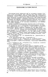
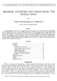
Text-Fig. 4. A. Nilssoniopteris-Yike bract with hairy petiole, Shin-Khuduk, la, x 1,5; В—E. Nilssoniopteris denticulata sp. nov., holotype,<br />
Bon-Tsagan, 45—19, basal portion of a leaf, x 1, marginal teeth, x 10, stomata in the abaxial intercostal band, x 300, and a stoma as seen<br />
from inside with SEM, x 2000; F. Nilssoniopteris denticulata sp. nov., narrow leaf with looping veins, Bon-Tsagan, 45—19, x 7.<br />
G. Pterophyllum cf. burejense PRYNADA, leaf fragment, Kholbotu-Gol, 197—27, x 1; H. Spongy mesophyll of the same leaf, x 300;<br />
I. Pterophyllum cf. angulatum HEER, irregularly segmented leaf, Gurvan-Eren, 236—29, x 1,5; J. Stoma of the same leaf, x 1200; K. Pterophyllum<br />
cf. sutchanense PRYN., leaf fragment, Modon-Usu, 2, x 1; L. Otozamites sp., detached pinnule, Gurvan-Eren, 236—29, x 2;<br />
M. Cycadolepis sp., bract showing hairy base and finely serrate margin above the middle, Bon-Tsagan, 23—22, x 4; N. Neozamites<br />
verchojanicus Vachr., detached pinnule, Bon-Tsagan, 45—19, x 1,5; O. Stomata of the same specimen, x 500.