EARLY CRETACEOUS FLORA OF MONGOLIA
EARLY CRETACEOUS FLORA OF MONGOLIA
EARLY CRETACEOUS FLORA OF MONGOLIA

You also want an ePaper? Increase the reach of your titles
YUMPU automatically turns print PDFs into web optimized ePapers that Google loves.
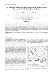
<strong>EARLY</strong> <strong>CRETACEOUS</strong> <strong>FLORA</strong><br />
<strong>OF</strong> <strong>MONGOLIA</strong><br />
BY<br />
VALENTIN KRASSILOV, Vladivostok")<br />
With 20 Plates, 11 Figures and 2 Tables in the Text<br />
Abstract<br />
The Lower Cretaceous lacustrine beds of Mongolia are rich in faunal remains, but the fossil plants are scarcely known.<br />
Rich plant localities were discovered recently by the Soviet-Mongolian geological expedition in the Central Mongolia (Bon-Tsagan<br />
Lake), Gurvan-Eren Range to the west, Mongolian Altai (Erdeni-Ula), Gobi Altai (Knolbotu-Gol, Khuriltu), and eastern Gobi<br />
(Tsagan-Tsab, Modon-Usu, Shin-Khuduk and Manlaj). In the central region, the plant-bearing beds belong in the Undur-Ukhin,<br />
Anda-Khuduk and Khulsyn-Gol Formations, while their correlatives in the western and eastern areas are assigned to the Gurvan-Eren,<br />
Tsagan-Tsab and Shin-Khuduk Formations.<br />
Four assemblage zones are recognized: (I) Baiera manchurica, (2) Otozamites lacustris — Pseudolarix erensis, (3) Baierella<br />
hastata — Araucaria mongolica, and (4) Limnothetis gobiensis — Limnoniobe insignis, Zones (I) and (2) are assigned to the<br />
Neocomian, (3) and (4) to the Aptian. These age assignments essentially agree with the invertebrate data.<br />
Plant megafossils came from the tuffaceous beds and aleuropelitic paper shales — varvites, deposited in meromictic lakes.<br />
Varvites contain abundant pollen grains in the lighter coloured lamellae. There are also coprolites and caddis fly cases with plant<br />
remains. Aquatic Lycopsida are abundant in the upper horizons, possibly evidencing the eutrophication of the lakes.<br />
At the beginning of the Cretaceous, the fern marshes have been drastically reduced (hence the paucity of ferns in the<br />
Cretaceous assemblages as contrasted with the Jurassic ones). Mud flats around the lakes were colonized by the hydrophylic<br />
bennettites (Otozamites) and presumably also by the primaeval reed- and sedge-like monocots. Other angiosperms represented by<br />
winged fruits (Gurvanella, Erenia) might grow on slopes occupied by the Pseudolarix forests. Araucaria — Brachyphyllum lowland<br />
community is conceived as the eastern extension of the European — Central Asian brachyphyllous forests, while some Siberian<br />
refugees, suposedly confined to the slopes, have given the Mongolian flora an ecotonal aspect.<br />
The Mongolian flora is peculiar in the unique association of archaic plants, such as Darneya and Swedenborgia, with early<br />
angiosperms. There are also endemic genera of the Lycopsida. Additional morphological information with some phylogenetic<br />
implications is provided for bennettites, Karkenia, Leptostrobus, conifers and pollen cones. Winged fruits and other angiosperm-like<br />
fossils may evidence a major radiation centre for angiosperms.<br />
The described species belong to the fungi, horse-tails, ferns, Lycopsida, Bennettitales, Ginkgoales, Czekanowskiales, conifers,<br />
angiosperms and angiosperm-like plants. New species are: Muscites ostracodiferus, Limnothetis gobiensis, Limnoniobe insignis,<br />
Nilssoniopteris denticulata, Otozamites lacustris, Ginkgoites mongolensis, Baierella hastata, Karkenia mongolica, Hartzia multifolia,<br />
Swedenborgia junior, Araucaria mongolica, Brachyphyllum densiramozum, B. sulcatum, Pseudolarix erensis, Schizolepis<br />
drepanoides, Pityospermum amplexum, Samaropsis aurita, S. sagitttata, Darneya angusta, Williostrobus latisaccus, Pityanthus<br />
microsaccus, Graminophyllum primum, Gurvanella dictyoptera, Erenia stenoptera, and Typhaera fusiformis. Five of them are also<br />
new genera: Limnothetis, Limnoniobe (lycopods), Gurvanella, Erenia (angiosperms) and Typhaera (incertae sedis). Lycopsida;<br />
Gymnospermae; Angiospermae; Plant Palaeoecology; Early Cretaceous; Mongolia.<br />
Preface<br />
Geological setting<br />
Contents<br />
2 Phytostratigraphy<br />
2 Palaeoecology . .<br />
') Address of the author: Institute of Biology and Pedology, Far Eastern Scientific Centre, 690022, Vladivostok 22, USSR.<br />
Palaeontographica Abt. B. Bd. 181 1<br />
4<br />
4
Phylogenetic comments<br />
Description<br />
Fungi .<br />
Bryophyta<br />
Horse-tails and ferns<br />
Lycopsida 8<br />
5 Bennettitales<br />
Ginkgoales<br />
6 Czekanowskiales<br />
6 Coniferales<br />
8 Angiosperms and angiosperm-like plants<br />
Preface<br />
The Lower Cretaceous beds of Mongolia, especially the paper shale facies are famous for their exceptionally<br />
rich fauna of crustaceans, insects and fishes. These beds have been extensively studied by the joint Soviet-<br />
Mongolian geological expedition in the area ranging from the Great Lakes in the west to the eastern Gobi. The<br />
principal outcrops are in the piedmonts of the parallel, north-west — south-east trending ranges of Khangai,<br />
Mongolian Altai and Gobi Altai. Plant fossils sporadically found in the paper shales have been mentioned in<br />
paleontological and stratigraphical papers (COCKERELL, 1924; SHUVALOV, 1975; MARTINSON, 1975). BALLA<br />
(1972) has listed 32 Mesozoic plant localities seven of which are presumably Cretaceous. A few plants have<br />
been properly described and illustrated (JAHNICHEN & KAHLERT, 1972), and these also are fragmentary. Much better<br />
preserved material was collected by palaeontologists of the Soviet-Mongolian expedition. In 1975 V. V. JAKOVLEV<br />
gathered numerous plant remains at Bon-Tsagan, Shin-Khuduk, Modon-Usu and Manlaj. In 1976 and<br />
1977 a party headed by Ju. A. POPOV, A. G. PONOMARENKO and S. M. SINITSA made fairly large collection at about<br />
twenty rich localities comprising several plant-beds each (PONOMARENKO & POPOV, 1976). A. G. PONOMARENKO<br />
and S. M. SINITSA kindly provided me with geological information.<br />
As the collections of the Soviet-Mongolian expedition were accumulated and studied, it became clear that<br />
we were dealing with a new major fossil flora which deserved detailed treatment. Plants from the Lower<br />
Cretaceous localities are described in this paper. The Jurassic and Late Cretaceous plants will be described<br />
elsewhere.<br />
Geological setting<br />
The Lower Cretaceous fossiliferous beds lay on the coarse-grained Upper Jurassic red beds. In the eastern<br />
Gobi, the lower horizons — the Tsagan-Tsab Formation — are tuffaceous conglomerates, sandstones, shales<br />
and marls interbedded with andesitic and basaltic tuffs. They are overlain by the bituminous aleuropelitic paper<br />
shales, marls and dolomitic lacustrine limestones — the Shin-Khuduk Formation (MARTINSON, 1975). In the<br />
central Mongolia (Khangai Range, Mongolian Altai, Bon-Tsagan Lake), the lower horizons are known as the<br />
Undur-Ukhin Formation, correlated with the Tsagan-Tsab. Thick sequences of the bituminous paper shale<br />
cyclites with occasional coal beds constitute the Anda-Khuduk Formation (SHUVALOV, 1975), obviously<br />
corresponding to the Shin-Khuduk in the east. The uppermost part of the Lower Cretaceous sequence — the<br />
Khulsyn-Gol Formation — is again coarse-grained, lighter coloured and tuffaceous. In the western region, the<br />
tuffaceous Tsagan-Tsab — Undur-Ukhin beds and the paper shales are replaced by monotonous sequence of<br />
gray, variagated or red sandstones, clays and marls — the Gurvan-Eren Formation (KHOSBAYAR, 1973).<br />
On the evidence of invertebrates and fishes, the Tsagan-Tsab level is assigned to the Neocomian, the<br />
Shin-Khuduk — to the late Neocomian-Aptian and the Khulsyn-Gol — to the Aptian-Albian.<br />
The most complete sequence of plant beds is in the central region, to the south of the Bon-Tsagan lake.<br />
The sequence is as follows (the dominant plants are marked with an asterisk):<br />
1. Sandstones, locality 2, with Baiera manchurica YABE et OISHI, Podozamites sp. and Brachyphyllum<br />
densiramosum sp. nov.<br />
2. Aleuro-argillites, locality 23-22, with Onychiopsis psilotoides (STOKES & WEBB) WARD, Otozamites<br />
lacustris sp. nov., ;: ' Cycadolepos sp., Podozamites sp., Brachyphyllum densiramosum sp, nov.,"' B. sulcatum<br />
sp. nov., Pseudolarix erensis sp. nov. (shoots and seeds), Pityanthus microsaccus sp. nov. Schizolepis drepanoides<br />
sp. nov.<br />
3. Green-gray and black aleuropelitic shales interbeded with marls and limestones, localities 73—3,9; 74—3,<br />
11<br />
16<br />
20<br />
22<br />
31
75—5, 87—8, 193—3, 194—1 with cf. Cyathea tyrmensis (SEW.) KRASSIL., Cladophlebis sp., Podozamites sp.,*<br />
Baierella hastata sp. nov.,* Darneya angusta sp. nov., Brachyphyllum densiramosum sp. nov.,* Araucaria monglica<br />
sp. nov.* (shoots and cones), Williostrobus latisaccus sp. nov., Samaropsis aurita sp. nov., Pityanthus microsaccus<br />
sp. nov., Pityospermum amplexum sp. nov.,* Typhaera fusiformis sp. nov.<br />
4. Gray aleuropelites and sandstones, principal locality 45—19, with Equisetum cf. laterale PHILL.,<br />
cf. Osmunda diamensis (SEW.) KRASSIL., cf. Dicksonia concinna HEER, Limnothetis gobiensis sp. nov.,* Limnoniobe<br />
insignis sp. nov.,* Nilssoniopteris denticulata sp. nov., Neozamites verchojanicus VACHR., Sphenobaiera ikorfatensis<br />
(SEW.) FLORIN, Hartzia multifolia sp. nov., Phoenicopsis angustifolia HEER, Podozamites sp., Darneya angusta<br />
sp. nov., Brachyphyllum densiramosum sp. nov.,* Araucaria mongolica sp. nov.*, Pityantus microsaccus sp. nov.,<br />
Pityolepos sp., Pityospermum amplexum sp. nov.*. Minor localities in these beds, 33, 34 and 60 contain: cf.<br />
Dicksonia concinna HEER, Limnothetis gobiensis sp. nov. and putative gametophytes of this plant,* Karkenia<br />
mongolica sp. nov., Sphenobaiera ikorfatensis (Sew.) Florin, Hartzia multifolia sp. nov., Leptostrobus sp.,<br />
Brachyphyllum densiramosum sp. nov.,* Araucaria mongolica sp. nov.,* Pityospermum amplexum sp. nov.*<br />
Beds (1) — (2) are assigned to the Undur-Ukhin Formation, (3) — to the Anda-Khuduk Formation, and (4) —<br />
to the Khulsyn-Gol Formation.<br />
To the south, in the Gobi Altai, fossil plants came from the Anda-Khuduk Formation outcropping at the<br />
Kholbotu-Gol River and Khuriltu. Localities 196—75, 197—27, 199—75, 210—29 216—12 in the gray auleropelitic<br />
shales contain cf. Osmunda sp., Limnothetis gobiensis sp. nov. Pterophyllum burejense PRYN., Podozamites sp.<br />
Brachyphyllum densiramosum sp. nov.,* Araucaria mongolica sp. nov.,* Samaropsis aurita sp. nov., Pityophyllum sp.,<br />
Pityanthus microsaccus sp. nov., Pityospermum amplexum sp. nov.,* Typhaera fusiformis sp. nov.<br />
To the south-west of the Bon-Tsagan lake, in Mongolian Altai, near the Erdeni-Ula Mountain, the gray<br />
aleurolites and yellowish-white tuffites representing lower horizons of the Khulsyn-Gol Formation contain<br />
localities 213—23, 25, 28 which yielded Muscites ostracodiferus sp. nov.,* cf. Dicksonia concinna Heer, Baierella<br />
hastata sp. nov.,* Karkenia monglica sp. nov.,* Podozamites sp., Brachyphyllum densiramosum sp. nov. (twigs<br />
and cones), Araucaria mongolica sp. nov., Pityophyllm sp.<br />
Further to the west, in the Gurvan-Eren Range, yellow shales of Gurvan-Eren Formation, locality 236—29,<br />
yielded a fungus cap, the gametophytes of unknown plants, Eqiusetum sp., Otozamites sp., Pterophyllum<br />
cf. acutilobum HEER, Brachyphyllum densiramosum sp. nov.,* B. sulcatum sp. nov., Pseudolarix erensis sp. nov.*<br />
(twigs, cone scales and seeds), Pityolepis sp., Problematospermum sp., Tyhpaera fusiformis sp. nov., and the<br />
angiosperm fruits Gurvanella dictyoptera sp. nov. and Erenia stenoptera sp. nov.<br />
In the eastern Gobi, a few plants have been collected by YAKOVLEV from the tuffaceous Tsagan-Tsab shales<br />
at Tsagan-Tsab. They are mostly Baiera manchurica YABE & OISHI* and Hartzia multifolia sp. nov. Other<br />
localities of presumably Tsagan-Tsab level are at the Modon-Usu Creek and Manlaj. Yellow sandstones at<br />
Modon-Usu, locality 2, contain Nilssoniopteris denticulata sp. nov., Pterophyllum cf. sutschanense PRYN.,<br />
Baiera manchurica YABE & OISHI* (in the fish bed), Podozamites sp.,* Swedenborgia cf. tyttosperma STANISL.<br />
Manlaj is especially important because of the angiosperm-like fossils Graminophyllum primum sp. nov.,*<br />
Cyperacites sp., Sparganium-like and Potamogeton-like fructifications which came from the gray shales with<br />
chironomid remains in the localities 2, 3 and 95—37. Other plants from localities 92—12, 14, 16, 95—10b, are<br />
Equisetostachys sp., Selaginella sp., cf. Pachypteris sp., Otozamites lacustris sp. nov.,* Ginkgoites sp.,<br />
Sphenobaiera sp., Leptostrobus sp., Podozamites sp., Pseudolarix erensis sp. nov.,* (a cone scale and seeds),<br />
Samaropsis aurita sp. nov. (a single germinated seed) and a pollen cone with unripe pollen grains.<br />
The Shin-Khuduk paper shales and their sandy interbeds at the Shin-Khuduk Creek, localities I, la, 117,<br />
118—1,6 8a-c, 119—3c,d, 7a, 8a and Modon-Usu Creek, locality 132—34 contain Equisetum sp., Osmunda sp.,<br />
Cladophlebis cf. denticulata (BRONGN.), cf. Cyathea tyrmensis (SEW.) KRASSIL., Neozamites verchojanicus<br />
VACHR., Cycadolepis sp. (cf. Nilssoniopteris denticulata sp. nov.), Ginkgoites mongolicus sp. nov.,* Sphenobaiera<br />
ikorfatensis (SEW.) FLORIN, Baierella hastata sp. nov.,* Karkenia mongolica Krassil.,* Podozamites sp.,*<br />
Swedenborgia junior sp. nov., S. cf. longiloba STANISL., Araucaria mongolica sp. nov.,* Brachyphyllum<br />
densiramosum sp. nov.,* Pityophyllum sp., Williostrobus latisaccus sp. nov., Pityanthus microsaccus sp. nov.,<br />
Stenomischus sp., Samaropsis aurita sp. nov.,* S. sagittata sp. nov., Pityospermum amplexum sp. nov.,*<br />
Problematospermum sp. (a pappus only). There are also abundant pollen grains, mostly belonging to Williostrobus<br />
latisaccus, Pityanthus and Darneya.
Phytostratigraphy<br />
In Mongolia, the Early Cretaceous localities contain sparse fern fragments only while the Jurassic floras are<br />
dominated by ferns. These types of assemblages appear to be easily distinguishable, but the fern criterion needs<br />
further practical corroboration. The fern beds may comprise also the lowermost Cretaceous. Czekanowskia is<br />
abundant in the Jurassic, but has not hitherto been recorded from the Cretaceous.<br />
Four phytostatigraphic units can be provisionally recognized in the Cretaceous:<br />
(1) The Baiera manchurica zone represented at Tsagan-Tsub, Modon-Usu and Bon-Tsagan. Baiera<br />
manchurica is common in the Urgal Formation (Valanginian) of the Bureja basin (KRASSILOV, 1972).<br />
(2) The Otozamites — Pseudolarix erensis zone, comprising the localities Bon-Tsagan, 23—22,Gurvan-Eren<br />
236—29 and Manlaj 2,3 and 92. Apart from Otozamites and Pseudolarix, the correlation of Bon-Tsagan, 23—22,<br />
and Gurvan-Eren is supported by Brachyphyllum sulcatum, while Gurvan-Eren and Manlaj contain the angiosperm<br />
fossils. This community has no close analogues outside Mongolia. The flora is of generalized Wealden<br />
aspect and does not seem younger than Neocomian.<br />
(3) The Baierella hastata (and its cones Karkenia mongolica) — Araucaria mongolica zone includes localities<br />
of the Shin-Khuduk — Anda-Khuduk level, mostly in the paper shale facies at Bon-Tsagan, 73, 74, 75, 87, 193,<br />
194, Kholbotu-Gol 196, 197, 199, 216, Khuriltu, 210, Erdeni-Ula 213, Shin-Khuduk, 1, 117, 118, 119, and<br />
Modon-Usu, 132. Baierella hastata is closely related to B. uninervis (SAMYL.) KRASSIL. from the Tschemchuco<br />
(Aptian) beds of the Bureja basin and Ginkgoites mongolica also represent a leaf morphotype common in the<br />
Tschemchuco beds. Neozamites occurs in the Aptian of Jacutia and Primorye. This community is comparable<br />
with the Aptian flora of Primorye (KRASSILOV, 1967) on account of Araucaria and other conifers.<br />
(4) The Limnothetis-Limnoniobe zone is confined to the Khulsyn-Gol Formation at Bon-Tsagan<br />
45—19. It is destinctive on account of the mass occurrence of endemic cryptogamous plants (new genera).<br />
However the gymnospermous species are the same as in tne third zone, suggesting the age not younger than<br />
Aptian.<br />
Palaeoecology<br />
The Early Cretaceous flora of Mongolia is ecotonal between the temperate (Siberian) and subtropical<br />
vegetation comprising such genera as Phoenicopsis, dominant in the Siberian realm, and Otozamites, typical<br />
representative of the Ptilophylletum communities. Judging by their frequencies, Siberian plants might have grown<br />
on slopes, while subtropical plants dominated the lowland vegetation.<br />
The Mongolian flora differs from both Bureja (temperate) and Primorye (subrtropical) floras by much lower<br />
fern content. Irrespective of their actual frequence in a flora, ferns are rare fossils except in the fern marshes. The<br />
abundance of ferns in the Jurassic localities of Mongolia suggests fairly extensive fern marshes on the deltaic mud<br />
flats. At the beginning of the Cretaceous, the fern marshes had been reduced and the fern fossils became rare.<br />
This happened sumultaneously with the development of meromictic lakes and deposition of seasonal varvites<br />
— the paper shales.<br />
Some reduction of forest vegetation can also be assumed. In the Manlaj localities, conifers are represented<br />
mostly by winged seeds and cone scales. It appears that a conifer forest was at some distance from the lake,<br />
presumably on slopes, while on the flat shores grew the primaeval reed- and sedge-like monocots. They possibly<br />
colonized the mud flats exposed when the lake shranked during a dry season. The appearance of angiosperm<br />
fossils in the lower horizons while they are lacking in the overlain strata, seems paradoxical, but it can be related<br />
to the reduction of indigenous Mesozoic vegetation. A bennettite Otozamites lacustris accompanied them, and<br />
judging by the leaf anatomy (see below), it was adapted to periodical submergence. In Bon-Tsagan, 23—22, the<br />
conifers are better represented, and in Gurvan-Eren, Pseudolarix erensis is most aundant and Brachyphyllum is<br />
also common. The Pseudolaix forest grew perhaps on nearby slopes. An angisperm Gurvanella might also occur<br />
on slopes, because the winged fruits only were fossilized. At the close of the Neocomian, the<br />
Otozamites-angiosperm community was replaced by Baierella-Podozamites shrubs, and the forest vegetation also<br />
underwent considerable changes. Araucaria mongolica and Brachyphyllum densiramosum are abundant in<br />
practically all Aptian localities. They are represented by shoots, microstrobili Williostrohus latisaccus,<br />
Stenomischus sp., megastrobili and seeds (Samaropsis). The conifer forests dominated by Brachyphyllum and
iraucaria were apparently proximal to the sites of deposition. These communities can be conceived as a continuaon<br />
of the European-Central Asian brachyphyllous forests (i.e., characterized by the Brachyphyllum shoot morhology<br />
assumed convergently by conifers of different affinites, mostly the Araucariaceae, Taxodiaceae and Chei-<br />
Dlepidiaceae). The brachyphyllous forests extended through Mongolia to Primorye and Japan. The northern limit<br />
f Araucaria was at the Tyrma River, 50° North (KRASSILOV, 1978 a). The expansion of brachyphyllous forests in<br />
Lptian time can be related to the putative amelioration of climate.<br />
Another type of the forest vegetation is represented by prolific samaras Pityospermum amplexum of Abietasous<br />
affinities. There are also less numerous leaves (PityophyHum) and microstrobili (.Pityanthus), presumably of<br />
le same plant. The abundance of samaras and their even distribution in major localities suggest an extensive slope<br />
)rests dominated by an Abietaceous conifer mixed with a few representatives of the Siberian floristic realm, such<br />
> Phoenicopsis angustifolia which appeared in the fossil record at the close of the Aptian.<br />
In the Shin-Khuduk varvites, the lighter coloured lamellae (spring layers) are densely covered with pollen<br />
rains distinguishable under low magnification. I picked these pollen grains by needle and after a few minutes<br />
sposure to HCL and HN03 counted them to obtain a rough estimate of the contribution of various conifers to<br />
le pollen rain (the figures obtained by this technique are presumably less biased than in the standart palynoloical<br />
procedure dealing with volumes rather than surfaces). The numerical representation per 100 pollen grains is<br />
> follows:<br />
ytyanthus microsaccus (Pinaceae) — 40,<br />
yilliostrobus latisaccus (Araucaria mongolica) — 36,<br />
darneya angusta (Podozamites) — 6.<br />
For the remaining 18, the megafossils were not recognized. The rise of aquatic or semiaquatic Limnothetis<br />
id Limnoniobe at the close of the Aptian (Bon Tsagan, 45—19) can be related to eutrophication of the lakes,<br />
lass occurrence of the Limnothetis gametophytes suggests that the megaspores germinated in water and the<br />
imetophytes floated before settling on a muddy ground.<br />
There is much evidence of the animal-plant interactions. The most interesting are the caddis fly cases built of<br />
arkenia seeds (Text-Fig. 7J). According to SUKATSHEVA (in press), these cases rank among the most sophisticated<br />
ructures ever built by caddis flies. The inner tube is made of fusenized plant tissues, resin bodies of Karkenia seeds<br />
id occasional megaspores of Limnothetis. The outer sheath is built of the Karkenia seeds which are very carefully<br />
id, their micropyles facing outside. The Karkenia seeds have been produced by a plant with Baierella hastata leaves,<br />
is conceivable that Baierella grew in thick bushes along the shores, shedding abundant seeds in water. Other caddis<br />
ies have occasionally used the anthers of Pityanthus (PL 17, fig. 228).<br />
The cuticles and rasin bodies of Karkenia found in coprolites evidence the feeding of birds or small reptiles<br />
i these fleshy seeds. The samaras appear less attractive for seed-eaters, but there is a single coprolitic body<br />
Э mm in diameter with four damaged seeds of Samaropsis aurita (PL 14, fig. 174).<br />
Plant fossils resembling aquatic angiosperm fructifications are known from the Manlaj locality only. This<br />
>cality is also especially rich in chironomid remains which literally cover the bedding planes. Some functional<br />
Jations between the unusual diversity of aquatic plants and the abundance of chironomids may be<br />
iggested by the role of chironomid larvae in the phosphorus cycle. In modern eutrophic lakes, the phosphorus<br />
mtent of water is increased by chironomids (GALEPP, 1979).<br />
Phylogenetic comments<br />
In the Early Cretaceous flora of Mongolia, Swedenborgia and Darneya, hitherto recorded mostly from the<br />
riassic, associate with primaeval angiosperms and peculiar endemic plants. One can assume that the environlents<br />
in Mongolia were favorable for both survival of relics and production of new forms in various plant<br />
-oups.<br />
Limnothetis with solitary sporangia terminal or pseudoterminal on specialized short-shoots constitute another<br />
sample (complimentary to Synlycostrobus KRASSILOV, 1978 b) of evolutionary experimentation in the Mesozoic<br />
ycopsida, leading to reproductive structures analogous to those of conifers. Limnoniobe represents hitherto<br />
irecorded group of Mesozoic heterosporous plants possibly related to the Pennsylvanian Polysporia.<br />
New data on the leaf anatomy of a bennettite Otozamites lacustris with aerenchymous spongy mesophyll suggest
hydrophytic adaptation. In the vascular bundles of the leaves there are members with perforated ends, suggesting<br />
the development of vessels in bennettites (PL 6, figs. 62—65).<br />
The association of the Karkenia megastrobili with several Ginkgoalean leaf morphotypes is supported by the<br />
new finds in Mongolia. Now Karkenia is known to associate with Ginkgoites, Sphenobaiera and Baierella.<br />
Leptostrobus (Czekanowskiales) with papillate marginal fringe is found for the first time outside the Bureja<br />
basin in the Far East of the USSR. There is evidence of a secretory function of the papillae, supporting my interpretation<br />
of the marginal fringe as a stigmatic structure. Stomatal apparatus in the Czekanowskiales might be of<br />
mesoperigenous type.<br />
Swedenborgia and Cycadocarpidium, associating with Podozamites in the Triassic and Liassic localities, were<br />
not hitherto recorded from younger Mesozoic stata, and the reproduction in the younger Podozamites conifers<br />
remained enigmatic. In Mongolia at least three kinds of Swedenborgia associate with Podozamites as well as the<br />
pollen cones of the genus Darneya. The latter are also prominent in the Triassic. GRAUVOGEL-STAMM (1978) has<br />
assigned Darneya to the Voltziaceae while Podozamites is conventionally referred to the Cycadocarpidiaceae.<br />
However Cycadocarpidium and Swedenborgia are sufficiently similar to the Voltziaceous seed-scale complexes to<br />
keep them in the same family. At least in one line of this family, the male and female reproductive structures have<br />
been fairly constant through 100 m.y.<br />
The conifers with Brachyphyllum leaves present a difficult classification problem. In some European<br />
localities, most of them belong to the Classopollis-producing Cheirolepidiaceae. Some of the dominant Mongolian<br />
brachyphylls are assigned to the Araucariaceae (on the evidence of microstrobili with pollen grains as well as<br />
megastrobili). Other may belong in the Classopollis-producing group or the Taxodiaceae.<br />
The cone scales of Pseudolarix erensis occasionally resemble those of Schizolepis suggesting possible phylogenetic<br />
links. Otherwise P. erensis is fairly modern-looking. Mongolia was also the home of other progenitors of<br />
the modern Pinaceae.<br />
Gurvanella and Erenia rank among the earliest angiosperms supporting the idea of the Central Asian origin<br />
of this group (KRASSILOV, 1977). The angiosperm-like fossils from Manlaj may evidence the initial radiation of<br />
aquatic monocots in which the reproductive structures characteristic of various orders have been created by a few<br />
megaevolutionary steps. However caution is needed in dealing with these fossils which are so poorly known at<br />
present.<br />
Fungi<br />
PL 1, fig. 15<br />
Remarks: A single specimen of a cap, 1 cm in diameter, show the undulating margin and numerous radial<br />
lamellae joined to the centrally attached stalk. Systematic position of this specimen is unclear, yet it is of some<br />
interest as the first find of Hymenomycetalean fungi in the Mesozoic.<br />
Locality : Gurvan-Eren, 236—29.<br />
Bryophyta<br />
Muscites ostracodiferus sp. nov.<br />
PI. 1, figs. 7—11, Text-Fig. 1A—С<br />
Holotype: Erdeni-Ula, 213—25, N 3787/500, PL 1, Fig. 5.<br />
Diagnosis : Shoots with a slender axis (0,3 mm), branching at acute angle, side branches reflexed, alternate, at<br />
interval of 8—10 mm, up to 30 mm long, mostly branching at acute or right angle. Leaves are sparse on the main<br />
Text-Fig. 1. A—C. Muscites ostracodiferus sp. nov., leafy shoots in association with ostracod shells, Erdeni-Ula, 213—25, x 7; D. Equisetostachys<br />
sp., polygonal shields of the sporangiophores, Manlaj, 92—12b, x 7; E, F. Equisetum sp., leaf whorls, Bon-Tsagan, 45—19, x 2.<br />
G, H. Equisetum sp., stems showing leaf commissures, Bon-Tsagan, 45—19 and Gurvan-Eren, 236—19, x 2; I. cf. Cyathea tyrmensis<br />
(SEW.) KRASSIL., pinna fragment, Shin-Khuduk, 118—8c, x 2; J. cf. Onychiopsispsilotoides (STOKES et WEBB) WARD, apical portion of a<br />
pinna, Bon-Tsagan, 23—22, x 2; K. cf. Cladophlebis denticulata serrate pinnules, Shin-Khuduk, 118—8c, x 2; L. cf. Osmunda diamensis<br />
(SEW.) KRASSILOV., pinnules slightly constricted at the base, Bon-Tsagan, 45—19, x 2; M. OsmundaAike pinnule, Shin-Khuduk, 119—3 c,<br />
x 2; N. Sphenopteroid pinnules, Shin-Khuduk, 118—8b, x 7.
axis, dense on the side branches, with internodes 0,7 mm (measured by leaf scars), spiral, decurrent, obliquely<br />
spreading, straight or reflexed, crowded and appressed at the apex, about 5—6 x 0.5—0.6 mm. Distal leaves are<br />
longer than the proximal. The leaf blade is narrow-lanceolate, expanded and sheathing at the base, gradually<br />
tapering to the apex, folded, margin entire. The costa is narrow, filling the apex. An apical hair is about 2 mm<br />
long.<br />
Remarks : Abundant remains of this moss associate with the spiny ostracod shells which look as if they were<br />
attached to the shoots by their spines. The moss is comparable to Muscites fontinalioides KRASSILOV (1973) from<br />
Bureja, but larger.<br />
Locality : Erdeni-Ula, 213—25.<br />
Horse-tails and ferns<br />
PL 1, figs. 12—14, Text-Fig. IE—N<br />
Remarks : The horse-tail and fern fossils are too fragmentary for systematic treatment. A horse-tail with long<br />
leaves of Equisetum laterale type (HARRIS, 1961) is found in Bon-Tsagan, 45—19, while smaller specimens with<br />
shorter leaves occur in Shin-Khuduk 118—8c and Gurvan-Eren, 236—29. A single specimen of Equisetostachys<br />
came from Manlaj (PL 1, fig. 12).<br />
Sphenopteroid ferns from Bon-Tsagan 23—22 are comparable with Onychiopsis psilotoides (STOKES & WEBB)<br />
WARD (Text-Fig. 1J) while a few fragments from Bon-Tsagan 45—19, 87—8 and Shin-Khuduk 118—86 with<br />
delicate pinnules showing unbranched veins resemble Cyathea tyrmensis (SEW.) KRASSILOV (1978b).<br />
Cladophleboid ferns with basally constricted pinnules, cf. Osmunda diamensis (SEW.) KRASSILOV came from<br />
Bon-Tsagan, 45—19, 87—8 and Shin-Khuduk, 119—8a (Text-Fig. 1L). Another Osmunda-like pinnule with<br />
oblique, forking and occasionally anastomosing veins was found in Shin-Khuduk 119—3c (Text-Fig. 1M). Serrate<br />
pinnules with once-forking veins, cf. Cladophlebis denticulata (BRONGN.) are from Shin-Khuduk 118—8c (Text-<br />
Fig. 1L).<br />
Lycopsida<br />
Limnothetis, gen. nov.<br />
Type species: Limnothetis gobiensis sp. nov.<br />
Name: Alluding to the extra care for a single offspring.<br />
Diagnosis : Shoots creeping, forking equally at base, unequally above, pseudomonopodial. Lateral branches<br />
distichous, alternate. Leaves ligulate, on the main axis monomorphous, two-ranked, on lateral branches<br />
dimorphous, four-ranked; the ventral leaves broadly ovate, imbricate; the dorsal leaves small, pointed.<br />
Adventitious roots in the axils of the ventral leaves. Fertile zones of crowded short shoots bearing a single<br />
terminal sporangium protected by imbricate bracts. Sporangia dimorphous, 1.2 x 1 mm and 1 x 0.6 mm.<br />
Microspores trilete, smooth.<br />
Remarks : The dimorphism of sporangia suggests heterospory, but spores were extracted from smaller<br />
sporangia only. The megaspores are found in the rock together with fertile shoots, but not in situ.<br />
Sterile shoots of this plant resemble Selaginella. In some species of Selaginella, sporangia are also in fertile<br />
zones. But in Limnothetis the sporangia seem terminal on fertile brachyblasts with a few bracts. Actually they<br />
might be pseudoterminal, in the axils of apical bracts. In either case, the unisporangiate brachyblasts differ from<br />
any sporangiophores of Selaginella or other members of the lycopsida.<br />
Limnothetis, gobiensis sp. nov.<br />
PI. 2, figs. 16—24, PI. 3, figs. 25—31, Text-Fig. 2<br />
Holotype: Bon Tsagan, 45—19, N 3559/10001. PL 2, fig. 17.<br />
Diagnosis: As for the genus.<br />
Description: A massive body with root scars in PL 2, fig. 16 is a rhizome bearing 6 shoots, their basal<br />
portions straight, 1 mm thick, with sparse leaves. About 30 mm above the base, the shoots fork, giving rise to
lsely leaved branches. Second branching is at interval of 1—3 mm, so that the branches seem whorled.<br />
sequent forking is unequal, giving the shoot a pseudomonopodial aspect, with the main axis straightened<br />
1 the lateral branches distichous. The lateral branches are at acute angle to the main axis, some of them bent<br />
:kwards, at interval of 10 mm in the middle portion of a shoot, but more crowded at the apex. Leaves on the<br />
in axis and at the angle of branching are broadly ovate or rhomboidal, about 3—4 x 2—2.5 mm, imbricate,<br />
:h the obliquely decurrent, clasping base and the attenuate, hair-like apex, funnel-shaped or folded along the<br />
n, flabellately striated, showing the ligular scar as a minute pit at the base (PL 2, fig. 24). Leaves on the lateral<br />
nches are dimorphous, four-ranked (PL 2, figs. 17, 18): those in the ventral ranks like on the main axis, the<br />
:sal ones small, about 1 mm long, not always discernible. Adventitious roots, when preserved, attached in the<br />
Is of the ventral leaves. Rounded bodies, 2—7 mm in diameter, with a thick folded cuticle associated with the<br />
>ots, might be tubers.<br />
;t-Fig. 2. Limnothetis gobiensis sp. nov., reconstruction of a fertile zone.<br />
In the fertile portion of a shoot, the specialized fertile brachyblasts (sporangiophores) are in the axil of each<br />
ltral leaf, crowded, overlapping, arranged distichously, alternate. The sporangiophores are elliptical, about<br />
nm long, consisting of a short axis bearing a single terminal sporangium and about 10 appressed bracts which<br />
se above the sporangium (PL 2, figs. 20, 21).<br />
Sporangia are mostly elliptical, 1 x 0.6 mm, containing numerous small spores (10 sporangia were macerated<br />
spores). Occasional sporangia are considerably larger, 1.2 x 1 mm, rounded and appear more bulky (PL 2,<br />
. 22). They are limonized, not yielding to maceration.<br />
Spores are about several hundred per sporangium, trilete, amb rounded, diameter 44—60 mm, leasurae<br />
sed, arching, extending to the amb. Exine thin, trilete, folded. Many spores are folded along two leasurae so<br />
.t they seem elliptical, with pointed ends (PL 3, fig. 31).<br />
Dispersed organs associated with Limnothetis gobiensis sp. nov.<br />
Megaspores found on the rock surface in close association with the shoots (PL 3, figs. 25, 26, 29) are all of one<br />
id, spherical, diameter 600-800 jLtm, amb rounded, fringe (arcuate lamellae) about 50 /лт wide, trilete ridges<br />
ending to equator. Exine is reticulate, with polygonal meshes 7—8 /лт wide, thickened at the corners, elongate<br />
i passing into concentrical ridges in the fringe. The exine is also ornamented with spines, more numerous on the<br />
tal face than on the proximal. The spine stumps are rounded, set on the corners of the reticulum, occasionally<br />
ising concentrical arrangement of the meshes.<br />
There are also numerous reniform bodies, sometimes covering the rock surface, about 1 mm in diameter,<br />
lvex, transversed by a median groove (PL 1, figs. 1—6). On transfer preparations these bodies show polygonal<br />
;ontographica Abt. B. Bd. 181 2
meshes 7—8 mm wide, resembling the reticulum of the megaspores. I believe these bodies to be floating<br />
megagametophytes of Limnothetis.<br />
Locality : Bon Tsagan.<br />
Limnoniobe, gen. nov.<br />
Type species: Limnoniobe insignis, sp. nov.<br />
Name: Alluding to so many sacrificed offsprings — the abundant fossil megaspores.<br />
Diagnosis: Plants with thick (30 mm) stem showing spirally arranged leaf- and root-scars. Leaves<br />
spreading radially from the stem, ribbon-shaped, 70 x 7 mm, venation immersed, stomata absent. Subepidermal<br />
parenchyma of very large polygonal cells. Megasporangia supposedly in fertile zones, large, globose. Megaspores<br />
about 800 fim in diameter, reticulate, distally spinose, proximally verrucose, showing triradiate mark with<br />
auriculae.<br />
Remarks:- Sporangia were not found in organic connection with the leafy stems. However they are<br />
reasonably attributable to the same plant on the evidence of close association of the shoots, sporangia and<br />
megaspores, often superimposed on the leaves. Sporangia and megaspores crowded around the transversly broken<br />
stems suggest fertile zones of the type known in Lycopodium, Selaginella, Spencerites, Sporangiostrobus and Polysporia.<br />
The Mongolian plant is comparable with the Nathorstiana — Stylites — Isoetes group on one hand and the<br />
Paleozoic Spencerites — Polysporia on the other. It is more close to Polysporia in the stem diameter, flattened leaves<br />
and auriculate megaspores. The leaf structure is remarkably similar showing large-celled mesophyll and much<br />
smaller tabular epidermis cells. The Polysporia shoots often break at fertile zones (DIMICHELE et al., 1979). In the<br />
light of these similarities, Limnoniobe can be conceived as the Mesozoic descendant of the Polysporia group<br />
differing from all other Lycopsid gernera in ribbon-shaped leaves. The mode of preservation of the very long leaves<br />
spreading radially from the stems suggests an aquatic plant.<br />
Limnoniobe insignis sp. nov.<br />
PL 4, figs. 32—42; PL 5, figs. 43—53; Text-Fig. 3<br />
Holotype : Bon-Tsagan, 45—19, N 3559/10000, PL 4, fig. 39.<br />
Diagnosis: As for the genus.<br />
Description : Stem fragments are up to 50 mm long (entire length unknown), 30 mm in diameter. They<br />
are preserved as compression or transverse cuts. The stem epidermis, preserved on a single specimen (PL 4, fig. 37),<br />
shows polygonal cells (of the same dimensions as in the leaf epidermis), transverse whrinkles and circular root<br />
scars, 0.3 mm in diameter, arranged spirally at 1 mm in parastichies. Leaf scars are variable, their form presumably<br />
depending on the portion of a stem preserved. They are either rounded-rhomboidal, 2.5—3 mm wide, irregularly<br />
spaced, or elongate, in more regular parastichies. Some of them show a single leaf trace. In the upper portion of a<br />
stem, the leaves are more crowded, imbricate, forming a shallow funnel. The leaves are ribbon-shaped more than<br />
70 mm long, 5—7 mm wide, margins entire, parallel, converging gradually to the base. The leaves are seemingly flat,<br />
Text-Fig. 3. Limnoniobe insignis sp. nov., a tentative reconstruction.
ut might be fleshy, with immersed veins, normally invisible. A specimen in which the epidermis is partially decayed<br />
exposing subepidermal tissues shows a mid vein 0.5 mm thick, bordered by the narrow sheating cells. The parenchymous<br />
tissue forms two rows of meshes along the midvein (PL 5, figs. 47, 48).<br />
The calcite incrustations of the leaves were studied with SEM (PL 5, fig. 45). They show epidermis and<br />
subepidermal spongy tissue. The epidermal cells are rectanguloid, squarish or polygonal, stretched transversely or<br />
isodiametrical, about 80 /лm wide. The transversely stretched and isodiametrical cells are in alternating zones. The<br />
subepidermal tissue is a honeycomb of very large polygons 250 /лт wide suggestive of aerenchyma.<br />
Hand-specimens with stems and leaves are often strewn with megaspores (PL 4, figs. 38, 39). Close to the<br />
transverse break of a stem there are globose megaspore masses 7 mm in diameter which are apparently the fills of<br />
megasporangia each containing about 60 megaspores. There are also spherical bodies of the same dimensions<br />
showing radial rows of rectanguloid and polygonal cells similar to the epidermal cells of the leaves (PL 4, fig. 42).<br />
These bodies might be megasporangia, but no megaspores were found in situ. The megaspores, preserved as casts<br />
and compressions, are spherical, amb rounded-triangular, diameter 700—1000 /лт, fringe about 200 /лт wide. The<br />
leasurae are raised, straight or somewhat undulating, reaching to equator and extending on the fringe as low ridges.<br />
The ends of triradiate mark are expanded into the bulbous auriculae as in Valvisisporites and Minerisporites. A few<br />
specimens show reticulum with rounded-polygonal meshes (PL 5, fig. 52). In most megaspores the reticulum is<br />
obliterated leaving verrucose sculpturing of the contact facets. The distal wall is spinose, showing a few large spines,<br />
about 30 /лт in diameter, and numerous smaller spinules between them. The fringe is radially folded.<br />
Some megaspores show numerous microspores stuck to their surface (PL 5, fig. 50, 51). In Selginella microspores<br />
often stick to the megaspores. One can surmise that in Limnoniobe the same was the case. Masses of similar<br />
microspores have been obtained by bulk maceration of the rock containing megaspores.<br />
Locality : Bon-Tsagan, 45—19.<br />
Gymnospermae<br />
Bennettitales<br />
Nilssoniopteris denticulata sp. nov.<br />
PL 7, figs. 71—74, Text-Fig. 4В—F<br />
Holotype : Bon-Tsagan, 45—19, N 3559/10002, PL 7, fig. 71.<br />
Diagnosis : Leaf blade attached adaxially leaving the median portion of a rachis exposed. Margin toothed.<br />
Veins mostly simple, opposite, straight, upturned abruptly near the margin, ending in the teeth, occasionally<br />
looping. Stomata transverse or oblique, paracytic, about 27 /лт wide.<br />
Description: The largest specimen (holotype) is the posterior portion of a leaf 40 mm wide, narrowing<br />
gradually to a short petiole. Rachis is stout, 3.5 mm wide, its median stripe 1 mm wide is exposed between the<br />
halves of the leaf blade. Lateral veins are thick, fairly distinct, opposite, arising at interval of 2 mm, at right angle to<br />
the rachis, simple. Close to the margin, the veins are upturned terminating in minute teeth (Text-Fig. 4C).Another<br />
leaf from the same locality is only 4.5 mm wide, with the blade halves nearly converging over the rachis. The lateral<br />
veins arise at interval of 0.8 mm, occasionally anastomosing and forming loops (PL 7, fig. 72). A specimen from<br />
Modon-Usu shows more frequent veins, 8 per 5 mm, forked at the base. The marginal teeth are of unqual size. The<br />
cuticle is thin. Costal zones cosist of 4—5 rows of narrow cells. Intercostal cells are irregular, with sinuous walls.<br />
Stomata are oriented transversely or obliquely to the veins. The width of the paracytic stomatal apparatuses is fairly<br />
constant — 27—28 /лт, but the width of subsidiary cells is variable. They can be only 5 jam wide.<br />
Remarks: These leaves are similar to Nilssoniopteris amurensis (NOVOPOKR.) KRASSIL. in the mode of the<br />
blade attachment to the rachis and prominent lateral veins. They differ in the marginal teeth and occasionally<br />
looping veins.<br />
A petiolate bract from Shin-Khuduk (PL 7, fig. 70) may belong in this species. The blade is lanceolate, acute,<br />
with a midvein wide at the base, tapering upward, lateral veins simple. The petiole is longer than the blade (20 mm),<br />
inflated at the base, pubescent.<br />
Locality : Bon-Tsagan, 45—19, Modon-Usu, 2, Shin-Khuduk, 1 a.
Text-Fig. 4 (Legend see p. 13)
Otozamites lacustris sp. nov.<br />
PL 6, figs. 64—68<br />
Holotype : Bon-Tsagan, 23—22, N 3559/10003. PL 6, fig. 58.<br />
Diagnosis: Pinnules small, about 10 x 5 mm, tongue-shaped, petiolulate, auriculate, veins thick,<br />
prominent, repeatedly forked, occasionally anastomosing. Upper epidermis cells with deeply sinuous walls. Stomata<br />
immersed in the mesophyll consisting mostly of spongy aerenchyma-like tissue.<br />
Description: Numerous detached pinnules are preserved in Bon-Tsagan and Manlaj. Most of them are<br />
incrustations or thin compressions, but a single specimen (PL 6, fig. 59) is apparently uncompressed, very thick, with<br />
convex adaxial surface, abaxially prominent veins and well preserved mesophyll resistent to maceration. Microscopic<br />
preparations of mesophyll and vascular bundles were made from this pinnule, while the cuticle is better<br />
preserved in thin compressions.<br />
The pinnules are tongue-shaped, with entire margins, rounded apex and auriculate base, the petiolule is 1 mm<br />
long. The veins are dense, flabellate, terminating at the margin, occasionally joined. The vein entering the basal<br />
auricule extends along its inner margin giving off short acroscopic branches. In some pinnules from Bon-Tsagan,<br />
two median veins are more prominent than the rest.<br />
Both upper and lower cuticles are very delicate. The upper epidermal cells are irregularly shaped with a central<br />
thickening and deeply sinuous anticlinal walls. The wall sinuosities are fungiform, 20 /xm wide. In the lower<br />
epidermis, the cell walls are less sinuous. Stomata are mostly transverse to the veins, occasionally oblique or<br />
longitudinal, seperated by one or two ordinary cells. The stomatal apparatuses with paracytic subsidiary cells are<br />
42 /mm wide, the guard cells are 25 \xm wide (PL 6, figs. 67, 68).<br />
The stomata are immersed in the mesophyll which consists mostly of a thick honeycomb spongy tissue<br />
resembling the aerenchyma of aquatic plants. The cells of spongy tissue are parallel to the leaf surface, 12—14 /xm<br />
wide, branched and joined by variously shaped appendices. The cell walls are thick, transversely striated. On the<br />
lower side of a leaf, the spongy parenchyma is just below the epidermis, while on the upper side there might be a thin<br />
layer of pallisade tissue.<br />
The vascular bandies consist of fusiform elements with tapered ends and predominantly helical-reticulate<br />
thickenings. Pits of the reticulum are elliptical or rounded. On the lateral wall, there are small prominently raised<br />
pores resembling the intervessel pitting (PL 6, figs. 60, 61). The ends seen in a few elements are either pointed,<br />
with a small rounded terminal pore (PL 6, fig. 62) or obliquely truncated showing a porous perforation (PL 6,<br />
figs. 63, 64, 65 upper end).<br />
Remarks : Otozamites falsus HARRIS has similar pinnules and very sinuous cell walls in the upper epidermis.<br />
A tissue with polygonal lumina described by MARCINKIEWICZ (1973, PL 3, fig. 4) as lower epidermis, is rather like the<br />
spongy parenchyma. O. lacustris differs from this and other species by petiolulate pinnules and occasional<br />
anastomoses. The pinnules are remarkably like those of the Paleozoic pteridosperm Reticulopteris (REIHMAN &<br />
SCHABILION, 1978), and the leaf anatomy is also convergently similar.<br />
Such anatomical features as the aerenchyma-like spongy tissue suggest periodical submergence of the<br />
leaves. The detachment of pinnules and their high frequency in the lacustrine deposits may also be related to<br />
submergence. The xylem elements consist mostly of tracheids but a few of them are interpreted as vessel members<br />
having porously perforated ends.<br />
Locality: Bon-Tsagan 23—22, Manlaj, 92—16.<br />
Text-Fig. 4. A. Nilssoniopteris-Yike bract with hairy petiole, Shin-Khuduk, la, x 1,5; В—E. Nilssoniopteris denticulata sp. nov., holotype,<br />
Bon-Tsagan, 45—19, basal portion of a leaf, x 1, marginal teeth, x 10, stomata in the abaxial intercostal band, x 300, and a stoma as seen<br />
from inside with SEM, x 2000; F. Nilssoniopteris denticulata sp. nov., narrow leaf with looping veins, Bon-Tsagan, 45—19, x 7.<br />
G. Pterophyllum cf. burejense PRYNADA, leaf fragment, Kholbotu-Gol, 197—27, x 1; H. Spongy mesophyll of the same leaf, x 300;<br />
I. Pterophyllum cf. angulatum HEER, irregularly segmented leaf, Gurvan-Eren, 236—29, x 1,5; J. Stoma of the same leaf, x 1200; K. Pterophyllum<br />
cf. sutchanense PRYN., leaf fragment, Modon-Usu, 2, x 1; L. Otozamites sp., detached pinnule, Gurvan-Eren, 236—29, x 2;<br />
M. Cycadolepis sp., bract showing hairy base and finely serrate margin above the middle, Bon-Tsagan, 23—22, x 4; N. Neozamites<br />
verchojanicus Vachr., detached pinnule, Bon-Tsagan, 45—19, x 1,5; O. Stomata of the same specimen, x 500.
Otozamites sp.<br />
Text-Fig. 4L<br />
Remarks : There are detached pinnules 25—27 x 8—9 mm, straight, with cordate base and rounded apex.<br />
Venation is flabellate, rather dense. The cuticle is delicate, with moderately sinuous walls and paracytic stomata.<br />
These pinnules are much larger than in O. lacustris, more like conventional Otozamites, e. g., O. klipsteinii (DUNKER)<br />
SEW.<br />
Locality : Gurvan-Eren, 236—29.<br />
Neozamites verchojanicus VAKHRAMEEV<br />
PL 7, fig. 69, Text-Fig. 4N, О<br />
Neozamites verchojanicus VAKHRAMEEV, 1962, p. 124, PL 12, figs. 1—5.<br />
Description: Pinnules about 30 x 9 mm, base cordate, apical portion flabellate. The marginal projections are<br />
simple or compound serrations at the base, narrow lobes 4 mm long in the median portion and dentations of unequal<br />
length at the apex. The lobes are notched, with the apical teeth of equal or unequal (the posterior one shorter) size.<br />
Veins dense, 8 per 5 mm, flabellate, terminating at the marginal projections. Cuticle thin, showing sinuous cell walls<br />
and syndetocheilic stomata with scarsely specialized subsidiary cells. The orientation of stomata is irregular. The<br />
guard cell thickenings are 20 /лт wide.<br />
Locality: Bon-Tsagan, 45—19; Shin-Khuduk, 118—8c.<br />
Pterophyllum spp.<br />
Text-Fig. 4G—К<br />
There are at least three species: (1) cf. P. burejense PRYNADA (see VAKHRAMEEV & DOLUDENKO, 1961) from<br />
Knolbotu-Gol, 197—27 (Text-Fig. 4G, H); segments linear-lanceolate, 40 x 5—8 mm, expanded at the base,<br />
gradually tapering to the apex, veins simple, parallel, 10 per segment. Cuticle delicate, stomata frequent, irregularly<br />
oriented. Mesophyll consists of spongy parenchyma with rounded-polygonal cells; (2) cf. P. sutchanense PRYNADA<br />
(see KRASSILOV, 1967) from Modon-Usu, 2 (Text-Fig. 4K); segments rectanguloid, 19 x 7 mm, apex rounded, veins<br />
10 per segment; (3) cf. P. angulatum HEER from Gurvan-Eren, 236—29, leaves irregularly divided into rectanguloid<br />
or trianguloid segments, 10 x 5—10 mm, margin thickened, veins simple, about 15 per 5 mm, cuticle delicate,<br />
stomata irregularly oriented (Text-Fig. 41, J).<br />
Cycadolepis sp.<br />
Text-Fig. 4M<br />
Description : Bract obovate 21 x 10 mm with median groove, apex broadly rounded, slightly notched, base<br />
cuneate, margin serrate in the distal portion, serrations small, unequal, irregularly spaced. Veins dense, thin,<br />
diverging from the base terminating at the margin. Base pubescent, hairs about 1 mm long, dense, forming tomentose<br />
cover. Sparse marginal hairs occur also in the distal half of the bract.<br />
Remarks: This bract may belong to the plant with Otozamites lacustris leaves. It differs from other Cycadolepis in<br />
median groove and marginal projections.<br />
Locality: Bon-Tsagan, 23—22.<br />
Text-Fig. 5. A—C. Ginkgoites mongolensis sp. nov., three leaf morphotypes, Shin-Khuduk, 118—1, 8c, 119—Зс, x 1; D. Lower<br />
epidermis of a leaf shown in Text-Fig. 5B, x 170; E. Stomata and hair bases of the same specimen, x 300. F. G. Baiera manchurica YABE et<br />
OISHI, resin bodies are shown by dashed lines, Modon-Usu and Tsagan-Tsab, x 1; H. Lower cuticle of a leaf shown in Text-Fig. 5F,<br />
x 300; I—K. Sphenobaiera cf. ikorfatensis (SEW.) FLORIN, leaf fragments, Shin-Khuduk, 118—8 a, 117, x 1. L, M. Resin bodies along the<br />
margin of a lobe, x 10, and a stoma, x 500, of a leaf shown in Text-Fig. 51.
Ginkgoites mongolensis sp. nov.<br />
PL 7, fig. 78, Text-Fig. 5 A—E<br />
Holotype: Shin-Khuduk, 118—8c, N 3665/5001, PL 7, fig. 78.<br />
Diagnosis: Leaves about 20 x 30 mm, blade divided into 2 or 3 primary lobes which are indented Vi of the length<br />
or less. Terminal lobes typically 6. Veins 9 per 5 mm. Stomata scattered, subsidiary cells papillate, lower epidermis<br />
pubescent.<br />
Description : Leaves are broadly cunneate or flabellate, 18—22 x 25—33 mm. There are two modes of the leaf<br />
division: 1) by a median sinus nearly to the base and 2) by two deep sinuses diverging at 45°. The resulting two<br />
or three primary segments are normally indented once or twice up to x h of their length giving rise to 3—6 terminal<br />
lobes. Occasional leaves are but slightly indented. The terminal lobes are 2—4 mm wide, truncate or slightly<br />
tapering and notched, with 4 to 7 veins. Lower cuticle showing costal zones about 100 jam wide, of three cell rows,<br />
with rounded hair bases. The intercostal zones are about 250 jLtm wide, with stomata widely spaced, irregularly<br />
oriented, monocyclic. Subsidiary cells, 4—6, with prominent papillae overhanging the guard cells, sometimes joined<br />
to form a ridge around stomatal pit. Less than х /ъ of the guard cell width is exposed in the pit which is about 30 /mm<br />
long.<br />
Remarks : Among the comparable leaf types, G. sibiricus (HEER) SEW. has typically 8—10-lobed leaves. In<br />
G. tapkensis DOLUD. et RASSK. leaves are larger, with densely papillate epidermis (DOLUDENKO & RASSKASOVA,<br />
1972). In G. jampolensis E. LEBEDEV the most frequent morphotype is 4-lobed (Krassilov, 1972). All these types of<br />
many-lobed leaves constantly associate with entire or bilobed leaves usually classified as different species. The<br />
ensuing species pairs (one for the many-lobed morphotype, the other for the entire) may evidence balanced leaf<br />
polymorphism in many Mesozoic Ginkgoalean plants.<br />
Locality: Shin-Khuduk, 118—1, 118—8c, 119—3c, 119—7a, Manlaj, 92—14.<br />
Baiera manchurica YABE & OISHI<br />
PL 7, fig. 79, Text-Fig. 5F—H<br />
Baiera manchurica — KRASSILOV, 1972, p. 34, PL 7, figs. 1—6; PL 8, figs. 6—11.<br />
Description: The dominant leaf type is flabellate, about 40 x 40 mm, divided into 6 primary lobes which fork<br />
to produce 12 terminal lobes, about 3 mm wide, with 4—5 veins each. Larger leaves from Tsagan-Tsab (blade 100<br />
mm wide, terminal lobes up to 55 mm long) show elongate resin bodies between the veins.<br />
Lower cuticle is thin, showing narrow costal zones of 4—5 cell rows and much wider intercostal zones with<br />
2—3 rows of stomata each. The costal cells are elongate, with ridged, somewhat undulating anticlinal walls. The<br />
intercostal cells are irregular, with prominent hemispherical papillae. Stomata are in broken rows, rather numerous,<br />
mostly longitudinal, with 5—6 subsidiary cells, sometimes with 1 or 2 encircling cells. The subsidiary cells are<br />
distinguishable by their radial arrangement and large proximal papillae covering most of the guard cells, often<br />
completely filling the stomatal pit.<br />
Remarks : These leaves show more prominent papillae then the Burejan specimens (KRASSILOV, 1972), otherwise<br />
they are similar.<br />
Locality: Modon-Usu, 2, Bon-Tsagan, 2, Tsagan-Tsab, 10.<br />
Baiera sp.<br />
Text-Fig. 6<br />
Description : Leaf blade is almost circular, 25 mm wide. Stalk is more than 7 mm long, forking at obtuse angle,<br />
each branch forking repeatedly at short interval, giving rise to 8 primary lobes. These are idented to V2 or V3 their<br />
length. The resulting 16 terminal lobes are 1 mm wide, acute, with 2 veins each. The marginal lobes overlap the<br />
stalk.
Remarks : The leaf is more deeply divided than B. manchurica YABE & OISHI. It differs also from B. furcata<br />
(LINDL. & НИТТ.) BRAUN (see HARRIS et al., 1974) in circular outline, with marginal lobes overlapping the stalk.<br />
Locality: Shin-Khuduk, 119—8a.<br />
Sphenobaiera cf. ikorfatensis (SEWARD) FLORIN<br />
Text-Fig. 51—M<br />
Description : Leaf is cuneate, margins diverge at 30°, basal portion stalk-like, callous along the oblique line of<br />
detachment. Entire portion of the blade is 25 mm long. It is divided above into 4 lobes diverging at a very acute<br />
angle. The lobes are 6—10 mm wide, indented 1/3 their length. The terminal lobes are about 40 mm long, acute or<br />
notched. Veins are thick, mostly simple, about 10 per lobe. A specimen from Shin-Khuduk, 118—8a shows fusiform<br />
resin bodies in files between the veins and along the margins (Text-Fig.5 L).<br />
The lower cuticle is moderately thick, showing ill-defined costal and intercostal zones of equal width. The<br />
costal cells are in rows, rectanguloid, about 60 x 20 ^um. The intercostal cells are somewhat broader, papillate.<br />
Stomata are longitudinal, with 6 subsidiary cells which are not thickened except at the borders of stomatal pit, with<br />
proximal papillae overhanging the guard cells. The stomatal pit is irregularly shaped, about 40 /лт long. The guard<br />
cells show prominent crescent-shaped ledges.<br />
Remarks : These leaves are similar to S. ikorfatensis (SEWARD, 1926; see also KRASSILOV, 1972) ranging from<br />
Greenland and Canada to Siberia. The stomata are smaller than in the Siberian specimens.<br />
Locality: Shin-Khuduk, 1, 117, 118—8a; Bon-Tsagan, 45—16, 60.<br />
Baierella hastata, sp. nov.<br />
PI. 7, figs. 75, 76, Text-Fig. 7 A—E<br />
Holotype : Shin-Khuduk, 119—8a, N 3664/5002. PI. 7, fig. 75.<br />
Diagnosis : Leaves typically 4-lobed, but also 2-lobed and 8-lobed, lobes acuminate, single-veined. Stomata on<br />
both sides, scattered, mostly longitudinal, amphicyclic, with 5—7 subsidiary cells. Stomatal pit borders ridged or<br />
papillate.<br />
Description : Leaves from Shin-Khuduk are mostly 4-lobed, broadly cuneate, about 60 mm long, forking<br />
10—20 mm above the base at 25°. Each of the primary lobes fork 10—15 mm above the first dichotomy at an acute<br />
angle. The terminal lobes are long, straight or slightly bending. Occasional leaves are bilobate. In Bon-Tsagan, the<br />
bilobate leaves predominate, while the leaves from Erdeni-Ula are mostly 8-lobed: each primary lobe forks<br />
repeatedly at short intervals. The leaf base is callous along the oblique line of detachment.<br />
Palaeontographica Abt. B. Bd. 181 3
The cuticle is equally thick on both sides. The costal zone is not marked on the cuticle. Cells are in longitudinal<br />
rows, rectanguloid, 30—50 ^m long in the basal portion, rounded-polygonal, often stretched transversely,<br />
25—30 /лт long in the lobes. Anticlinal walls are ridged, thickened at the corners. Stomata are scattered uniformly<br />
all over the leaf surface, occasionally forming broken rows. There are about 8 stomata across the lobe, longitudinal<br />
spacing 250—320 /лт. Stomata are mostly longitudinal, but in certain areas, more often near the margins, they are<br />
predominantly trans verse. Two median rows of transverse stomata have been observed occasionally in the Erdeni-<br />
Ula specimens. Stomatal apparatuses are amphicyclic, with 5—7 radially arranged subsidiary cells. Stomatal pit is<br />
rounded, 16—18 /лт long, bordered by a thick ridge or papillae. Guard cells showing rounded central thickenings,<br />
poles are long, cutinized (Text-Fig. 7E).<br />
The' abaxial and adaxial stomata are alike, except that the latter have more circles of encircling cells. Also the<br />
underdeveloped and unfunctional stomata, completely covered by papillae occur more frequently on the upper<br />
side.<br />
Remarks : Leaves of this type have been described as Stenopteris, but the type species, S. desmomera SAP.<br />
belongs in Pachypteris (BARALE, 1970). POTONIE (1933) has established the genus Baierella for Ginkgoalean<br />
leaves with single-veined terminal lobes. This genus was overlooked, and the appropriate leaves from Siberian<br />
localities were described as Sphenobaiera. In Baierella uninervis (SAMYLINA) KRASSIL. from Siberia the adaxial<br />
stomata are significantly less frequent than the abaxial (KRASSILOV, 1972), while the Mongolian leaves are isolateral<br />
in respect to stomata. The 4-lobed and 8-lobed morphotypes may belong in different species, but they share such<br />
characters as the hastate lobe apices and the isolateral distribution of stomata.<br />
Locality: Bon-Tsagan, 74—3, 87/8 Shin-Khuduk, 118—8c, 119—8a, 119—3d; Erdeni-Ula, 216—13.<br />
Karkenia mongolica KRASSILOV<br />
Text-Fig. 7F—I<br />
Karkenia mongolica — KRASSILOV and SUKATCHEVA, 1979, p. 120, PI. 25, figs. 1—8.<br />
Description: A strobilus from Erdeni-Ula (Text-Fig. 7F) is cylindrical, 20 mm wide, with stout axis 2,5 mm<br />
thick, bearing about 30—40 ovules on long (5 mm) stalks attached at acute angle and pointing upward. Ovules are<br />
5x4 mm, chalazal end rounded, micropylar end tapered and notched. Many specimens show median suture and<br />
longitudinal striation. The integument is thick, with numerous resin bodies which are seen on compressions as<br />
densely packed tubercles. The resin bodies are oval, 0.7—1.4 mm long.<br />
Outer cuticle is thick, showing polygonal epidermal cells, scattered stomata and the gland openings. The cells<br />
are irregularly oriented, about 40—42 x 30—36 /лт, with inconspicuous median thickening. Stomata are widely<br />
spaced, amphicyclic, with 5—7, typically 6 radially arranged subsidiary cells. The stomatal pit is elliptical, about<br />
18—20 /лт long, bordered by a prominent cuticular ridge.<br />
Remarks: The Mongolian species differs from the Siberian K. asiatica KRASSILOV, 1972, by larger ovules,<br />
stomata and more numerous resin bodies.<br />
The strobili and seeds of Karkenia mongolica occur abundantly in the same localities as Baierella hastata.<br />
The seeds and leaves have similar epidermal cells and stomata. The seeds and resin bodies were used by caddis<br />
flies (Text-Fig. 7J).<br />
Locality: Shin-Khuduk, 117, 118—8a, 118—8b, 118—8c, 119—3c, 119—8a; Erdeni-Ula, 213—28;<br />
Modon-Usu, 132—34.<br />
Text-Fig. 7. A—C. Baierella hastata sp. nov., leaves crowded on a bed-plane, Erdeni-Ula, 213-13, x 1, hastate leaf lobes, Shin-Khuduk,<br />
119—8 a, x 3, and bilobate leaf, Bon-Tsagan, 87—8, x 1; D. E. Distribution of stomata in the lower epidermis and a stoma, SEM inside<br />
view, of unfigured four-lobed specimen of Baierella hastata sp. nov. from Erdeni-Ula, 213—13, x 1500; F—I. Karkenia mongolica<br />
KRASSILOV, strobilus, Erdeni-Ula, 213—28, x 2, a seed showing resin bodies, x 10, a stoma, x 300, and a resin duct opening in the seed<br />
cuticle, x 200, Shin-Khuduk; J. Caddis fly larvae case from the same locality, with Karkenia mongolica KRASSILOV seeds in the outer<br />
sheath and resin bodies (black) in the inner tube, x 2.
Czekanowskiales<br />
Phoenicopsis angustifolia HEER<br />
PI. 7, fig. 83, Text-Fig. 8 А—С<br />
Phoenicopsis angustifolia — DOLUDENKO and RASSKASOVA, 1972, p. 28, PI. 41—46.<br />
Description: Leaves 8 per short shoot, ribbon-shaped, margins almost parallel, converging gradually to the<br />
base, apices rounded or truncate. The whole length was presumably about 100 mm, maximal width 3.5 mm, width at<br />
the base 1 mm. Veins are prominent, simple, 6—7 per leaf.<br />
Cuticle is moderately thick on both sides, the upper cuticle showing rows of elongate cells about 60 /лт long,<br />
with oblique transverse walls. Lower epidermis is differentiated into costal zones of elongate rectangular or<br />
fusiform cells and narrower intercostal zones of shorter rectanguloid cells. Stomata are in two Vows in each<br />
intercostal zone, longitudinal, occasionally oblique or transverse, widely spaced along the rows, with two ordinary<br />
cells between them. Stomata of ajacent rows close, alternate. Stomatal apparatuses monocylic, general outline<br />
polygonal or rhomboidal, about 55 /лт long, occasionally with one or two encircling cells. Subsidiary cells 4—5, in<br />
the transverse stomata up to 7, thickened, proximally bulging, radially striated, convex over the guard cells which<br />
are attached by the polar and lateral cutinized lamellae (PL 7, fig. 83). Occasionally one or two subsidiary cells<br />
remain unthickened. In a single stoma one of the subsidiary cells is paired to the guard cell initial, suggesting<br />
mesogenous origin of some subsidiary cells while the whole apparatus might be mesoperigenous.<br />
Remarks : These leaves are comparable to the narrow-leaved morphotype of the variable Phoenicopsis angustifolia<br />
HEER. FLORIN (1936) has treated Phoenicopsis as a collective genus divisible on the base of cuticular characters<br />
into three genera — Stephenophyllum, Windwardia and Culgoveria. The latter are often considered as<br />
subgenera of Phoenicopsis (e.g., DOLUDENKO & RASSKASOVA, 1972). However in the FLORIN'S genera the leaves<br />
are at least partially amphistomatic, while in Phoenicopsos angustifolia, which is the type species of Phoenicopsis,<br />
they are hypostomatic throughout. Thus, Phoenicopsis differs from Stephenophyllum, Windwardia and Culgoveria<br />
in cuticular structure.<br />
Locality: Bon-Tsagan, 45—19.<br />
Hartizia multifolia sp. nov.<br />
PI. 7, figs. 77, Text-Fig. 8G, H.<br />
Holotype: Bon Tsagan, 45—19, N 3559/10006, Text-Fig. 8 G.<br />
Diagnosis : Short shoots bearing about 14 simple linear leaves 1 mm wide. Stomata on both sides, widely<br />
spaced. Scale leaves broadly obovate, about 3x3 mm.<br />
Description : Short shoots are massive, about 6 mm long, bearing rather large scale leaves (3x3 mm). There<br />
are up to 14 linear leaves in the most complete specimens. The linear leaves are seemingly flat, up to 90 mm long,<br />
1 mm wide. No leaf dichotomy has been observed.<br />
Cuticle is moderately thick on both sides, showing elongate rectanguloid cells which are somewhat<br />
broader in the lower epidermis. Longitudinal cell walls are ridged, somewhat undulating, transverse walls often<br />
oblique. Stomata are more frequent in the lower epidermis, longitudinal, widely spaced, with 4—5<br />
subsidiary cells, of which 2 or 3 are polar, the other lateral. Stomatal apparatus is elongate-rhomboidal or elliptical,<br />
55—60 jLtm long.<br />
Remarks : The number of leaves per short shoot in Hartizia (and also in Czekanowskia and Phoenicopsis) is<br />
typically 7—8, while in the Mongolian species they are twice as numerous. The scale leaves are larger than in other<br />
species of Hartizia or Czekanowskia, and the stomata are fewer. The linear leaves are flat and not three-sided as<br />
in Czekanowskia aciculata KRASSILOV (1972).<br />
Locality : Bon-Tsagan, 45—19, Tsagan-Tsab, 10.<br />
Text-Fig. 8. A, B. Phoenicopsis angustifolia HEER, leaf clusters, Bon-Tsagan, 45—19, x 1; C. Intercostal band of two stomatal rows of a<br />
specimen shown in Text-Fig. 8B, x 500; D. Leptostrobus sp., valve of a capsule Bon-Tsagan, 34—3, x 2,5; E. F. Papillate fringe („stigma"),<br />
x 180, and a stoma from the central part of the same valve, x 550; G. H. Hartzia multifolia sp. nov., short shoot, x 2, and stomatal row<br />
in the lower cuticle of a linear leaf, x 550, holotype, Bon-Tsagan, 45—19.
Text-Fig. 8 (Legend see p. 20)
Leptostrobus sp.<br />
PL 7, figs. 80, 81, Text-Fig. 8D—F<br />
Description: The detached valves of bivalvate capsules are obovate, 8x6 mm, convex, bordered by a flat<br />
fringe, 1 mm wide. Cuticle of the convex central part is fairly thick, folded, showing elongate pointed cells, about<br />
75 x 37 jam. Stomata scattered, mostly longitudinal, occasionally contiguous, with incomplete ring of encircling<br />
cells and 5—7 radially arranged subsidiary cells, forming irregular polygon 50—85 ^m wide. The subsidiary cells<br />
are considerably thickened. The encircling cells are developed asymmetrically, lacking at one of the poles.<br />
Stomatal pit is elliptical, small, 12.5 /лт long, cuticle of the marginal fringe is much thinner, densely papillate.<br />
Papillae are conical or clavate, with saucer-shaped apeces depressed in the centre, inclined to the margin, arranged<br />
in files or clustered around irregular cavities. When broken, the papillae show a central canal evidencing secretory<br />
function (PI. 7, fig. 82).<br />
Remarks : The valve with papillae fringe is similar to L. stigmatoideus KRASSIL. from Bureja (KRASSILOV, 1972),<br />
but the latter species has paired papillae and more distinct cavities on the fringe. The secretory function of the<br />
papillae is in agreement with my interpretation of marginal fringes as primitive stigmas.<br />
L о с a 1 i t e s : Bon-Tsagan, 34—3, Manlaj, 12.<br />
Coniferales<br />
Podozamites spp.<br />
PL 8, figs. 88, 90, 92—94<br />
Remarks : Shoots and leaves of Podozamites are common in many localities, especially at Shin-Khuduk. There<br />
are broadly elliptical leaves with rounded apeces, narrow liniar leaves up to 150 mm long and a variety of<br />
transitional forms. In the larger leaves, the hypodermal strands are often seen between the veins (Pi. 8, fig. 93).<br />
Swedenborgia junior sp. nov.<br />
PL 8, figs. 84—88<br />
Holotype : Shin-Khuduk, 118—8c, N 3664/5005. Pi. 8, fig. 84.<br />
Diagnosis : Cone elongate-ovate, seed-scales trilobate, about 10x6 mm, lobe apices rounded, seeds 3x2 mm,<br />
wingless.<br />
Description: A single cone from Shin-Khuduk is elongate-ovate, 30 x 13 mm, with imbricate scales. The<br />
individual scales are hardly discernible except at the base where they are torn apart. They consist of a stalk 5 mm long<br />
and a trilobate blade 10 x 6—7 mm. There are also isolated scales, some of them no bigger than 3 x 2.5 mm. The lobes<br />
are rounded, occasionally truncate, not thickened above the seed pits which are on the midvein below the middle of<br />
a lobe. Seeds are anatropous, ovate, pointed. No traces of a bract are discernible.<br />
Remarks : The seed-scales resemble Swedenborgia megasperma STANISL., but the lobe apeces are rounded, not<br />
pointed, and the seeds are smaller. According to Stanislavsky (1976), the triovulate Swedenborgia and<br />
Cycadocarpidium are much similar except that the bract is reduced in the former genus.<br />
Localities: Shin-Khuduk, 118—6, 118—8c.<br />
Swedenborgia spp.<br />
PL 8, figs. 89—91<br />
Remarks : There are isolated bracts different from S. junior. Some of them are five-lobed, 15 mm wide. The lobes<br />
are narrow, attenuate, twisted, 5—7 mm long. These scales are comparable with S. longiloba STANISLAVSKY (1971).<br />
Scales of another type are trilobate 5x7 mm, deeply divided, with linear lobes, 3x1 mm, thickened above the seed<br />
pits, acuminate. They resemble S. tyttosperma STANISLAVSKY (1976).<br />
Localities : Shin-Khuduk 119—3c, Modon-Usu, 2
Araucaria mongolica sp. nov.<br />
PL 9, figs. 95—107, PL 10, figs. 117—120, PL 11, figs. 123—126.<br />
Holotype : Shin-Khuduk, 119—7a, N 3664/5008, PI. 9, fig. 95.<br />
Diagnosis: Leaves polymorphous, triangular, subulate, 3—4 mm long or linear-lanceolate bifacial, 15 mm<br />
long, epi- or amphistomatic. Adaxial stomatal bands with 2—4 rows of stomata, abaxial bands with 1—2 rows.<br />
Stomata mostly oblique, but also longitudinal and transverse, with 5—7 subsidiary cells. Guard cells with T-shaped<br />
thickenings, 60 jLtm long. Megastrobili ovate, 12 x 16 mm, scales nut-like, wingless, awned.<br />
Description : The shoots are branching in one plane. Lateral branchletes arise at 45—60°, at intervals of<br />
10—15 mm, more crowded near the shoot apex. At the base of the branchlets, there is a rounded pit 1 mm in<br />
diameter, filled with anthracitic coal: an opening of a resin duct (PI. 9, figs. 105, 106).<br />
Leaves on the main axis and lateral branchlets are spiral, whorled at the apices. The worled leaf arrangement<br />
is related to the resting buds. A single specimen (Pi. 9, figs. 102, 103) shows three buds, one terminal on the<br />
main axis and two on short lateral branches.<br />
Leaves are variable, and the shoots with extreme leaf forms can easily be assigned to different species.<br />
However there are transitional forms, and the cuticles are similar. The leaf forms are as follows:<br />
(1) subulate, acute, strongly curved, imbricate, at acute angle to the axis, 3—4 mm long, carinate, often<br />
compressed along the keel and showing a single stomatal band only, 3—4 mm long;<br />
(2) short-lanceolate, arranged distichously at 75° to the axis, decurrent, shortly pointed, carinate, 4—5 mm<br />
long;<br />
(3) lanceolate, straight, imbricate or spreading in one plane, bluntly pointed, up to 10 mm long;<br />
(4) linear-lanceolate, bifacial, straight or curved at the apex, attenuate, dagger-shaped, spreading radially<br />
at obtuse angle. In the shoots with subulate leaves (1) there are usually a few straight leaves (3). The shoots<br />
with long leaves (4) comprise also short leaves (2) or (3) at the base.<br />
The leaves are amphistomatic or (in the type 1) epistomatic. The adaxial stomatal bands are close to the<br />
margins (PI. 11, fig. 123) with a twice as wide costal band between them. The stomatal bands are ill-defined,<br />
not sunken, of 4 stomatal rows at the base, tapering to 2 stomatal rows in the upper part. The cells are<br />
rectanguloid or square in the stomatal bands, elongate above the vein, straight-walled.<br />
Abaxial stomatal bands of 1—2 stomatal rows. About 55 per cent of the stomata are oblique, 35 per cent<br />
longitudinal and 10 per cent transverse. Stomata are evenly spaced, monocyclic, with 5—7 subsidiary cells<br />
which are thickened, forming a ring about 100 /xm in diameter. The polar subsidiary cells are somewhat<br />
smaller than the lateral. Stomatal pit is rounded or angular. Guard cells are concave, with T-shaped polar<br />
thickenings, 60 /лт long.<br />
Megastrobili are found attached terminally on the shoots with long leaves (PI. 10, fig. 117). There are also<br />
detached cones, all of them closed, about 25 x 16 mm, with imbricate, broadly obcuneate scales, 8x6 mm. In<br />
split cones, the seeds are seen enclosed in the scale tissue. The seed-scale complexes yielded a scale cuticle<br />
and rather tough inner cuticle, possibly of a seed integument, showing clearly marked rectangular cells.<br />
Remarks : This species resembles some extant members of Eutacta in unreduced leaves on the cone-bearing<br />
shoot and stalkless cones, while the wingless cone scales are like those of Columbea, but much smaller. Leaves<br />
similar to one or another morphotypes of Araucaria mongolica can be found in both Eutacta and Columbea<br />
sections. The microstobili Williostrobus latisaccus (see below) might belong to this species.<br />
Localities: Bon-Tsagan, 35—5,7, 45, 60, 87—8, 193—3, 194—1; Shin-Khuduk, 119—3g, 119—7a,<br />
119—8a; Kholbotu-Gol, 197—27, 30, 199—2, 199—5,6; Khuriltu, 210—24; Erdeni-Ula, 213—25.<br />
Brachyphyllum densiramosum sp. nov.<br />
PL 10, figs. 108—114, PL 11, figs. 121, 122, Text-Fig. 9L—O.<br />
Holotype: Shin-Khuduk, 1. PI. 10, fig. 109.<br />
Diagnosis : Shoots copiously branching in one plane, ultimate branchlets cylindrical, of unequal length (average<br />
length 30—40 mm). Phyllotaxis 2/5, leaves small, appressed, amphistomatic, with median stomatal band on
adaxial side and a few abaxial stomata. Margins pilose. Stomata longitudinal or oblique, monocyclic, with 6—7<br />
subsidiary cells. Abaxial cuticle papillate. Megastrobili pendulous on lateral branchlets.<br />
Description: There are branching shoot fragments up to 120 mm long. A shoot 5 mm thick (PL 10, fig. 110)<br />
represents the proximal portion of a twig branching asymmetrically, the branches on one side are much shorter<br />
than on the other. The branches are widely spaced. In less thick shoots, the ultimate branchlets are more<br />
crowded, at interval of about 5 mm or less. In some shoot fragments, there are as much as 30 branchlets per<br />
60 mm. They arise at acute angle, alternate, occasinally whorled, mostly simple. Forking of a branchlet was<br />
observed in a single specimen only. The branchlets 30—40 mm (up to 60 mm) long sometimes alternate with<br />
short branchlets no more than 10 mm long.<br />
Phyllotaxis of 2 /5 was observed in compressions removed from the rock. The leaves are triangular,<br />
appressed or (in thicker shoots) slightly spreading, acute, abaxially convex, straight or slightly curved. On the<br />
thick axes the free portion is 3 mm long, the cushion 2 mm long. On the ultimate branchlets they are 2 mm<br />
and 1.5 mm respectively. Leaf margins are hairy. In most cases the hair bases only are preserved giving the<br />
margin a microdendate aspect.<br />
The abaxial (outer) cuticle is thick, showing stomata crowded at the base and occasional apparatuses<br />
scattered all over the surface. The adaxial cuticle is delicate except along the margins, showing stomata in a<br />
median band extending to the apex. Leaf cushions show frequent stomata, mostly monocyclic, longitudinal or<br />
oblique, occasionally transverse. Subsidiary cells, 6—7, radially arranged around the elliptical or angular<br />
stomatal pit, thickened, proximally bulging or papillate. The guard cells are only slightly cutinized. The ordinary<br />
cells are irregular — rectangular, about 40 x 17 /тт. Anticlinal walls are slightly undulating, unevenly<br />
thickened. Surface wall is flat, with small papillae in staggered rows of 3—5 (Text-Fig. 9N). The papillae are not<br />
always discernible. Cleared shoot compressions show longitudinal resin ducts.<br />
A cone from Erdeni-Ula (PL 10, fig. 114) is attached to the downward bending shoot, ovate, 12 x 8 mm, of<br />
about 30 scales. The scales are imbricate, flat, broadly rhomboidal, 4 mm wide, with a median groove. Minute<br />
pits along the margin of a scale represent resin ducts. The scale cuticle is like that of a leaf. There are also<br />
detached cones, up to 22 x 12 mm.<br />
Remarks : The shoots are distinguishable by copious branching and small appressed leaves with a few abaxial<br />
stomata. They constantly associate with Samaropsis aurita. In some specimens of the cones, the outlines of<br />
winged seeds are discernible in the scale casts. The cones resemble Elatides ovalis HEER which also<br />
contained Samaropsis seeds. Stenomischus sp. (see below) might be a male cone of this species.<br />
Localities: Bon-Tsagan, 23—22, 35—3,7, 45, 60, 87—8, 193—3, 194—1; Shin-Khuduk, 1, 118—1,<br />
118—6, 118—8a, 118—8b, 118—8c, 119—3c, 119—3d, 119—8a; Modon-Usu 132—34: Kholbotu-Gol,<br />
196—75, 199—2; Khuriltu, 210—24; Erdeni-Ula, 213—25; Gurvan-Eren, 236.<br />
Brachyphyllum sulcatum sp. nov.<br />
PL 10, figs. 115, 116, Text-Fig. 9К<br />
Holotype : Gurvan-Eren, 236—29, N 3149/3000, PL 10, fig. 115.<br />
Diagnosis: Shoot thick, cylindical, leaves triquetrous, fleshy, spreading, with numerous abaxial stomatal<br />
grooves.<br />
Text-Fig. 9. A. Pityolepis sp., cone scale, abaxial aspect with a bract, Gurvan-Eren, 236—29, x 2; B. Pityolepis sp., awned bract, Gurvan-<br />
Eren, 236—29, x 5; C. Pityospermum amplexum sp. nov., samara with seed locule clasped by the wing showing marginal hairs, holotype,<br />
Bon-Tsagan, 45—19, x 10; D. Pityanthus microsaccus sp. nov., anther, Bon-Tsagan, 45—19, x 15. E. F. Stamen and detached anther with<br />
broader connectives than in the typical Pityanthus microsaccus sp. nov. Shin-Khuduk, 119—3c, and Kholbotu-Gol, 199—2, x 10. G. Pityophyllum<br />
sp., leaves from Shin-Khuduk, 119—8a and Manlaj, 95—10c, x 1; H, I. Pseudolarix erensis sp. nov., long shoot with brachyblasts,<br />
x 1, and a branching brachyblast, x 3, Gurvan-Eren, 236—29; J. Samaropsis saggitata sp. nov. showing halazal thickening and<br />
forking micropyle, holotype, Shin-Khuduk, 117, x 3; K. Brachyphyllum sulcatum sp. nov., shoot with radially spreading leaves showing<br />
stomatal grooves, Gurvan-Eren 236—29, x 10; L. Brachyphyllum densiranosum sp. nov., branchlet with appressed leaves showing<br />
stomatal areas (dotted) and resin ducts (broken lines), Gurvan-Eren, 236—29, x 7; M, N. Leaf margin showing minute teeth, and abaxial<br />
cuticle showing papillae, of the same specimen, x 600; O. Brachyphyllum densiramosum sp. nov., stoma with univenly thickened subsidiary<br />
cells, Bon-Tsagan, 60, x 2000.
i<br />
w<br />
'S-;:'-<br />
.У<br />
Palaeontographica Abt. В. Bd. 181<br />
\ I ...<br />
\ I<br />
\ !<br />
"Y<br />
N \<br />
V\<br />
- - }<br />
/ /<br />
ч<br />
/ s<br />
•Ч'<br />
: Ч<br />
• V лчм<br />
1,1<br />
I > > дч i<br />
• 1<br />
I t ><br />
14 1<br />
I<br />
: \ < «л, ('.г<br />
л » 1 ;• г .,»1<br />
ДО<br />
•n nN \\ л ОМУ' 1 > Ч<br />
/.- >n vN i vi'jy.-i. , . /,v<br />
Г: 0 V \<br />
$ V \ Vy я''-;-,*<br />
v" !.<br />
"" ; J\ И.<br />
Ч\ ... "" -/!<br />
А<br />
\<br />
N?" \\<br />
г л VV П/v<br />
\\<br />
N
Description: The largest shoot from Gurvan-Eren is 9 mm thick, cylindrical, abruptly consticted at the apex.<br />
Leaves, 4 x 3.5 mm, straight or slightly curved, thick, carinate, acute, longitudinally striated. The striation<br />
corresponds to stomatal grooves, better seen in the specimen from Bon-Tsagan (PL 10, fig. 116). There are<br />
about 10 abaxial grooves per leaf. Some of them wedging out or joining below the apex.<br />
Remarks : The stomatal grooves are exceptional in the Mesozoic brachyphylls. They are characteristic of some<br />
Paleozoic conifers.<br />
Localities : Gurvan-Eren, 236—29; Bon-Tsagan 23—22.<br />
Pseudolarix erensis sp. nov.<br />
PL 12, figs. 130, 131; PL 13, figs. 143—160<br />
Holotype : Gurvan-Eren, 236—29, N 3149/3001, PL 13, fig. 147.<br />
Diagnosis : Cone scales deciduous, broadly-ovate, about 11x8 mm, shortly stalked, rounded, bluntly pointed<br />
or notched, occasionally split along the median groove. Bract broadly cuneate, reaching to */з of the seed<br />
scale. Samaras 10 x 5—6 mm, wing lanceolate, bluntly pointed or rounded, outer margin arched, clasping 2 /з<br />
of the seed.<br />
Description: The diagnosis applies to numerous cone scales from Gurvan-Eren. They associate with twigs,<br />
short shoots, leaves and seeds presumably belonging to the same species. Judging by the abundance of<br />
detached cone scales, they were decidious. The scales show a minute stalk and occasionally also small basal<br />
projections (auricules). Some of them are split along the median groove. The shape of the apex is variable —<br />
rounded, triangular, bluntly pointed or notched (PL 13, figs. 145—151). The abaxial face is flabellately striated.<br />
The average dimensions are 11 x 8 mm (6x5 — 14x10 mm). Bracts were seen at the base of several<br />
scales (PL 13, fig. 144). They reach to no more than ] /з of the scale length.<br />
Some scales are apparently divided into two halves which were dispersed with the seeds attached. Such<br />
half-scales with intact seeds (PL 13, fig. 156) helped to recognize the detached samaras of this species found<br />
in Gurvan-Eren as well as in Manlaj and Bon-Tsagan. They are of variable dimensions, 6 x 4—15 x 6 mm,<br />
average 10 x 5—6 mm. The seed is 2—4 mm long, average 3,5 mm, typically oblique to the wing, micropyle<br />
pointing outward, but occasionally longitudinal. The wing is lanceolate, finely striated, the inner (sutural) margin<br />
straight, the outer margin arched, clasping about 2/3 of the seed, the apex is narrow, bluntly pointed or<br />
rounded (PL 13, figs. 157—160).<br />
Twigs are 5—6 mm thick, with longitudinal leaf scars. The brachyblasts are paired or in groups of 3. There<br />
are also detached brachyblasts which are cylindrical or barrel-shaped, about 5—20 mm long, 7 mm thick,<br />
covered with scales, the annual constrictions not clearly defined. Some brachyblasts are preserved with their<br />
leaves still attached (PL 13, fig. 164). In addition to the Gurvan-Eren material, there is a single brachyblast<br />
from Bon-Tsagan (PL 12, fig. 131) bearing 15 leaves which are 20 mm long, 2 mm wide, apparently soft, with<br />
shortly acuminate apices.<br />
Cuticles were obtained from detached leaves (PL 13, figs. 166—167) which are somewhat broader (2.5 mm)<br />
than those on brachyblasts. Their attribution to P. erensis remains questionable. They show a resin duct<br />
along the vein and two stomatal bands. The stomatal bands are narrow, 200 [xm wide, separated by a twice as<br />
wide costal zone, sunken, papillate, of 4 stomatal rows each. Stomata are not clearly marked, apparently<br />
monocyclic, with a ring of 5-6 subsidiary cells.<br />
Remarks : The Early Cretaceous species of Pseudolarix — P. dorofeevii SAMYL., P. bacharevii VACHR. et<br />
LEBEDEV, P. nipponica KIMURA & HORIUCHI and also Mesolarix from Mongolia (JANICHEN & KAHLERT, 1972)<br />
are founded on brachyblasts and leaves of Pityocladus and Pityophyllum types. Some of them are similar to<br />
brachyblasts and leaves from Gurvan-Eren (see KIMURA & HORIUCHI, 1978). Scbizolepis dahuricus PRYN.<br />
from the Baikal Lake region (PRYNADA, 1962) resembles the split Schizolepis-like scales of P. erensis while<br />
Pityospermum turgense PRYN. is similar to the seeds from Gurvan-Eren and Manlaj. It is conceivable, that in<br />
the Baikal area there was another species of Pseudolarix, closely related to P. erensis, but retaining some<br />
atavistic Scbizolepis characters.<br />
Localities : Gurvan-Eren 236—29, Manlaj, 92—14, Bon-Tsagan, 23—22.
Pityolepis sp.<br />
Text-fig. 9A, В<br />
Remarks: A single scale from Gurvan-Eren is spathulate, 20 x 14 mm, cuneate at the base, expanded and<br />
broadly rounded at the apex. A bract marked at the base of the scale is x h of the scale length. There are also<br />
detached bracts, obcuneate, awned, flabellately striated. They resemble the bracts of Abies (e.g., A. alba) more<br />
than those of any other extant genera.<br />
Localities: Gurvan-Eren 236—29; Bon-Tsagan 45—19.<br />
Scbizolepis drepanoides sp. nov.<br />
PL 12, figs. 127—128<br />
Holotype : Bon-Tsagan, 23—22, N 3559/10 009, PL 12, fig. 127.<br />
Diagnosis : Loose strobilus of bilobate asymmetrical scales, with one of the lobes reduced. Seed locule in<br />
the proximal part of a scale. Bract suturing at a prominent transverse ridge above the locule.<br />
Description: A single specimen shows an axis 1,5 mm thick. Scales are arranged spirally (those not in<br />
the bedding plane detached, leaving rounded scars) at intervals of about 3 mm, at 45—60° to the axis. They consist<br />
of a stalk 3 mm long and a blade which is divided to 2 /з of its length. The lobes are of unequal width, one of them,<br />
presumably sterile, cramped and often buried in the rock, as if folded at an angle to the normally developed lobe.<br />
The latter is ovate, 71 x 13.5 mm, basally expanded, tapering to the rounded apex, sometimes constricted at the<br />
middle. The locule occupies the proximal half of a lobe. Above it, there is a prominent transverse ridge which might<br />
correspond to a suture between the seed scale and bract. Lobes are longitudinally striated.<br />
Remarks: Most Siberian species of Scbizolepis are like S. liasokeuperianus BRAUN, revised by HARRIS<br />
(1979). They constantly associate with the Pityocladus shoots. There is little doubt that these species belonged in<br />
the Pinaceae. They might be ancestral to Pseudolarix. The Mongolian species can be conceived as transitional<br />
between Scbizolepis with symmetrical bilobate scales and Drepanolepis with entire scales. The detached scales<br />
are like samaras of Pityospermum type, e. g., Pityospermum pachypteron PRYNADA (1962).<br />
Locality : Bon-Tsagan, 23—22.<br />
Pityospermum amplexum sp. nov.<br />
PL 12, figs. 132—142, Text-fig. 9C<br />
Holotype : Bon-Tsagan, 45—19, N 3589/10 0010, PL 12, fig. 132.<br />
Diagnosis : Samaras large, about 20 x 8 mm, up to 35 x 10 mm, wing distally expanded, clasping the<br />
whole perimeter of a seed except the micropyle. Seed locule oblique.<br />
Description : Samaras of this species are rather large, average dimensions 20 x 8 mm, the largest<br />
specimens 35 x 10 mm, the wing 4—5 times longer than the seed. The wing is lanceolate or spathulate tapering to<br />
the base, broadly rounded at the apex, margins diverge at 45°, straight, the outer margin arching abruptly in the<br />
apical portion. The blade is thickened at the inner (sutural) margin, attenuating outward. Polygonal cells are seen<br />
along the outer margin which is slightly undulating, apparently one cell thick. Surface of the wing is finely striated,<br />
the striations, studied with SEM (PL 12, fig. 142), consist of broken resinous microtubes. The seed is ovate , about<br />
5x5 mm, oblique to the sutural margin of a wing, showing a median ridge and fine longitudinal striation. The<br />
micropyle is short, truncate, sometimes protruding, tubular (PL 12, fig. 139). The seed locule is fringed by the wing<br />
tissue which is folded over the seed. In favorably preserved specimens, short hairs are seen on the fringe (Textfig.<br />
9C). They appear also as small pits on the locule.<br />
Remarks: These samaras are the most abundant fossils in many localities. They asociate with the<br />
brachyblasts and leaves of Pityocladus and PityophyHum (PL 12, fig. 129) resembling those of Pseudolarix<br />
erensis. However the brachyblasts are more massive, the leaves longer and the samaras larger than in the latter<br />
species. The samaras differ also in relatively longer wing clasping most of a seed. They are comparable with<br />
Pityospermum pachypteron PRYNADA (1962) from the Baikal region, but differ in the wing having maximum width<br />
near the apex.
Localities: Bon-Tsagan, 23—22, 34—5,7, 45—19, 60, 75—5, 87—8; Kholbotu-Gol, 197—30, 199—75;<br />
Erdeni-Ula, 213—25; Shin-Khuduk, 118—6, 118—8c 119—3c, 119—3d, 119—7a, 19—8a; Modon-Usu,<br />
132—34.<br />
Samaropsis aurita sp. nov.<br />
PL 14, figs. 168—174.<br />
Holotype: Shin-Khuduk, 119—3d, N 3664/5009. PL 14, fig. 170.<br />
Diagnosis : Samaras about 7 x 5 mm, wing membraneous, smooth, forming asymmetrical auricles,<br />
bordering chalaza but interrupted at micropyle tube which is short, knob-like.<br />
Description : The seed is elongate-ovate, 3—6 mm long (in 80% of the seeds the length is about 4—5<br />
mm), longitudinally striated, rounded or slightly cordate at the chalazal end, acuminate at the micropyle which<br />
appears as a small knob. Many seeds show lingitudinal groove marking the raphe. Wing is developed as<br />
membraneous projections on both sides of a seed expanding from the micropyle to the chalazal end where they<br />
form prominent auricles. The chalaza is fringed by a narrow stripe of a wing 0.5 mm wide. The auricles are rounded<br />
or angular, usually of unequal size. In exceptional samaras, the wing extends above the micropyle which occurs in<br />
the notch. There are also occasional samaras with underdeveloped wing, wedging out just above the middle of a<br />
seed. In a single specimen, the wing is reduced to small chalazal auricles (PL 14, fig. 172).<br />
Five haphazardly oriented seeds are included in a rounded body, presumably a coprolite of a bird (PL 14, fig.<br />
174). Fragments of these seeds were cleared showing the apex of a nucellus with lense-shaped pollen chamber<br />
100 /лт wide and narrow micropylar canal. The cells of the nucellus are narrow, elongate. The cells of the<br />
integument are shorter, rectangular, with straight walls.<br />
A single germinated seed from Manlaj is rather small, with narrow wing forming asymmetrical auricles. It<br />
shows four cotyledones about 4 mm long (PL 14, fig. 174).<br />
Remarks : These samaras are comparable with S. rotundata HEER (1976) in which, however, the<br />
micropyle is forked, bordered by the wing. Heer depicted several germinating specimens of S. rotundata. He<br />
attributed them to Leptostrobus laxiflora HEER , but actually they might belong in Elatides ovalis HEER. The<br />
Mongolian species associates with Brachyphyllum densiramosum.<br />
Locality: Bon-Tsagan, 87/8, Knoblotu-Gol, 197—28; Shin-Khuduk, 118—8b, 118—8c, 119—3d,<br />
119—7a; Manlaj, 92—14.<br />
Samaropsis sagittata sp. nov.<br />
PL 14, figs. 175—178, Text-Fig. 9J.<br />
Holotype: Shin-Khuduk, 117, N 3664/50010, PL 14, fig. 177.<br />
Diagnosis: Samaras about 8x8 mm, seed ovate, acuminate, 5 x 2.5 mm, micropyle protruding as a short<br />
tube, occasionally forking, chalaza broad, thick, appearing in compressions as a small semicircular area. Wing<br />
reniform, membraneous, thin, bordering the micropyle but interupted at the chalaza, broadly rounded at the<br />
micropylar end, saggitate at the base. Auricles broad, slightly asymmetrical, acute or rounded. Wing surface folded<br />
and striated, striations radiating from the chalaza.<br />
Remarks : These samaras are larger than S. aurita and differ also in the thinner, radially striated wing<br />
continuous over micropylar end but intrupted at the chalaza, and longer micropyle tube, occasionally forking, with<br />
both branches extending along the apical notch of a wing.<br />
S. saggitata is rather rare species, and I have no guesses at its parental plant.<br />
Locality : Shin-Khuduk, 117, 118—8a, c, 119—3d, 119—8a.<br />
Darneya angusta sp. nov.<br />
PL 15, figs. 179—190.<br />
Holotype: Bon-Tsagan, 45—19, N 3559/100 012, PL 15, fig. 179.
Diagnosis : Pollen cone elongate, 27 x 7 mm, with a pericone of a few bracts. Stamens arise spirally at 75°<br />
to the axis. Filament slender, 1.5 mm long. Anther relatively massive, with scale-like connective and adaxial cluster<br />
of about 5 sporangia attached at the base of a connective and extending over it. Connective rhomboidal, 3 mm<br />
wide, flabellately striated, cuticle delicate, showing narrow rectanguloid or wedge-shaped cells. Sporangia<br />
elongate-clavate, tapering to the base, shortly stalked, about 2.5 x 0.7 mm. Pollen grains with 2 small distal<br />
bladders, mean diameter 75 /лт.<br />
Description: A single cone from Bon-Tsagan 45—19 is elongate, with rounded apex, a whorl of bracts at<br />
the base (a pericone) and imbricate scale-like connectives resembling the bracts in flabellate striation, but smaller.<br />
Some stamens show a slender filament and adaxial sporangia clustered at the base of a connective. The<br />
sporangia are protruding finger-like over the connective or pendant (PL 15, fig. 181), some of them torn apart,<br />
narrowly clavate, with a short stalk. Macerated stamens yielded the cuticle of a connective showing rows of<br />
elongate rectangular or wedge-shaped cells, 13—18 fxm wide. No pollen grains were extracted from sporangia.<br />
A cone from Bon-Tsagan 87 (PL 15, fig. 185) is broken transversely, showing scale-like conectives and<br />
adaxial sporangia with immature pollen grains, about 75 fxm in diameter. Distal sacci are elongate-eliptical showing<br />
infrasexinal reticulate pattern and transverse bars. Microspore nucleus is seen as a dark body near the proximal<br />
wall (PL 15, figs. 186—188).<br />
Remarks: D. angusta sp. nov. is similar to D. dentata GRAUVOGEL-STAMM (1978), but much smaller. Other<br />
Triassic species have forking sporangiophores. GRAUVOGEL-STAMM tentatively assigned these pollen-cones to the<br />
Voltziaceae. Since Podozamites is the only Late Mesozoic representative of the Voltziaceae (s. 1., including Swedenborgia,<br />
see above) it is conceivable that D. angusta was the pollen-cone of a Podozamites plant.<br />
Pollen grains showing infrasaccate structural elements are comparable with Podosproites RAO, Parvisaccites<br />
COUPER and other protosaccate pollen genera. Occasional grains with the cappus folded over the central body<br />
resemble Trisaccites COOKSON & PIKE.<br />
In the Triassic Darneya, the pollen grains also show the rounded dark bodies which look like metamorphosed<br />
microspore nuclei. In spruce, the exine of pollen grains is thickened and the patterning of sacci is developed just<br />
before the first division of the microspore nucleus (OWENS & MOLDER, 1979). The pollen grains of Darneya are<br />
presumably at this stage.<br />
Dispersed pollen grains with small transversely patterned sacci (PL 15, fig. 19) may belong in this species.<br />
Locality: Bon-Tsagan, 45—19, 87—8.<br />
Williostrobus latisaccus sp. nov.<br />
PL 16, figs. 192—201, 204, 206<br />
Holotype : Bon-Tsagan, 87—8, N 3559/100 Oil, PL 16, fig. 198.<br />
Diagnosis : Pollen-cones up to 20 mm long, with a pericone of thick triangular bracts. Stamens with small<br />
squamulose conective and an abaxial cluster of 3—4 large sporangia. Pollen grains large, about 100 fim in<br />
diameter, spherical, exine folded, infragranulate.<br />
Description: A cone from Bon-Tsagan 87 (PL 16, fig. 198), 15 x 8 mm, with an axis 0.7 mm thick shows a<br />
whorl of bracts at the base. Bracts are fleshy, triangular, 2 mm long. Stamens are arranged loosely (presumably<br />
due to elongation of the axis at pollination), at right angle to the axis. Filaments are slender, 1.5—2 mm long,<br />
0.2 mm thick, somewhat thickened at the base and decurrent. Connective appears as a small reflexed squamule, not<br />
always discernible. Abaxial clusters of sporangia show up to 6 separate items, but some sporangia are probably<br />
split into valves. The actual number of sporangia was three, occasionally four. Sporangia are elliptical, 3x1.5 mm.<br />
Similar cones from Shin-Khuduk (PL 16, figs. 192—194) shows somewhat smaller sporangia, 2—2.5 mm long. In<br />
Shin-Khuduk, there are also more compact, presumably immature cones up to 20 mm long, with rather short<br />
stamens and rounded sporangia 1.2 x 0.8 mm (PL 16, fig. 195). Pollen grains were obtained from several<br />
Shin-Khuduk specimens. They are spherical, 85—100 /лт in diameter, exine irregularly folded, often showing<br />
thick circumpolar folds, granulate. There are also abundant dispersed pollen grains of this type, some of them up to<br />
130 fxm in diameter.
Remarks: GRAUVOGEL-STAMM & SCHAARSCHMIDT (1978) have established the genus Williostrobus for<br />
the pollen-cones with more than two sporangia per anther. Compact and loose strobili appear dissimilar, but they<br />
presumably represent different developmental stages. Terminal pollen-cones provided by a pericone of bracts are<br />
characteristic of Araucaria sect. Eutacta. The pericone bracts of W. latisaccus are like scale-leaves of A. mongolica<br />
sp. nov. Pollen grains are like Araucariacites COOKSON ex COUPER, 1953.<br />
Locality: Bon-Tsagan, 87—8, Shin-Khuduk, 118—8c, 119—3c.<br />
Stenomischus sp.<br />
PL 20, figs. 260—261<br />
Description: A pollen cone, 20 x 8 mm, ovate, compact, showing slender filaments and anthers<br />
consisting of a scale-like connective and two rounded sporangia 1 mm in diameter. Pollen grains are poor preserved,<br />
rounded, apparently inaperturate.<br />
Remarks: In Siberian localities, strobili of this type associate with Elatides. They might belong to<br />
Brachyphyllum densiramosum sp. nov.<br />
Locality: Shin Khuduk, 118—3c.<br />
Holotype: Bon-Tsagan, 45—19, N 3559/100 015.<br />
Pityanthus microsaccus sp. nov. 1 '*<br />
PL 17, figs. 207—216, 219, 226; Text-Fig. 9D<br />
Diagnosis: Pollen cone elongate-ovate, 25 x 7 mm, of numerous (100 or more) stamens. Filaments<br />
slender, 0,3 mm thick. Anther rather massive, consists of a connective and two appressed sporangia. Connective<br />
scale-like, semicircular, flabellately striated, 1,5—1,8 mm wide. Sporangia ovate, smaller than connective, 0.8x<br />
0.5 mm. Pollen grains bisaccate, haploxylonoid, about 80—85 /лт wide, sacci slightly pendant, finely<br />
infrareticulate.<br />
Description: A single cone from Bon-Tsagan, 23—22 associates with the Pseudolarix brachyblasts. It<br />
is compact, showing imbricate connectives (PL 17, fig. 207). Some connectives were removed revealing paired<br />
sporangia. In Shin-Khuduk, there is a disintegrated cone showing stamens distinctly (PL 17, figs. 208—212).<br />
Anthers are at right angle to the slender filaments, depressed at the centre, where filaments were attached.<br />
Connectives are relatively large, flabellate, with undulating margin. Sporangia are firmly appressed, occasionally<br />
diverge at acute angle.<br />
Some of the anthers were prepared for SEM, revealing cuticular structures (PL 17, fig. 212). Cells of a<br />
connective are narrow elongate, arranged in radial rows. Cells of sporangia are rounded — polygonal or<br />
elliptical, bulging, thick-walled.<br />
Detached anthers are common in Shin-Khuduk localities. They were occasionally used in the caddis fly<br />
cases (PL 17, fig. 228).<br />
Pollen grains from microsporangia are bisaccate, mostly haploxylonoid, about 80—85 /лт wide,<br />
rounded-elliptical in polar view. Sacci are rather thick, often one thicker than another, hemispherical or<br />
bean-shaped, smaller or equal to the body, in the smaller grains somewhat pendant distally, in the larger grains<br />
less so, flattened in the equatorial plane, finely infrareticulate, with coarser pits caused by corrosion. Cappus is<br />
moderately arched, in polar view circular, marked by strong folds beyond the lines of the proximal saccus<br />
attachment (PL 17, figs. 216, 220) Distal saccus bases are distinct, smooth, straight and parallel or slightly arched<br />
and diverging, leaving a narrow mesosaccate area where the exine is attenuated or lacking (PL 17, fig. 226).<br />
Remarks : Pityanthus scalbiensis VAN KONIJNENBURG — VAN CITTERT, the only species in the genus<br />
known in detail (HARRIS, 1979) differs from the Mongolian species in the shape of the connectives. The pollen grains<br />
1) GRAUVOGEL-STAXMM & SCHAARSCHMIDT (1968) proposed rejection of Pityanthus as based on inadequate material and replaceable<br />
by Masculostrohus. However P. scalbiensis and Mongolian species differ from typical Masculostrobus in attachment of sporangia.
have strongly arched cappus and rather small, distally pendant sacci. The pollen-cones of Pityanthus type associate<br />
with Pseudolarix erensis and Pityospermum amplexum. They may belong in more than one species, but the<br />
variation is continuous. In Shin-Khuduk, 119—3c and Kholbotu-Gol, there are detached stamens with larger<br />
connectives, 2,5 mm wide (Text-Fig. 9E, F).<br />
They are otherwise similar to Pityantus microsaccus. Bisaccate pollen grains are abundant in Shin-Khuduk.<br />
Some of them are indistinguishable from the pollen grains obtained from sporangia, but for the most part they are<br />
100—138 /лт> relatively broader, with bladders equal to the body or somewhat longer, only slightly pendant. The<br />
mesosaccate area is broader than in the in situ grains. Occasional grains are papillionate (PL 17, fig. 225). I believe<br />
that the smaller pollen grains with the distally pendant sacci are unripe, belonging to the same plant as the larger<br />
specimens.<br />
In many cases, the smaller grains stick to the larger (PL 17, figs. 221, 224) as if they were from the same<br />
sporangium, but dispersed at different developmental stages. Smaller pollen grains resemble Protohaploxypinus<br />
differing in less pendant sacci and finer reticulation. Other conventional coniferous bisaccates — Pityosporites,<br />
Protodiploxipinus, Abietineaepollenites — have more thickened cappus and strongly pendant sacci while in<br />
Podocarpidites the sacci are relatively larger. Larger grains with the equatorially flattened sacci are comparable<br />
with the Alisporites — Falcisporites — Klausipollenites — Cuneatisporites group. Alisporites is described by<br />
JANSONIUS (1971) as sulcate, with the sulcus indistinctly expressed by the short sexinal folds and reflected by the<br />
nexinal folds. JANSONIUS has applied Alisporites to the "pteridospermoid" pollen while excluding the paratypes of<br />
coniferoid aspect. SRIVASTAVA (1975) has mentioned that the "sulcus" in Alisporites appears to be a<br />
misinterpretation of sexinal folds.<br />
Pteruchipollenites and Falcisporites are often considered as synonymous to Alisporites. According to<br />
JANSONIUS (1971), in Pteruchipollenites (x Protoconiferus) sulcus is lacking while Falcisporites has a distal<br />
leptoma delineated by distinct saccus bases. Other authors describe the sulcus in Falcisporites as<br />
dumb-bell-shaped. The cappus is somewhat thicker, the sacci smaller and more coarsly reticulate than in Alisporites.<br />
In Klausipollenites, the saccus bases and leptoma are indistinctly marked.<br />
Cuneatisporites differs in mostly diploxylonoid sacci (though in larger grains they are haploxylonoid) and distinct<br />
radial folds at the saccus bases (SCHEURING, 1978). POTONIE & SCHWEITZER (1960) have described the<br />
developmental transition from haploxylonoid to diploxylonoid saccus attachment in Ulmannia frumentaria.<br />
A few grains of Platysaccus type are probably abortive, with contracted central body (cf. SCHEURING, 1978).<br />
Locality : Bon Tsagan, 23—22, 45—19, 87—8; Shin-Khuduk, 119—3; Kholbotu-Gol, 216.<br />
A pollen cone<br />
PL 20, figs. 256—259<br />
Description: A pollen cone is narrow-ovate, 13 x 5 mm, showing densely packed sporangia which appear<br />
to arise directly from the axis, but on closer inspection reveal clustering by 4 or 5 on the very short filaments. There<br />
are no traces of connectives. Sporangia are elongate, 3x1 mm, acute or bluntly pointed, slightly reflexed, with a<br />
median groove. Macerated sporangial walls show small polygonal cells. Unripe pollen grains, probably at the<br />
meiospore stage just after the release from the callose wall, are enclosed in a cellular membrane. Individual retrads<br />
are still discernible (PL 20, fig. 258).<br />
Locality: Manlaj, 95—10c.<br />
Angiosperms<br />
Gurvanella, gen. nov.<br />
Type species: Gurvanella dictyoptera sp. nov.<br />
Diagnosis : Fruits winged, stalked, bilocular, symmetrical. Endocarp spherical, rather thick, concave at<br />
attachment. Stigma persistent, funnel-shaped, on a short style. Wing membraneous, reticulate, bordering the<br />
stigma and extending to the stalk.
Gurvanella dictyoptera sp. nov.<br />
PL 18, figs. 229—237, Text-Fig. 10A.<br />
Holotype: Gurvan-Eren, 236—29, N 3149/3005. Pi. 18, fig. 230.<br />
Diagnosis: As for the genus.<br />
Description : There are two fruits, each preserved as a diptych in the aleurolitic shale. They are about<br />
6 mm in diameter, with a stalk 1,5 mm thick, producing a deep concavity at the attachment. Endocarp is spherical,<br />
about 1 mm thick, divided by a stout median septa into two symmetrical locules. Endocarp is crowned by a very<br />
short style bearing the funnel-shaped stigma 1,5 mm wide. In one of the specimens, short hairs are seen on the<br />
stigma (Pi. 18, fig. 236). Wing is membraneous, reticulate, symmetrical, 2 mm wide, bordering the stigma and<br />
extending down to the stalk. A few upper veins radiating into the wings are raising parallel to the stigma. Other<br />
veins are bending downward, forming elongate-polygonal meshes. The interior of a locule shows large transversly<br />
stretched polygonal cells and fine longitudinal striation. In one of the locules, there is an elongate body, probably<br />
the cast of a single seed. The cuticle of endocarp is smooth, cells not clearly marked. Two vascular bundles are seen<br />
in the stalk. They diverge at the base of the endocarp, entering the locules.<br />
Remarks : Winged fruits of this type are known in Eucommiaceae, Ulmaceae, Juglandaceae (Pterocaryopsis)y<br />
Rutaceae (Ptelea, Pteleaecarpum), Zygophyllaceae (Zygophyllum), Sapindaceae (Dadonaea, Koelreuteria,<br />
Sinoradlkofera), Chenopodiaceae (Abronia), Aceraceae (Dipteronia) and Euphorbiaceae (Hymenocardia). In the<br />
biloculate fruits of Eucommia, one of the locules is reduced. It is conceivable, however, that in ancestral stock both<br />
locules have been equally developed. Endocarp is elongate, tapering into a long style with stigmatic arms bending<br />
outward. Wing extends from the stigmatic arms to a slender stalk. Reticulate venation with elongate meshes is<br />
seen above the endocarp, while the wing shows transverse striations only.<br />
In the Ulmaceae, the ovary is unilocular, occasionally bilocular. The stigma arms in Ulmus and Holoptelea are<br />
bending inward. Fruit surface shows coarse reticulum above the locule and repeated forking veins in the wing.<br />
In the Juglandaceae the wing is oblique to the endocarp, transversely or radially wrinkled. However the fossil fruits<br />
Pterocaryopsis Chandler are described as bisymmetrical, flattened, winged, showing a deep attachment concavity<br />
between the lobes of endocarp which are almost completely separated. The wings are opaque, filled with<br />
parenchymatous tissue (CHANDLER, 1961, 1978). Gurvanella differs from these fruits mainly in the complete<br />
separation of the locules and the reticulate wing.<br />
Winged fruits of the Rutaceae — Zygophyllaceae — Sapindaceae plexus are not easily distinguishable as<br />
fossils. Ptelea has bi- or tricarpellate indehiscent samaras with a circular reticulate wing which can be much reduced<br />
as in P. aptera PARRY (BAILEY, 1962). Endocarp is oval, thin, hard, divided by rather stout septa into two<br />
symmetrical biovulate locules, one of which is usually sterile, the other contains a single elongate seed. Pericarp is<br />
leathery, partially extending into the wing. My survey of herbarium specimens has shown that the apical portion of<br />
В<br />
Text-Fig. 10. Gurvanella dictyoptera sp.<br />
nov. (A), compared with the extant Ptelea<br />
trifoliata L. (B).
the fruits is variable: the style is encompassed by the wing or protruding about 1 mm above it. Occasionally the<br />
apex is slightly emarginate. The stigma is bifid, rarely persistent in the ripe fruits. A disclike gynophore is seen at<br />
the base of the fruit. Externally the endocarp area shows a longitudinal groove interrupted in the middle and<br />
prominent transverse ridges. Reticulum of the wing consist of radial veins which form meshes deminishing toward<br />
the margin. There is a marginal vein. The meshes are filled with veinlets. The shape of endocarp, locules and<br />
probably also the seed are similar to those of Gurvanella, but venation pattern is different and the bifid stigma is not<br />
bordered by the wing.<br />
Fossil fruits of Ptelea from the Oligocene and Miocene of North America are similar to extant P. trifoliata (see<br />
BECKER, 1972). European records of Ptelea are less convincing. At least some of the fruits referred to Ptelea,<br />
Pteleaecarpum and Abronia may belong in Ulmus and Koelreuteria (see KIRCHHEIMER, 1957; MAI, 1963;<br />
KNOBLOCH, 1969). Fruits of Koelreuteria are trilocular, with the septae not extending above the middle of<br />
endocarp. External ridges corresponding to the septae are connected by veins forming transverse meshes. In<br />
Dadonaea, another Sapindaceae, the number of locules ranges from two to six, more often three. Endocarp is<br />
larger relative to the wing than in Ptelea and Gurvanella. The wing is thin, almost smooth. Fossil Dadonaea from<br />
the Tertiary of North America (BERRY, 1916) is more like Ptelea.<br />
The samaras of Dipteronia, also known as fossil (MCGINITIE, 1953) differ from Gurvanella in the circular,<br />
somewhat asymmetrical wing showing flabellate venation, those of Abronia — in the elongate endocarps and the<br />
straight radial veins of the wing looping around the endocarp and along the margin. In Hymenocardia, the fruits are<br />
dehiscent into valves, the stigmatic arms are slender and the wings do not show distinct reticulum.<br />
In conclusion, Gurvanella is unlike any of the winged fruits. There are, however, some points of resemblance<br />
with Eucommia, Ptelea and Pterocaryopsis.<br />
Locality : Gurvan-Eren, 236—29.<br />
Erenia gen. nov.<br />
Type species: Erenia stenoptera sp. nov.<br />
Diagnosis : Fruits small, winged, stalked. Endocarp elliptical, bilocular. Stigma persistent, sessile. Wing<br />
narrow, smooth, slightly oblique, avoiding the stalk.<br />
Remarks : Erenia differs from Gurvanella in smaller size, elliptical endocarp, sessile stigma and relatively<br />
narrow smooth circular wing. It is comparable with the same winged fruits as Gurvanella (see above) but closer to<br />
the Juglandaceae on account of the slightly oblique wing.<br />
Erenia stenoptera sp. nov.<br />
PL 18, figs. 238—239<br />
Holotype : Gurvan-Eren, 236—29, N 3149/3006, PL 18, fig. 238.<br />
Diagnosis: As for the genus.<br />
Description : There are two fruits each preserved as a diptych. Both are elliptical, 2x2 mm, showing<br />
median septa and a stalk 1 mm long. Stigma is persistent, funnel-shaped, sessile. Wing is membraneous, 0,6 mm<br />
wide, smooth, circular and flattened in the median plane, but avoiding the stalk.<br />
Locality: Gurvan-Eren, 236—29.<br />
Angiosperm — like fossils<br />
Graminophyllum primum sp. nov.<br />
PL 19, figs. 244—246<br />
Holotype: Manlaj, 2, N 3568/3018. PL 19, fig. 245.<br />
Diagnosis: Stem articulate, 4 mm thick, leaves sheathing, sessile, ligulate, blade 10 mm wide with<br />
10 parallel veins, sheath open, with conspicuous auricules.<br />
Description : There are fragments of articulate stems 4 mm thick. The internodes are about 55 mm long,<br />
the nodes are considerably thickened, bearing sheathing leaves. The sheath is apparently open, the blade is sessile,<br />
Palaeontographica Abt. B. Bd. 181<br />
5
flat, with parallel margins, but slightly expanded at the base, 10 mm wide, more than 45 mm long (whole length<br />
unknown). Veins are parallel, broad, not clearly defined, obscured by longitudinal striation which might correspond<br />
to sclerenchymous fibres.<br />
Border line between the sheath and the blade is marked by the arching groove (PL 19, fig. 245) which is<br />
interpreted as the trace of a ligule. Occasionally preserved auricles at the margin of a sheath are thin, reflexed,<br />
3 mm long, bearing short bristles.<br />
Remarks : Graminophyllum is considered here as a collective genus comprising sheathing leaves with a<br />
linear blade and parallel venation. This leaf morphotype is shared by a number of the monocotyledonous families,<br />
i.e. the Gramineae, Cyperaceae, Juncaceae, Restionaceae and Typhaceae. Auriculate sheaths are also known in<br />
several families, but the reflexed bristled auricles are more like those of the bambusoid grasses, e.g., Sasa<br />
kurilensis (RUPR.) MAKINO. Otherwise Graminophyllum primum does not correspond to the bambusoid morphotype<br />
of stalked leaves with a midvein.<br />
Locality: Manlaj 2, 95—37.<br />
Cyperacites sp.<br />
PL 19, figs. 240—243, Text-Fig. 11 A<br />
Description: A single specimen of a stem 46 mm long, 0,8 mm thick, bearing a terminal cyme of three<br />
spike-like structures. At the base of the cyme there are three hair-like appendages. The spikes are stalked, elliptical,<br />
9 mm long. Stalk of the median spike is 4 mm long, continuous with the stem, but thinner. Stalks of the lateral spikes<br />
are 2 mm long, subopposite, diverging at 20°. Spikes show a central body apparently formed of appressed glumes,<br />
and numerous long bristles.<br />
Text-Fig. 11. Cyperacites sp. (A) and the Sparganium-like fossil (D) compared with fructifications of the extant species Bulbostylis<br />
capillaris NEES (B), Juncus articulatus Висн (С), Rhynchospora macrostachya TORR. (F), and Sparganium simplex HUDS. (F).
Remarks: Spikelets resemble those of Eriophorum with long tufts of hypogynous bristles. However in<br />
Eriophorum vaginatum L. the spikelets are solitary, terminal on the stems. Superficially similar terminal cymes of<br />
three spikelets are known in Bulbostylis, Bulboschoenus and other cyperaceous genera. In Bulbostylis, the glumes<br />
are spiral and the inflorescence bracts are hair-like. In Kylinga, Kylingiella, Marisculus and some other genera the<br />
capitate inflorescences are occasionally divided into three lobes or spikes and the inflorescence bracts are often<br />
hair-like (e.g., Marisculus peteri: GOETGHEBEUR, 1977).<br />
Locality: Manlaj, 3.<br />
Potamogeton-\\ke spike<br />
PL 20, figs. 253—255<br />
Description: A single specimen of loose cylindrical spike-like fructification 13 mm long, 5 mm broad<br />
showing five whorls of nutlets and an incomplete peduncle 5 mm long, 1 mm thick. The whorls are about<br />
2—2,5 mm apart consisting of two (in the upper whorls) or three nutlets each. The nutlets are oval, about<br />
3 x 1,8—2 mm, ventrally convex, with a short beak continuous with the ventral margin. There are also imprints of<br />
long filiform leaves overlapping the spike.<br />
Remarks : This fossil is similar to the loose few-flowered spikes of Potamogeton pectinatus L. (normally with<br />
five whorls of nutlets), P. crispus L. and some other species. The leaves are like submersed leaves in, e.g.,<br />
P. gramine us L.<br />
Locality: Manlaj, 95—37.<br />
Sparganium-like heads<br />
PL 20, figs. 262—264, Text-Fig. 11 D<br />
Description: A single specimen 18 mm long showing an axis with two compressed heads. The axis is<br />
straight, smooth, 1 mm thick. The heads are 5 mm apart, sessile, 10 /лт in diameter consisting of more than<br />
10 nutlets each. The nutlets are sessile, mucronate, straight or slightly curved, 3—3,2 mm long, 1—1,3 mm wide,<br />
tapering into the mucro which is about 3 mm long, apparently stiff. At the base of the lower head, there is a leaf-like<br />
organ, presumably an inflorescence bract 2 mm wide, torn off 4 mm above the base.<br />
Remarks: The heads resemble those of Sparganium, though smaller than in the extant species.<br />
In S. (Xanth о sparganium) minimum HILL and some other species the fructifications consist of two or more heads<br />
sessile in the axils of the leaf-like bracts. The nutlets are fusiform or elliptical, with a beak longer or equal to the<br />
body.<br />
This fossil is superficially similar also to the capitate or corymbiform inflorescences in some Juncaceae and<br />
Cyperaceae. In the case of the Juncaceae, the heads would consist of capsules and tepals (both are<br />
conspicuously mucronate in such species as Juncus nipponensis Висн and Luzula forsteri (SM.) Dc), while in the<br />
case of the Cyperaceae there would be the heads or corymbiform clusters of spikelets dominated by the upper<br />
glumes subtending a mucronate achene, as in Rhynchospora. Both interpretations are more strained than the<br />
Sparganium version.<br />
Locality: Manlaj, 95—37.<br />
Ту p h a e r a gen. nov.<br />
Type species: Typhaera fusiformis sp. nov.<br />
Diagnosis : Diaspore fusiform, stalked, tapering into a long mucro, with a tuft of hairs arising from the<br />
stalk.<br />
Remarks : This diaspore resembles a follicle of Typha with a gynophore bearing several tiers of long<br />
multicellular hairs and a very long style. In the ripe follicles of Typha, the gynophore is much longer than the stalk<br />
of Typhaera. The style is expanded terminally into a ligular stigma. In sterile carpoids, the stigma is not developed<br />
(BRIGS & JOHNSON, 1968). In Typhaera, the tips of the beaks are not preserved and it is not known wether they<br />
were bearing stigmas. If conceived as a micropylar tube, the beak is exceptionally long.
Typhaera fusiformis sp. nov.<br />
PL 19, figs. 247—251<br />
Holotype : Gurvan-Eren, 236—29, N 3149/3004, PL 19, fig. 251.<br />
Diagnosis: As for the genus.<br />
Description: The largest diaspore from Gurvan-Eren is fusiform, 9x2 mm, tapering to the stalk 0,7 mm<br />
long and to the incompletely preserved beak more than 10 mm long. The body is longitudinally striated. Another<br />
diaspore from the same locality is 5 x 1,5 mm, with a tuft of hairs 12 mm long, arising from the stalk and surrounding<br />
the body (PL 19, fig. 247). Diaspores from Bon-Tsagan and Kholbotu-Gol are shorter and bulkier,<br />
3,5 x 1,8 mm, with a thick tomentum of hairs at the base (PL 19, fig. 249).<br />
Locality : Gurvan-Eren, 236—29, Bon-Tsagan, 87—8; Kholbotu-Gol, 197—30.<br />
Problematospermum sp.<br />
PL 19, fig. 252<br />
Remarks : Detached pappus is comparable with that of Problematospermum ovalis TURUTANOVA-KETOVA<br />
(see KRASSILOV, 1973). In the latter species the pappus often became detached from the seeds or achenes as in some<br />
extant members of the Compositae.<br />
Locality: Gurvan-Eren, 236—29; Shin-Khuduk, 119—7a.<br />
References<br />
BALLEY, V. L. (1962): Revision of the genus Ptelea (Rutaceae). — Brittonia, 14: 1—45.<br />
BALLA, Z. (1972): On the palaeontological age assignments of the Jurassic deposits in Mongolia (critical review of the literature data).<br />
— Acta Geol. Acad. Sci. Hung., 16: 163—183.<br />
BARALE, G. (1970): Contribution de la flore jurassique de France: paleoflore du gisement kimmeridgien de Creys (Isere). — Mem. Fac.<br />
Sci. Univ. Lyon (506): 1—133.<br />
BECKER, H. F. (1972): The Metzel Ranch flora of the Upper Ruby River Basin, Southwestern Montana. — Palaeontographica B, 141<br />
(1—2): 1—61.<br />
BERRY, E. W. (1916): The lower Eocene floras of Southeastern North America. Gov. Print. Office, Washington, 481 pp.<br />
BRIGGS, B. G. & JOHNSON, L. A. S. (1968): The status and relationships of the Australasian species of Typha — Contribs. New South<br />
Wales Nat. Herb., 4 (2): 57—69.<br />
CHANDLER, M. E. J. (1961): The Lower Tertiary floras of southern England, I. Brit. Mus. Nat. Hist., London, 354 pp.<br />
—,— (1978): Supplement to the Lower Tertiary floras of southern England. — Tertiary Res. Spec. Paper (4): 1—47.<br />
COCKERELL, T. D. A. (1924): Fossils in the Ondai Sair Formation, Mongolia. — Bull. Amer. Mus. Nat. Hist., 21: 129—144.<br />
COUPER, R. A. (1953): Upper Mesozoic and Cainozoic spores and pollen grains from New Zealand. — N.Z. geol. Surv. Paleont. Bull.,<br />
22: 1—77.<br />
DIMICHELE, W. A., MOHAFFY, J. & PHILLIPS, T. L. (1979): Lycopods of Pennsylvanian age coals: Polysporia. — Can. J. Bot., 57 (16):<br />
1740—1753.<br />
DOLUDENKO, M. P. & RASSKASOVA, E. C. (1972): Ginkgoalean and Czekanowskialean plants from the Irkutsk basin. — In Mesozoic<br />
plants of the East Siberia. — Trans. Geol. Inst. Acad. Sci. USSR, 230: 7—33.<br />
EITEN, L. T. (1976): Inflorescence units in the Cyperaceae. — Ann. Missouri Bot. Gard., 63: 81—112.<br />
FLORIN, R. (1936): Die fossilen Ginkgophyten von Franz-Joseph-Land, nebst Erorterungen iiber vermeintliche Cordaitales<br />
mesozoischen Alters, I. — Palaeontographica, B, 81: 1—173.<br />
GALLEPP, G. W. (1979): Chironomid influence of phosphorus release in sediment-water microcosms. — Ecology, 60 (3): 547—556.<br />
GILLY, CH. L. (1943): An Afro-South-American Cyperaceous complex. — Brittonia, 51 (1): 1—20.<br />
GOETGHEBEUR, P. (1977): Studies in Cyperaceae, I. Taxonomic notes on Ascolepis and Marisculus, a new genus of the tribe Cypereae.<br />
— Bull. Jard. bot. nat. Beige, 47: 435—447.<br />
GRAUVOGEL-STAMM, L. (1978): Le flore du gres a Voltzia (Buntsandstein superieur) des Vosges du Nord (France). Morphologie,<br />
anatomie, interpretations phylogenique et paleogeographique. — Mem. Sci. geol. Univ. Louis. Pasteur de Strasb. Inst. Geol. (50):<br />
1—225.<br />
GRAUVOGEL-STAMM, L. & SCHAARSCHMIDT, F. (1978): Zur Nomenklatur von Masculostrohus SEWARD. Sci. geol. Bull., 31 (2):<br />
105—107.<br />
HEER, O. (1876): Beitrage zur Jura-Flora Ostsibiriens und des Amurlandes — Mem. Acad. Imp. Sci., St. Petersb., 22: 1—122.<br />
HARRIS, Т. M. (1961): The Yorkshire Jurassic Flora, I. Brit. Mus. Nat. Hist., London, 212 pp.<br />
—,— (1979): The Yorkshire Jurassic Flora, 5. Brit. Mus. Nat. Hist., London, 166 p.
HARRIS, Т. M., MILLINGTON, W. & MILLER, J. (1974): The Yorkshire Jurassic Flora, 4. Brit. Mus. Nat. Hist., London, 150 p.<br />
JAHNICHEN, H. & KAHLERT, E. (1972): Ober eine mesozoische Flora aus der Mongolischen Volksrepublik. — Geologie, 21 (8):<br />
964—1001.<br />
JANSONIUS, J. (1971): Emended diagnosis of Alisporites DAUGHERTY 1941. — Pollen et Spores, 13 (2): 347—357.<br />
KHOSBAYAR, P. (1973): New data on the Upper Jurassic and Lower Cretaceous beds of the Western Mongolia. — Proc. Acad. Sci.<br />
USSR, 208 (5).<br />
KIMURA, T. & HURIUCHI, J. (1978): Pseudolarix nipponica sp. nov., from the Palaeogene Noda Group, North-East Japan. — Proc. Jap.<br />
Acad., 54 (8): 429—434.<br />
KIRCHHEIMER, F. (1957): Die Laubgewachse der Braunkohlenzeit. V. W. Knapp, Halle (Saale) 783 S.<br />
KNOBLOCH, E. (1969): Tertiare Floren von Mahren. Morav. Mus., Brno, 201 S.<br />
KRASSILOV, V. A. (1967): Early Cretaceous flora of South Primorie and its significance for stratigraphy. Nauka, Moscow, 364 pp.<br />
—,— (1972): Mesozoic flora of the Bureja basin (Ginkgoales and Czekanowskiales). Nauka, Moscow, 150 pp.<br />
—,— (1973): Jurassic disseminules with pappus and their bearing on the problem of angiosperm ancestry. — Geophytology, 3: 1—4.<br />
—,— (1977): The origin of angiosperms. — Bot. Rev., 43 (1): 143—176.<br />
—,— (1978a): Araucariaceae as indicators of climate and paleolatitudes. — Rev. Palaeobot. Palynol., 26: 113—124.<br />
—,— (1978b): Mesozoic lycopods and ferns from the Bureja basin. — Palaeontographica B, 166 (1—3): 16—29.<br />
KRASSILOV, V. A. & SUKATCHEVA (1979): Caddis fly cases from the Karkenia (Ginkgoales) seeds in the Lower Cretaceous of Mongolia.<br />
— In: Far-Eastern Palaeofloristics. Vladivostok.<br />
MAI, D. H. (1963): Beitrage zur Kenntnis der Tertiarflora von Seifhennersdorf (Sachsen.) —Jb. Staatl. Mus. Miner. Geol. Dresden,<br />
39: 1—114.<br />
MARCINKIEWICZ, T. (1973): Otozamites falsus (Bennettitales) from the Upper Liassic of the Holy Cross Mts, Poland. — Acta Palaeont.<br />
Polon., 18 (2): 173—177.<br />
MARTINSON, G. G. (1975): On the problem of fundamentals of stratigraphy and correlation of the Mesozoic continental deposits in<br />
Mongolia. — In: Stratigraphy of Mesozoic deposits of Mongolia. Trans. Joined Sov. — Mong. geol. exped. 13: 7—24.<br />
MCGINITIE, H. D. (1953): The fossil plants of the Florissant beds. — Carnegie Inst. Wash. Publ., 534: 1—178.<br />
OWENS, J. N. & MOLDER, M. (1979): Sexual reproduction of white spruce (Picea glauca). — Can. J. Bot., 57 (2): 152—169.<br />
PONOMARENKO, A. G. & POPOV, Ju. A. (1976): Localities of the insect fossils in the Mongolian People Republic. — In: Palaeontology<br />
and biostratigraphy of Mongolia. Trans. Joined Sov. — Mong. palaeont. exped. 3: 137—146.<br />
POTONIE, R. (1933): Ober einige Pflanzenreste aus dem Jura Persiens. — Arb. Inst. Palaobot. Petrogr. Brennst., 3: 247—250.<br />
POTONIE, L. & SCHWEITZER, H. J. (1960): Der Pollen von Ullmannia frumentaria. — Palaont. Zeitschr., 34 (1): 27—39.<br />
PRYNADA, V. D. (1962): Mesozoic flora of the East Siberia and Trans-Baikal area. State geol. techn. publ., Moscow, 311 pp.<br />
REIHMAN, M. A. & SCHABILION, J. T. (1978): Petrified neuropteroid foliage from a Middle Pennsylvanian coal ball. — Am. J. Bot., 65<br />
(8): 834—844.<br />
SCHEURING, B. W. (1978): Mikroflorer aus den Meridekalken des Mts San Gorgio (Kanton Tessin). — Schweitz. Palaont. Abh., 100:<br />
1—205.<br />
SEWARD, A. C. (1926): The Cretaceous plant-bearing rocks of Western Greenland. — Phil. Trans. Roy. Soc. Lond. (B), 215: 57—175.<br />
SHUVALOV, V. F. (1975): Mesozoic stratigraphy in the Central Mongolia — In: Stratigraphy of Mesozoic deposits of Mongolia. Trans.<br />
Joined Sov. — Mong. geol. exped., 13: 50—112.<br />
SRIVASTAVA, S. K. (1975): Microspores from the Fredericksburg group (Albian) of the southern United States. — Palaeobiologie<br />
continentale, Montpellier, 6 (2): 1—119.<br />
STANISLAVSKY, F. A. (1971): Fossil flora and stratigraphy of the Upper Triassic beds of the Donetsk basin (Rhaetian Rajsky flora).<br />
Naukova Dumka, Kiev, 132 pp.<br />
—,— (1976): Middle Keuperian flora of the Donetsk basin. Naukova Dumka, Kiev, 168p.<br />
VAKHRAMEEV, V. A.: New cycadophytes from the Cretaceous of Yakutia. — Paleont. J. (3): 123—129.<br />
VAKHRAMEEV, V. & DOLUDENKO, M. P. (1961): Late Jurassic — Early Cretaceous flora of Bureja basin and its stratigraphic<br />
significance. — Trans. Geol. Acad. Sci. USSR, 54: 136 pp.<br />
Explanation of Plates<br />
Plate 1<br />
Figs. 1—6. Reniform bodies (putative gametophytes of Limnothetis gobiensis sp. nov.) Bon-Tsagan, 34—3.<br />
Fig. 1. Transfer preparation showing cells, x 300.<br />
Fig. 2. Abundant reniform bodies on a bedding plane, x 1.<br />
Figs. 3—6. Reniform bodies showing median groove, x 7.<br />
Figs. 7—11. Muscites ostracodiferus sp. nov., leafy shoots and associated ostracod shells, holotype-fig. 9. Erdeni-Ula, 213—25. x 7.<br />
Fig. 12. Equisetostachys sp., strobilus; polygonal shields of sporangiophores are seen at the base. Manlaj, 92—12b, x 3.<br />
Figs. 13—14. Fern gametophytes (?) Gurvan-Eren, 236—29. x 7.<br />
Fig. 15. Fungus cap showing lamellae attached at the stalk. Gurvan-Eren, 236—29. x 7.
Plate 2<br />
Figs. 16—24. Limnothetis gobiensis, sp. nov. Bon-Tsagan, 45—19.<br />
Fig. 16. Rhizome bearing several leafy shoots, x 1,5.<br />
Fig. 17. Branching shoot, holotype. x 3.<br />
Fig. 18. Portion of the holotype showing minute dorsal leaves, x 10.<br />
Fig. 19. Pseudomonopodially branching shoot, x 1,5.<br />
Figs. 20, 21. Sporangiophores bearing a single microsporangium overtopped by bracts, x 10.<br />
Fig. 22. Sporangiophore with larger sporangium (megasporangium?). x 10.<br />
Fig. 23. Shoot with adventitions roots, x 2.<br />
Fig. 24. Leaves showing ligular pits, x 10.<br />
Plate 3<br />
Figs. 25—29. Megaspores tentatively assigned to Limnothetis gobiensis sp. nov. Bon-Tsagan, 45—19.<br />
Figs. 25, 26. Proximal and distal aspects, x 450, SEM.<br />
Fig. 27. Reticulum with the stumps of the spines, x 1150, SEM.<br />
Fig. 28. Stump of a spine with radially arranged reticulum meshes, x 4000, SEM.<br />
Fig. 29. Megaspore on the bedding plane associated with the shoots, x 10.<br />
Figs. 30, 31. Limnothetis gobiensis sp. nov. Microspores from sporangia. Bon-Tsagan, 45—19.<br />
Fig. 30. Proximal aspect, x 2000, SEM.<br />
Fig. 31. Folded microspores, x 1400, SEM.<br />
Plate 4<br />
Figs. 32—42. Limnoniobe insignis sp. nov. Bon-Tsagan, 45—19.<br />
Fig. 32. Transverse view of a shoot showing radially spreading leaves, x 2.<br />
Fig. 33, 34. Stem fragments showing leaf scars in p.irastichies. x 1.<br />
Fig. 35. Stem showing irregularly arranged leat scars and attached leaves in the upper portion, x 1,5.<br />
Fig. 36. Stem with the root scars, x 1,5.<br />
Fig. 37. Same specimen, the root scars, x 10.<br />
Fig. 38. Megaspore on the leaf, holotype. x 20.<br />
Fig. 39. Transversely broken stem, holotype, with radially spreading leaves, some of them detached. The rock surface is<br />
strewn with megaspores seen as small dots, x 1.<br />
Figs. 40, 41. Megaspore masses (contents of the megasporangia), holotype. x 7.<br />
Fig. 42. Spherical body (megasporangium?) showing cells similar to the leaf epidermal cells of Limnoniobe insignis sp. nov. x 7.<br />
Plate 5<br />
Figs. 43—53. Limnoniobe insignis sp. nov. Bon-Tsagan, 45—19.<br />
Fig. 43. Incrustation of a leaf showing epidermal cells, x 20.<br />
Fig. 44. Subepidermal spongy tissue, x 20.<br />
Fig. 45. Epidermal cells, incrustation, x 400, SEM.<br />
Fig. 46. Megaspores on a leaf, x 12.<br />
Figs. 47, 48. Leaf with decayed epidermis showing midrib bordered by two rows of meshes which are formed by spongy tissue,<br />
x 20.<br />
Fig. 49. Megaspore, proximal face showing the triradiate mark ends expanded into auricles, x 110.<br />
Figs. 50, 51. Microspores stuck to a megaspore. x 2500, SEM.<br />
Fig. 52. Megaspore, distal face showing polygonal meshes and stumps of the spines, x 450, SEM.<br />
Fig. 53. Imprints of megaspores on a bedding plane showing proximal (left) and distal faces, x 36.<br />
Plate 6<br />
Figs. 54—68. Otozamites lacustris sp. nov.<br />
Figs. 54—57. Detached pinnules and their bases enlarged to show venation. Manlaj, 92—16. x 3 and 7.<br />
Fig. 58. Pinnule, holotype. Bon-Tsagan, 23—22. x 7.<br />
Fig. 59. Compression of a pinnule. Bon-Tsagan, 23—22. x 7. Microscopical preparations are mostly from this pinnule.<br />
Figs. 60, 61. Vascular elements of the leaf veins showing helical-reticulate thickening and small raised intervessel pits, x 600.<br />
Figs. 62—65. Vascular elements with tapered ends showing perforation plates. The terminal porous perforation is conspicuous in<br />
fig. 63. x 300 and 600.<br />
Fig. 66. Wall sinuosities of the upper epidermal cells, x 600.<br />
Fig. 67. Stoma sunken in the subepidermal spongy tissue (aerenchyma). x 1000, SEM.<br />
Fig. 68. Spongy tissue with immersed stomata. x 450, SEM.
Plate 7<br />
69. Neozamites verchojanicus Vachr., pinnule showing lobato-serrate margin. Bon-Tsagan, 45—14. x 1,5.<br />
70. Bract, supposedly of Nilssoniopteris denticulata sp. nov. Shin-Khuduk, la. x 1,5.<br />
. 71—74. Nilssoniopteris denticulata sp. nov., Bon-Tsagan, 45—19.<br />
71. Basal part of a broad leaf, holotype. x 1.<br />
. 72, 73. Narrow leaf with looping lateral veins, x 2 and 7.<br />
74. Lower cuticle of the narrow leaf, x 800, SEM.<br />
. 75, 76. Baieralla hastata sp. nov. Shin-Khuduk, 119—8 a.<br />
75. Four-lobed leaf, holotype. x 1.<br />
76. Stoma of the upper epidermis, x 1100, SEM.<br />
77. Hartzia multifolia sp. nov., short shoot bearing 14 linear leaves. Tsagan-Tsab. 10. x 1.<br />
78. Ginkgoites mongolensis sp. nov., leaf divided into three primary segments, holotype. Shin-Khuduk, 118—8c. x 1.<br />
79. Baiera manchurica Yabe et Oishi, leaf in association with a fish skeleton. Modon-Usu. x 1.<br />
. 80—82. Leptostrobus sp. Bon-Tsagan, 34—3.<br />
80. Valve of a capsule showing marginal fringe, x 2,5.<br />
81. Papillate marginal fringe ("stigma"), x 500, SEM.<br />
82. Broken papilla of the marginal fringe showing central canal, x 1600, SEM.<br />
83. Phoenicopsis angustifolia HEER. Bon-Tsagan, 45—19, stomatal apparatus showing lateral subsidiary cells parallel<br />
to the gurard cells, x 2000, SEM.<br />
Plate 8<br />
84—88. Swedenborgia junior sp. nov.<br />
84. Strobilius, holotype. Shin-Khuduk, 118—8c, x 2.<br />
85. Scale of the holotype. x 10.<br />
86. Scale with an ovule attached to the right lobe. Shin-Khuduk, 118—8 c. x 10.<br />
87. Petiolate scale. Shin-Khuduk, 118—6. x 5.<br />
88. Small scale in association with a Podozamites leaf. Shin-Khuduk, 118—2c. x 2.<br />
89. Swedenborgia sp. (cf. S. longiloba Stanisl.) showing five lobes. Shin-Khuduk, 119—Зс. x 4.<br />
90. Swedenborgia sp. (cf. S. tyttosperma Stanisl.) in association with a Podozamites shoot. Modon-Usu, 2. x 1.<br />
91. Same scale as in fig. 203. x 10.<br />
92. Podozamites sp. Shin-Khuduk, 119—8a. x 1.<br />
93. Portion of the same specimen showing veins and hypodermal strands, x 10.<br />
94. Podozamites sp. leaf shoot. Shin-Khuduk, 118—8c, x 1.<br />
Plate 9<br />
. 95—107. Araucaria mongolica sp. nov.<br />
95. Shoot with subulate leaves, holotype. Shin-Khuduk, 119—7a. x 2.<br />
96. Shoot with short-lanceolate leaves at the base and longer, bluntly pointed leaves above. Shin-Khuduk, 119—8a. x 2.<br />
97. Epistomatic leaves compressed along the keel showing a single stomatal band. Shin-Khuduk, 119—7a. x 7.<br />
98. Shoot with linear-lanceolate acute leaves. Bon-Tsagan, 45—19. x 1.<br />
99. Shoot with polymorphous triangular and short-lanceolate leaves. Bon-Tsagan, 45—19. x 7.<br />
. 100, 101. Shoots with short-lanceolate leaves whorled at the resting buds. Shin-Khuduk, 119—8 a and Bon-Tsagan, 87—8, x 2.<br />
. 102, 103. Shoot showing resting buds on the short lateral branches. Kholbotu-Gol, 197—27. x 1 and 5.<br />
104. Shoot with scale-leaves. Kholbotu-Gol, 197—30. x 2.<br />
105. Shoot showing resin ducts at the base of each lateral branchlet. Bon-Tsagan, 87—8, x 1.<br />
106. Lateral branchlet with adaxial resin duct (black spot at the base), same specimen as in fig. 105. x 7.<br />
107. Branchlet with the leaves whorled at the apex. Bon-Tsagan, 87—8. x 5.<br />
Plate 10<br />
. 108—114. Brachyphyllum densiramosum sp. nov.<br />
108. Detached cone. Shin-Khuduk, 119. x 2.<br />
109. Shoot with the longest lateral branchlets, one of them forking, holotype. Bon-Tsagan, 1. x 1.<br />
110. Thick branching shoot. Bon-Tsagan, 45—19. x 2.<br />
111. Shoot bearing incomplete cone. Shin-Khuduk, 119. x 1.<br />
112. Shoot with dense lateral branchlets of unequal length. Shin-Khuduk, 119. x 1.<br />
113. Shoot branching in one plane. Bon-Tsagan, 45—19. x 1.<br />
114. Attached cone. Erdenu-Ula, 213—25. x 3.
Figs. 115, 116. Brachyphyllum sulcatum sp. nov.<br />
Fig. 115. Shoot suddenly constricted at the apex, holotype. Gurvan-Eren, 236—29. x 2.<br />
Fig. 116. Shoot fragment with sulcate leaves. Bon-Tsagan, 22—23. x 5.<br />
Figs. 117—120. Araucaria mongolica sp. nov.<br />
Fig. 117. Cone in terminal position on a shoot with unreduced leaves. Bon-Tsagan, 45—19. x 3.<br />
Fig. 118. Detached cone. Bon-Tsagan, 45—19. x 2.<br />
Fig. 119. Awned apophysis of a scale in the same cone, x 10.<br />
Fig. 120. Split cone showing ovules. Bon-Tsgan, 74—2, x 7.<br />
Plate 11<br />
Figs. 121, 122. Brachyphyllum densiramosum sp. nov.,<br />
Fig. 121 Papillae bordering stomatal pits, outside view. Bon-Tsagan, 87—8. x 800, SEM.<br />
Fig. 122 Stoma, inside view. Bon-Tsagan, 60. x 2000, SEM.<br />
Figs. 123-126. Araucaria mongolica sp. nov. cuticles from holotype (figs. 123, 125) and specimen shown in Pi. 10, fig. 117<br />
(figs. 124, 126), SEM.<br />
Fig. 123. Adaxial stomatal band of a subulate leaf, x 500.<br />
Fig. 124. Abaxial stoma showing a ring of thickened subsidiary cells, x 1500.<br />
Fig. 125. Adaxial stoma showing T-shaped polar beams, x 1500.<br />
Fig. 126. Part of the abaxial stomatal band with oblique stomata. x 500.<br />
Plate 12<br />
Figs. 127, 128. Scbizolepis drepanoides sp. nov. Bon-Tsagan, 23—22.<br />
Fig. 127. Strobilus. x 2,5.<br />
Fig. 128. Bilobate scale with the upper lobe reduced. A short transverse ridge is seen on the normally developed lobe, x 10.<br />
Fig. 129. Pityophyllum sp. Manlaj 95—10c. x 1.<br />
Figs. 130, 131. Pseudolarix erensis sp. nov.<br />
Fig. 130. Twig bearing brachyblasts. Gurvan-Eren, 236—29. x 1.<br />
Fig. 131. Brachyblast with 15 leaves. Bon-Tsagan, 23—22, x 2.<br />
Figs. 132—142. Pityospermum amplexum sp. nov.<br />
Figs. 132—137. Samaras showing variable shapes of the wing. Shin-Khuduk, 118—8c (fig. 134) and Bon-Tsagan, 45—19 (the rest);<br />
holotype is shown in fig. 132. x 2.<br />
Fig. 138. Seed locule showing cell rows. Shin-Khuduk, 119—8a. x 10.<br />
Fig. 139. Seed with protruding micropylar tube. Bon-Tsagan, 45—19. x 10.<br />
Fig. 140. Seed cast showing median ridge. Bon-Tsagan, 87—8. x 10.<br />
Fig. 141. Seed locule fringed by the wing folding over the seed. Bon-Tsagan, 87—8. x 10.<br />
Fig. 142. Wing striations appearing as broken resinous tubes. Bon-Tsagan, 87—8. x 500, SEM.<br />
Plate 13<br />
Figs. 143—167. Pseudolarix erensis sp. nov. Manlaj, 92—14 (fig. 154) and Gurvan-Eren, 236—29 (the rest).<br />
Fig. 143. Scale with auricles, holotype. x 10.<br />
Fig. 144. Stalked scale, adaxial aspect with a bract, x 10.<br />
Figs. 145—151. Scales showing variable shapes of the apeces. x 2,5.<br />
Fig. 152. Scale showing seed locules. x 5.<br />
Fig. 153. Split scale, x 5.<br />
Fig. 156, 159. Detached half-scales, x 5 and 10.<br />
Figs. 154, 155,<br />
157, 158. Samaras, x 3.<br />
Fig. 160. Seed locule of a samara, x 10.<br />
Fig. 161. Twig bearing brachyblasts. x 1.<br />
Figs. 162, 163. Branched and barrel-shaped brachyblasts. x 2.<br />
Fig. 164. Brachyblast with two leaves attached, x 2.<br />
165, 166. Leaf showing stomatal bands, x 2 and 7.<br />
Fig. 167. Part of a stomatal band, the same specimen, x 60.<br />
Figs. 168—174.<br />
Fig. 168, 169.<br />
Plate 14<br />
Samaropsis aurita sp. nov.<br />
Samaras showing various shapes of the auricles. Shin-Khuduk, 119—8a. x 10.
Fig. 170. Samara showing median ridge (raphe), holotype. Shin-Khuduk, 119—3d. x 10.<br />
Fig. 171. Micropylar end of a seed from the coprolite (fig. 174), showing pollen chamber and micropylar canal. Kholbotu-<br />
Gol, 199—2. x 170.<br />
Fig. 172. Samara with reduced wings. Shin-Khuduk, 119—8 a. x 10.<br />
Fig. 173.<br />
Germinated seed. Manlaj, 92—14. x 7.<br />
Fi g- 174 - Coprolite with five seeds. Kholbotu-Gol, 199—2. x 5.<br />
Figs. 175—178. Samaropsis saggitata sp. nov.<br />
Figs. 175, 176. Samara showing thickened chalaza. Shin-Khuduk, 119—3d. x 3 and 10.<br />
Fig. 177. Samara with forking micropyle, holotype. Shin-Khuduk, 117. x 3.<br />
Fig. 178. Saggitate samara. Shin-Khuduk, 119—8a. x 3.<br />
Plate 15<br />
Figs. 179—190. Darneya angusta sp. nov.<br />
Figs. 179—180. Pollen cone, holotype. Bon-Tsagan, 45—19. x 2 and 3.<br />
Figs. 181, 182. Parts of the holotype showing clusters of sporangia projecting over the connective, x 10.<br />
Fig. 183. Cuticle of a connective, holotype. x 170.<br />
Fig. 184. Detached sporangium, x 10.<br />
Fig. 185. Transversely broken pollen cone. Bon-Tsagan, 87—8. x 7. Figured pollen grains are from this cone.<br />
Figs. 186—190. Pollen grains showing distally pendant sacci. x 600, light microscope and 1400, SEM.<br />
Fig. 191. Dispersed pollen grain (the ripe grain of Darneya anugsta?). Shin Khuduk, 119—3d. x 600.<br />
Plate 16<br />
Figs. 192—206. Williostrobus latisaccus sp. nov.<br />
Figs. 192—194. Pollen cone showing split sporangia. Shin-Khuduk, 119—Зс. x 2,5 and 5.<br />
Figs. 195—196. Compact pollen-cone with short stamens. Shin-Khuduk, 118—8c. x 1 and 7.<br />
Fig. 197. Transfer preparation of a pollen cone. Shin-Khuduk, 118—8 c. x 10.<br />
Fig. 198. Pollen cone showing a whorl of bracts at the base, holotype. Bon-Tsagan, 87—8. x 2,5.<br />
Figs. 199—201. Stamens of the holotype showing relatively short, scale-like connectives and split sporangia, x 10.<br />
Figs. 202, 203,205. Dispersed pollen grains supposedly of Williostrobus latisaccus sp. nov. Shin-Khuduk, 119—3d. x 300.<br />
Figs. 204, 206. Pollen-grains from the cones shown in figs. 195—197. x 300.<br />
Plate 17<br />
Figs. 207—211. Pityanthus microsaccus sp. nov.<br />
Fig. 207. Closed pollen cone showing imbricate connectives. Bon-Tsagan, 23—22. x 3.<br />
Fig. 208. Desintegrated pollen cone, showing a portion of an axis and radially spreading stamens, holotype. Bon-Tsagan,<br />
45—19. x 10.<br />
Figs. 209—211. Detached anthers of the holotype. x 15.<br />
Fig. 212. Transfer preparation of an anther showing bulging cells of two sporangia and flabellate connective cell rows,<br />
holotype . x 110, SEM.<br />
Figs. 213—216,<br />
219,226. Pollen grains from sporangia of the holotype. x 300 light microscope, and 1700, SEM.<br />
Figs. 217,218,<br />
220—225. Dispersed bisaccatae pollen grains, supposedly of Pityanthus microsaccus sp. nov. Bon-Tsagan, 45—19 and<br />
Shin-Khuduk, 119—3d. x 300.<br />
Figs. 221, 224. Pollen grains of the same type dispersed with smaller, presumably unripe bisaccates stuck to them. Shin-Khuduk,<br />
119—3d. x 300.<br />
Fig. 225. An exceptional pollen grain with nearly spherical sacci from the same locality, x 300.<br />
Fig. 227. Compressed mass of bisaccate pollen grains (coprolite). Shin-Khuduk, 119—3d. x 100. .<br />
Fig. 228. Anthers in the sheath of a caddis fly case. Bon-Tsagan, 45—19. x 10.<br />
Plate 18<br />
Figs. 229—237. Gurvanella dictyoptera sp. nov. Gurvan-Eren, 239—29.<br />
Figs. 229—232. Two fruit incrustations and their counterparts, x 5 (fig. 230, holotype) and 3 (the rest).<br />
Fig. 233. Fruit enlarged to show the endocarp interior with two locules and the wing, x 10.<br />
Figs. 234—235. Apical parts of two fruits showing venation of the wing, x 10.<br />
Palaeontographica Abt. B. Bd. 181 6
Fig. 236. Stigma showing short hairs, x 10.<br />
Fig. 237. Endocarp showing parenchymous layer of polygonal cells, x 10.<br />
Figs. 238, 239. Erenia stenoptera sp. nov., biloculate fruits with membraneous wing, holotype — fig. 238. Gurvan-Eren, 236—29.<br />
x 10.<br />
Plate 19<br />
Figs. 240—243. Cyperacites sp., inflorescence (?) of three stalked spikes showing filamentous bracts at the node. Manlaj, 3.<br />
x 1, 2, 3 and 10 respectively.<br />
Figs. 244—246. Graminophyllum primum sp. nov.<br />
Fig. 244. Articulate stem with a sheathing leaf at the node. Manlaj, 95—37. x 2.<br />
Fig. 245. Leaf showing arcuate scar of a ligule at the top of a sheath, holotype. Manlaj, 2. x 2.<br />
Fig. 246. Bristled auricule at the edge of a sheath. Manlaj, 95—37. x 7.<br />
Figs. 247—251. Typhaera fusiformis sp. nov.<br />
Figs. 247, 248. Diaspore with a tuft of bristles arising from the stalk, holotype. Gurvan-Eren, 236—19. x 5 and 10.<br />
Fig. 249. Mucronate diaspore with bristles. Kholbotu-Gol, 197—30. x 10.<br />
Figs. 250, 251. Stalked diaspore with a long mucro. Gurvan-Eren, 236—29. x 3 and 10.<br />
Fig. 252. Problematospermum sp., detached pappus. Shin-Khuduk, 119—7a. x 10.<br />
Plate 20<br />
Figs. 253—255. Potamogeton-like spike of five whorls of nutlets associated with filiform leaves and chironomid remains. Manlaj,<br />
95—37. x 2, 3 and 7.<br />
Fig. 256. Pollen cone. Manlaj, 95—10c. x 3.<br />
Figs. 257—259. Microspores from the pollen cone shown in fig. 256. Several tetrads are discernible, x 1000, 2000 and 3000, SEM.<br />
Figs. 260, 261. Stenomischus sp. pollen cone showing stamens with slender filaments and scale-like connectives. Shin-Knuduk,<br />
118—3 c. x 3 and 7.<br />
Figs. 262—264. Sparganium-like fossil, an axis bearing two heads of the awned nutlets. Note a leaf-like bract at the base of the lower<br />
head. Manlaj, 95—37. x 3 and 7.<br />
Table 1. Provisional age assignments of the Mongolian Lower Cretaceous Formations and plant assemblage-zones.<br />
Age Rocks Plant assemblage-zones<br />
(major localities)<br />
Albian<br />
Aptian<br />
Barremian<br />
Khulsyn-Gol Formation: aleuropelites, sandstones, tuffites<br />
Anda Khuduk Formation: paper shales, marls, limestones<br />
Hauterivian Undur Ukhin Formation: sandstones, gray shales, tuffaceous<br />
Valanginian<br />
shales<br />
Berriasian U niUIllUI IlUiy ;<br />
Tithonian<br />
Red beds<br />
Limnothetis-Limnoniohe<br />
(Bon-Tsagan, 45)<br />
Baierella hastata Araucaria mongolica<br />
(Bon-Tsagan, 87, Kholbotu-Gol,<br />
Erdeni-Ula, Shin-Khuduk)<br />
Otozamites-Pseudolarix erensis<br />
(Gurvan-Eren, Bon-Tsagan, 23,<br />
Manlaj)<br />
Baiera manchurica (Tsagan-Tsab,<br />
Modon Usu, Bon-Tsagan, 2)
Table 2. Localities of the Early Cretaceous plants in Mongolia.<br />
Species<br />
Localities<br />
Muscites ostracodiferus<br />
Equisetum sp.<br />
Equisetostachys sp.<br />
cf. Onychiopsis psilotoides<br />
cf. Dicksonia concinna<br />
cf. Cyawea tyrmensis<br />
cf. Osmunda diamensis<br />
Osmunda sp.<br />
Cladophlebis cf. denticulata<br />
Selaginella sp.<br />
Limnothetis gobiensis<br />
Limnoniobe insignis<br />
Pachypteris sp.<br />
Nilssoniopteris denticulata<br />
Otozamites lacustris<br />
Otozamites sp.<br />
Neozamites verchojanicus<br />
Pterophyllum cf. burejense<br />
P. cf. sUtchanense<br />
P. cf. angulatum<br />
Cycadolepis sp.<br />
Ginkgoites mongolensis<br />
Baiera manchurica<br />
Baiera sp.<br />
Sphenobaiera cf. ikorfatensis<br />
Baierella hastata<br />
Karkenia mongolica<br />
Phoenicopsis angustifolia<br />
Hartzia multifolia<br />
Leptostrobus sp.<br />
Podozamites sp.<br />
Swedenborgia junior<br />
S. cf. longiloba<br />
S. cf. tyttosperma<br />
Araucaria mongolica<br />
Brachyphyllum densiramosum<br />
B. sulcatum<br />
Pseudolarix erensis<br />
PityophyHum sp.<br />
Pityolepis sp.<br />
Scbizolepis drepanoides<br />
Pityospermum amplexum<br />
Samaropsis aurita<br />
S. sagittata<br />
Darneya angusta<br />
Williostrobus latisaccus<br />
Stenomischus sp.<br />
Pityanthus microsaccus<br />
Gurvanella dictyoptera<br />
Erenia stenoptera<br />
Graminophyllum primum<br />
Cyparacites sp.<br />
cf. Potamogeton sp.<br />
cf. Sparganium sp.<br />
Typhaera fusiformis<br />
Problematospermum sp.<br />
•л S<br />
H ^<br />
ё Й<br />
6JD<br />
О<br />
T3<br />
О<br />
H S<br />
с<br />
n<br />
t<br />
О<br />
с<br />
о<br />
PQ<br />
С<br />
О ^<br />
PQ Г\ SDO тз оо<br />
rt оо rt NO<br />
Н to * гС<br />
с ^ В ^<br />
£ Й СЛ<br />
о ja<br />
£ ^<br />
о з<br />
^<br />
ш<br />
10 11