Floral Structure of Kirkia (Kirkiaceae) and its ... - Annals of Botany
Floral Structure of Kirkia (Kirkiaceae) and its ... - Annals of Botany
Floral Structure of Kirkia (Kirkiaceae) and its ... - Annals of Botany

Create successful ePaper yourself
Turn your PDF publications into a flip-book with our unique Google optimized e-Paper software.
542<br />
A<br />
H I<br />
Bachelier <strong>and</strong> Endress — Flowers <strong>of</strong> <strong>Kirkia</strong> <strong>and</strong> Position in Sapindales<br />
K L<br />
<strong>and</strong> Igersheim, 2000). Superficially, the entire gynoecium<br />
gives the impression <strong>of</strong> being apocarpous because the<br />
dorsal part <strong>of</strong> the carpels is conspicuously bulging<br />
(Fig. 4A, H). However, the gynoecium has a syncarpous<br />
superior ovary with a short stalk (gynophore) (Fig. 5A,<br />
I–N). Above the ovary, the gynoecium is apocarpous<br />
(Fig. 5A–H). However, the free parts <strong>of</strong> the carpels are contiguous,<br />
form a conical stylar part (Fig. 5A, E–H), <strong>and</strong> are<br />
distally postgenitally united for half <strong>of</strong> their length<br />
(Fig. 5A–D). They form an oblique <strong>and</strong> flattened fourlobed<br />
receptive plate (‘stigmatic head’), each lobe corresponding<br />
to the tip <strong>of</strong> a carpel (Figs 4A, H, I <strong>and</strong> 5A, B).<br />
B C D<br />
E F G<br />
J M N<br />
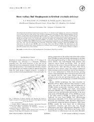
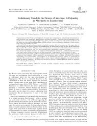
F IG.4. <strong>Kirkia</strong> wilmsii. Reproductive structures. (A) Preanthetic female flower, lateral view, perianth partly removed; arrowheads pointing to protruding<br />
petal bases. (B–G) Anthers: (B–D) sterile anther <strong>of</strong> anthetic female flower; (E–G) fertile anther <strong>of</strong> male flower bud; (B, E) ventral view; (C, F) lateral<br />
view; (D, G) dorsal view, filament attachment hidden between thecae. (H) Preanthetic gynoecium <strong>of</strong> the flower in (A), lateral view. (I) Same gynoecium,<br />
close-up <strong>of</strong> stigmatic head, lateral view. (J) Stigmatic papillae. (K) Hemispherical protrusion above the ovary, lateral view. (L) Sterile placenta (P) (with<br />
collapsed epidermal cells) appressed to the base <strong>of</strong> the (fertile) ovule (O). (M, N) Fertile ovule: (M) raphal side with arrowhead pointing to sterile placenta<br />
<strong>and</strong> arrow pointing to raphe; (N) antiraphal side with asterisk indicating collapsed enlarged cells <strong>of</strong> outer integument. Scale bars: A ¼ 500 mm; B, C, D, I,<br />
M, N ¼ 90 mm; E, F, G, H ¼ 200 mm; J, L ¼ 10 mm; K ¼ 50 mm.<br />
P<br />
O<br />
The free part <strong>of</strong> the carpels is plicate <strong>and</strong> has a ventral<br />
median longitudinal slit extending from the stigma down<br />
to the ovary (Fig. 5A–H). The (united) stigmas form an<br />
external compitum (Fig. 5A, B). The stigmatic surface<br />
has unicellular (spherical) <strong>and</strong> uniseriate multicellular<br />
(moniliform) papillae (Figs 4J <strong>and</strong> 6A) <strong>and</strong> is covered<br />
with secretion. Four pollen tube transmitting tracts differentiate<br />
downwards in the inner angle <strong>of</strong> the ventral slit <strong>of</strong> the<br />
carpels (Fig. 5A–H). Below the short compitum, they<br />
extend separately toward the base <strong>of</strong> the stylar canals <strong>and</strong><br />
the placentae (Fig. 5A, C–I). The gynoecium is entirely<br />
synascidiate in the ovary (Fig. 5A, I–M). A symplicate