Floral Structure of Kirkia (Kirkiaceae) and its ... - Annals of Botany
Floral Structure of Kirkia (Kirkiaceae) and its ... - Annals of Botany
Floral Structure of Kirkia (Kirkiaceae) and its ... - Annals of Botany

You also want an ePaper? Increase the reach of your titles
YUMPU automatically turns print PDFs into web optimized ePapers that Google loves.
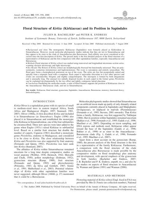
<strong>Annals</strong> <strong>of</strong> <strong>Botany</strong> 102: 539–550, 2008<br />
doi:10.1093/aob/mcn139, available online at www.aob.oxfordjournals.org<br />
<strong>Floral</strong> <strong>Structure</strong> <strong>of</strong> <strong>Kirkia</strong> (<strong>Kirkia</strong>ceae) <strong>and</strong> <strong>its</strong> Position in Sapindales<br />
JULIEN B. BACHELIER* <strong>and</strong> PETER K. ENDRESS<br />
Institute <strong>of</strong> Systematic <strong>Botany</strong>, University <strong>of</strong> Zurich, Zollikerstrasse 107, 8008 Zurich, Switzerl<strong>and</strong><br />
Received: 6 May 2008 Returned for revision: 11 June 2008 Accepted: 26 June 2008 Published electronically: 7 August 2008<br />
† Background <strong>and</strong> Aims The monogeneric <strong>Kirkia</strong>ceae (Sapindales) were formerly placed as Kirkioideae in<br />
Simaroubaceae. However, recent molecular phylogenetic studies indicate that they are not in Simaroubaceae <strong>and</strong><br />
they appear to be sister to the clade <strong>of</strong> Anacardiaceae plus Burseraceae. Such affinity was never considered or discussed<br />
since the first description <strong>of</strong> <strong>Kirkia</strong>. The present study is the first detailed analysis <strong>of</strong> the floral structure <strong>of</strong> a<br />
representative <strong>of</strong> <strong>Kirkia</strong>ceae <strong>and</strong> the first comparison with other sapindalean families, especially Anacardiaceae <strong>and</strong><br />
Burseraceae.<br />
† Methods <strong>Floral</strong> structure <strong>of</strong> <strong>Kirkia</strong> wilmsii was studied using transversal <strong>and</strong> longitudinal microtome section series,<br />
scanning electron microscopy <strong>and</strong> light microscopy.<br />
† Key Results The flowers <strong>of</strong> <strong>Kirkia</strong> wilmsii are morphologically bisexual but functionally unisexual. They are polysymmetric,<br />
isomerous (tetramerous) <strong>and</strong> haplostemonous. The ovary is syncarpous <strong>and</strong> entirely synascidiate. The<br />
floral apex forms a hemispherical protrusion on top <strong>of</strong> the ovary. The styles are free but postgenitally united <strong>and</strong><br />
apically form a stigmatic head with a compitum. Each carpel is uniovulate (biovulate in a few other species) <strong>and</strong><br />
ovules are crassinucellar, bitegmic <strong>and</strong> slightly campylotropous. The micropyle is formed by both integuments<br />
<strong>and</strong> is unusually long. The unusual two radially disposed locules in each carpel in the former genus Pleiokirkia<br />
can be explained developmentally by the two <strong>of</strong>fset <strong>and</strong> tightly contiguous lateral placentae.<br />
† Conclusions Paralleling the molecular results, a suite <strong>of</strong> floral features supports the position <strong>of</strong> <strong>Kirkia</strong>ceae close to<br />
the Anacardiaceae–Burseraceae clade, <strong>and</strong> not in Simaroubaceae.<br />
Key words: <strong>Kirkia</strong>ceae, floral structure, gynoecium, Sapindales, Anacardiaceae, Burseraceae, monoecy, functional dioecy,<br />
heterodichogamy.<br />
INTRODUCTION<br />
<strong>Kirkia</strong> Oliver is a sapindalean genus with six species <strong>of</strong> small<br />
to medium-sized trees in eastern tropical Africa, South<br />
Africa <strong>and</strong> Madagascar (Engler, 1897; Stannard, 1981,<br />
2007). Oliver (1868a, b) first described <strong>Kirkia</strong> <strong>and</strong> included<br />
it in Simaroubaceae (as Simarubeae). Engler (1896) also<br />
placed it in Simaroubaceae <strong>and</strong> established the monotypic<br />
tribe Kirkieae in Simarouboideae, one <strong>of</strong> the four subfamilies<br />
he circumscribed. Three new species were later added to the<br />
genus when Engler (1931c) raised Kirkieae to subfamilial<br />
level. Based on a similar fruit structure but double the<br />
number <strong>of</strong> carpels, Capuron (1961) described a monotypic<br />
genus Pleiokirkia, endemic to Madagascar, <strong>and</strong> considered<br />
it to be close to <strong>Kirkia</strong>. The close relationship between<br />
<strong>Kirkia</strong> <strong>and</strong> Pleiokirkia was also supported by fruit anatomy<br />
(Fern<strong>and</strong>o <strong>and</strong> Quinn, 1992). Pleiokirkia was later sunk<br />
into <strong>Kirkia</strong> (Stannard, 2007).<br />
The affinities <strong>of</strong> <strong>Kirkia</strong> within Simaroubaceae remained<br />
uncertain for a long time despite comparative studies on<br />
wood anatomy (Webber, 1936; Heimsch, 1942; Metcalfe<br />
<strong>and</strong> Chalk, 1950), pollen morphology (Erdtman, 1952),<br />
gynoecium structure (Ramp, 1988), fruit structure<br />
(Fern<strong>and</strong>o <strong>and</strong> Quinn, 1992) <strong>and</strong> phytochemistry<br />
(Polonsky, 1983; da Silva <strong>and</strong> Gottlieb, 1987; Simão<br />
et al., 1991; Mulholl<strong>and</strong> et al., 2003). Potential relationships<br />
<strong>of</strong> <strong>Kirkia</strong> with other sapindalean families were<br />
never suggested, although Oliver (1868b, p. 27) mentioned<br />
that it could be a Burseraceae.<br />
* For correspondence. E-mail julien.bachelier@systbot.uzh.ch<br />
Molecular phylogenetic studies showed that Simaroubaceae<br />
are an artificial taxon made up partly <strong>of</strong> only distantly related<br />
components currently placed in Sapindales <strong>and</strong> Malpighiales<br />
(Irvingiaceae), or unplaced in malvids (Picramniaceae)<br />
(Fern<strong>and</strong>o et al., 1995; Stevens, 2001 onwards). That <strong>Kirkia</strong><br />
forms a family, <strong>Kirkia</strong>ceae, was first suggested by Takhtajan<br />
(1966). But <strong>its</strong> position within Sapindales remained uncertain<br />
(Bakker et al., 1998; Fern<strong>and</strong>o et al., 1995; Gadek et al., 1996;<br />
Muellner et al., 2007). Depending on taxon sampling, <strong>and</strong><br />
DNA regions <strong>and</strong> methods used, <strong>Kirkia</strong>ceae either appear<br />
toward the base <strong>of</strong> the Sapindales (Gadek et al., 1996;<br />
Bakker et al., 1998) or as sister to the Anacardiaceae–<br />
Burseraceae clade (Fig. 1; Gadek et al., 1996; Fern<strong>and</strong>o<br />
et al., 1995; Muellner et al., 2007).<br />
Apart from a short account on the gynoecium (Ramp,<br />
1988), the present analysis <strong>of</strong> the floral structure is the first<br />
in a representative <strong>of</strong> the family <strong>Kirkia</strong>ceae. Furthermore,<br />
a comparison with the floral structure <strong>of</strong> the clade<br />
Anacardiaceae plus Burseraceae is made possible by the<br />
comparative studies on floral morphology plus anatomy<br />
<strong>and</strong> development, with special emphasis <strong>of</strong> the gynoecium<br />
in both families (Bachelier <strong>and</strong> Endress, 2007;<br />
J. B. Bachelier <strong>and</strong> P. K. Endress, unpubl. res.), <strong>and</strong> also by<br />
the studies on aspects <strong>of</strong> floral structure in Anacardiaceae<br />
by Wannan <strong>and</strong> Quinn (1991) <strong>and</strong> Wannan (2006).<br />
MATERIALS AND METHODS<br />
Flowering material <strong>of</strong> <strong>Kirkia</strong> wilmsii Engl. fixed in FAA was<br />
provided by Mrs D. Fourie (no collection number), National<br />
# The Author 2008. Published by Oxford University Press on behalf <strong>of</strong> the <strong>Annals</strong> <strong>of</strong> <strong>Botany</strong> Company. All rights reserved.<br />
For Permissions, please email: journals.permissions@oxfordjournals.org
540<br />
100<br />
51<br />
Outgroups<br />
100<br />
Biebersteiniaceae<br />
100<br />
Nitrariaceae<br />
100<br />
Sapindaceae<br />
87<br />
94<br />
<strong>Kirkia</strong>ceae<br />
69<br />
100<br />
Anacardiaceae<br />
100<br />
Burseraceae<br />
100<br />
51<br />
100<br />
Rutaceae<br />
100<br />
Simaroubaceae<br />
100<br />
Meliaceae<br />
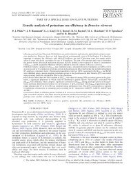
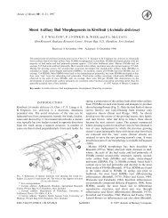
F IG. 1. Phylogenetic relationships in Sapindales, based on rbcL<br />
sequences (Bayesian posterior probabilities indicated above the branches;<br />
simplified from Muellner et al., 2007).<br />
Botanical Garden, Pretoria (South Africa), to E. Ramp in<br />
1987. The material was studied using light microscopy<br />
(LM) <strong>and</strong> scanning electron microscopy (SEM). For LM<br />
investigations, the material was embedded in Kulzer’s<br />
Technovit 7100 (2-hydroxyethyl methacrylate), following a<br />
protocol adapted from Igersheim (1993) <strong>and</strong> Igersheim <strong>and</strong><br />
Cichocki (1996). Serial microtome sections were made at<br />
5, 7 or 10 mm, using a Microm HM 355 rotary microtome<br />
<strong>and</strong> a st<strong>and</strong>ard microtome knife D. The sections were<br />
stained with ruthenium red <strong>and</strong> toluidine blue, <strong>and</strong><br />
mounted in Histomount (protocol adapted from Weber <strong>and</strong><br />
Igersheim, 1994). For SEM investigations, specimens were<br />
stained with 2 % osmium tetroxide, dehydrated in ethanol<br />
<strong>and</strong> acetone, critical-point dried <strong>and</strong> sputter coated with<br />
gold, <strong>and</strong> studied at 20 kV with a Hitachi S-4000 scanning<br />
electron microscope. The fixed material <strong>and</strong> permanent<br />
slides <strong>of</strong> serial microtome sections are deposited at the<br />
Institute <strong>of</strong> Systematic <strong>Botany</strong>, University <strong>of</strong> Zürich (Z).<br />
A B<br />
Bachelier <strong>and</strong> Endress — Flowers <strong>of</strong> <strong>Kirkia</strong> <strong>and</strong> Position in Sapindales<br />
C<br />
Morphology<br />
RESULTS<br />
The flowers are arranged in compound thyrsoids with the<br />
cymes dichasial <strong>and</strong> in higher branching orders monochasial.<br />
Although functionally unisexual, the flowers are<br />
always morphologically bisexual. They are polysymmetric<br />
<strong>and</strong> isomerous, <strong>and</strong> mostly tetramerous (Figs 2 <strong>and</strong> 3).<br />
Pentamerous or hexamerous flowers are also found on<br />
some low branching orders, <strong>and</strong> trimerous flowers on high<br />
branching orders.<br />
The flowers are relatively small (,1 cm in diameter).<br />
They have long, jointed pedicels <strong>and</strong> a broad floral base.<br />
They are haplostemonous, with the stamens alternipetalous<br />
<strong>and</strong> the carpels antepetalous (Figs 3 <strong>and</strong> 4A). A short floral<br />
cup is formed by congenitally united petal <strong>and</strong> stamen<br />
bases (Fig. 3C, G, H).<br />
Sepals are free <strong>and</strong> triangular (Fig. 2A–C). They are contiguous<br />
(valvate) in early stages <strong>of</strong> development but later<br />
the floral base <strong>and</strong> floral cup enlarge <strong>and</strong> thus their aestivation<br />
becomes open (Fig. 2A–D). The base <strong>of</strong> the sepals<br />
takes part in the floral cup but their extended margins<br />
remain free <strong>and</strong> overlap basally (Figs 2D <strong>and</strong> 3C, G, H).<br />
In tetramerous flowers, the sepals are arranged in pairs<br />
with the outer pair in median position (Figs 2A–C <strong>and</strong> 3).<br />
In pentamerous flowers, their aestivation is quincuncial at<br />
the base.<br />
Petals are free, linear <strong>and</strong> acute (Fig. 2A, F). Basally,<br />
they exp<strong>and</strong> between the sepal margins with a dorsal<br />
bulge (Fig. 4A). In contrast to the sepals, their aestivation<br />
is basally open but it is imbricate further up (Fig. 2A, E)<br />
<strong>and</strong> two patterns are observed in tetramerous flowers: (1)<br />
one petal inside, one petal outside, <strong>and</strong> two in between<br />
(Fig. 3A, B), or (2) two petals outside <strong>and</strong> two inside<br />
(Fig. 3E, F). The petals protect the inner floral organs in<br />
D<br />
F<br />
E G<br />
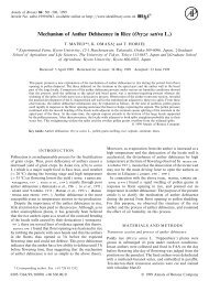
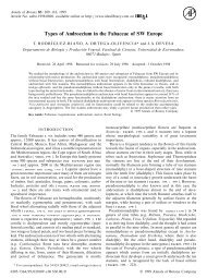
F IG.2. <strong>Kirkia</strong> wilmsii. Flower buds <strong>and</strong> parts <strong>of</strong> flower buds. (A) Bud, lateral view, arrowhead points to close-up in (D). (B) Same bud, from above, with<br />
sepals arranged in decussate pairs. (C) Another bud, from below, with sepals arranged in decussate pairs. (D) Bud shown in (A), lateral view, close-up on<br />
floral base <strong>and</strong> overlapping sepal margins (arrowhead). (E) Petal aestivation basally open <strong>and</strong> imbricate further up. (F) Petal tip, dorsal side. (G) Carpet <strong>of</strong><br />
secretory hairs on inner side <strong>of</strong> petal base. Scale bars: A, C ¼ 400 mm; B ¼ 200 mm; D, E, F, G ¼ 100 mm.
E<br />
A<br />
Bachelier <strong>and</strong> Endress — Flowers <strong>of</strong> <strong>Kirkia</strong> <strong>and</strong> Position in Sapindales 541<br />
F G H<br />
late bud when they become longer than the sepals or even<br />
earlier when sepal aestivation changes from valvate to<br />
open. Postgenital coherence between the overlapping<br />
margins <strong>of</strong> the petals is formed by interdentation <strong>of</strong> their<br />
papillate surface <strong>and</strong> striate cuticular ornamentation. At<br />
anthesis, the exp<strong>and</strong>ed petals are curved slightly inwards<br />
<strong>and</strong> their basal dorsal bulges push the sepal margins away<br />
from each other. The arrangement <strong>of</strong> the sepals in decussate<br />
pairs is more conspicuous because the outer pair appears<br />
inserted below the inner pair. Calyx <strong>and</strong> corolla are<br />
widely open <strong>and</strong> <strong>and</strong>roecium <strong>and</strong> gynoecium are thus<br />
exposed (Fig. 4A; see also figures in Immelman, 1984).<br />
Stamens have a broad <strong>and</strong> thick filament base that<br />
narrows <strong>and</strong> becomes more round further up, <strong>and</strong> a sagittate<br />
<strong>and</strong> slightly apiculate anther (Fig. 4A). Anthers are dorsally<br />
basifixed (Fig. 4A–G). The transition from filament to<br />
anther is hidden by the dorsal parts <strong>of</strong> the thecae, which<br />
curve backwards around the constricted tip <strong>of</strong> the filament<br />
<strong>and</strong> form a pseudopit (Fig. 4D, G; a pit open on one side,<br />
B C D<br />
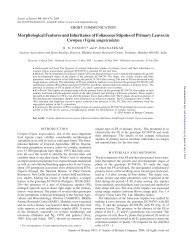
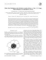
F IG.3. <strong>Kirkia</strong> wilmsii. Transverse microtome section series <strong>of</strong> two flower buds. Morphological surfaces drawn with thick continuous lines; secondary<br />
morphological surfaces drawn with thick dashed lines; vascular bundles drawn with thin continuous lines. (A–D) Male flower bud: (A) open sepal aestivation<br />
<strong>and</strong> imbricate petal tips; (B) contiguous (valvate) sepal aestivation <strong>and</strong> imbricate petal bases, showing two pairs <strong>of</strong> antesepalous stamens <strong>and</strong><br />
introrse anthers with a broad <strong>and</strong> thick connective; (C) valvate sepal bases <strong>and</strong> floral cup formed by fusion <strong>of</strong> the central part <strong>of</strong> sepal bases, <strong>and</strong><br />
petal <strong>and</strong> stamen bases, showing secretory hairs on the inner side <strong>of</strong> the petal bases <strong>and</strong> four antepetalous (delayed) sterile carpels; (D) floral base.<br />
(E–I) Sterile flower bud: (E) imbricate petal tips, arranged in pairs; (F) sepals <strong>and</strong> petals arranged in pairs, two pairs <strong>of</strong> antesepalous sterile stamens,<br />
with anthers dorsifixed basally <strong>and</strong> filament attachment hidden in a pseudopit (for term see Endress <strong>and</strong> Stumpf, 1991); (G) overlapping free sepal<br />
margins <strong>and</strong> floral cup formed by fusion <strong>of</strong> the central part <strong>of</strong> sepal bases, <strong>and</strong> petal <strong>and</strong> stamen bases, showing the carpet <strong>of</strong> secretory hairs on the<br />
inner side <strong>of</strong> the petal bases <strong>and</strong> four antepetalous carpels; (H) dorsal side <strong>of</strong> petal bases exp<strong>and</strong>ing between the free sepal bases <strong>and</strong> floral cup surrounding<br />
a (sterile) syncarpous <strong>and</strong> synascidiate ovary with four aborted ovules; (I) pedicel. Scale bars: A–D ¼ 200 mm; E–I ¼ 500 mm.<br />
here the dorsal side; for term see Endress <strong>and</strong> Stumpf,<br />
1991). The connective is thick <strong>and</strong> broad (Fig. 3B, F).<br />
Each anther has a shallow dorsal <strong>and</strong> a deep ventral<br />
median (longitudinal) furrow (Fig. 3B, F). The anther is<br />
broader on the dorsal than the ventral side, <strong>and</strong> is thus introrse<br />
(Figs 3B, F <strong>and</strong> 4B, D, E, G). The dehiscence lines<br />
extend from the tip <strong>of</strong> the thecae down to their base <strong>and</strong><br />
encompass their lower shoulders (Fig. 4B, C, E, F). In<br />
our material, the flowers <strong>of</strong> the low branching orders <strong>of</strong><br />
the inflorescence had sterile anthers <strong>and</strong> were thus functionally<br />
female (Fig. 4A). In contrast, in flowers terminating<br />
axes <strong>of</strong> higher branching orders, the anthers were more<br />
developed than the carpels, <strong>and</strong> thus were more likely functionally<br />
male (Fig. 3A–D). In some flowers, both sexes<br />
appeared abortive (Fig. 3E–I). A thick <strong>and</strong> lobed intrastaminal<br />
nectary disc is present but exp<strong>and</strong>s only late in development<br />
(Figs 4A <strong>and</strong> 5A).<br />
The gynoecium is <strong>of</strong> angiospermy type 4 (Fig. 5; carpels<br />
closed entirely by postgenital fusion; for term see Endress<br />
I
542<br />
A<br />
H I<br />
Bachelier <strong>and</strong> Endress — Flowers <strong>of</strong> <strong>Kirkia</strong> <strong>and</strong> Position in Sapindales<br />
K L<br />
<strong>and</strong> Igersheim, 2000). Superficially, the entire gynoecium<br />
gives the impression <strong>of</strong> being apocarpous because the<br />
dorsal part <strong>of</strong> the carpels is conspicuously bulging<br />
(Fig. 4A, H). However, the gynoecium has a syncarpous<br />
superior ovary with a short stalk (gynophore) (Fig. 5A,<br />
I–N). Above the ovary, the gynoecium is apocarpous<br />
(Fig. 5A–H). However, the free parts <strong>of</strong> the carpels are contiguous,<br />
form a conical stylar part (Fig. 5A, E–H), <strong>and</strong> are<br />
distally postgenitally united for half <strong>of</strong> their length<br />
(Fig. 5A–D). They form an oblique <strong>and</strong> flattened fourlobed<br />
receptive plate (‘stigmatic head’), each lobe corresponding<br />
to the tip <strong>of</strong> a carpel (Figs 4A, H, I <strong>and</strong> 5A, B).<br />
B C D<br />
E F G<br />
J M N<br />
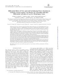
F IG.4. <strong>Kirkia</strong> wilmsii. Reproductive structures. (A) Preanthetic female flower, lateral view, perianth partly removed; arrowheads pointing to protruding<br />
petal bases. (B–G) Anthers: (B–D) sterile anther <strong>of</strong> anthetic female flower; (E–G) fertile anther <strong>of</strong> male flower bud; (B, E) ventral view; (C, F) lateral<br />
view; (D, G) dorsal view, filament attachment hidden between thecae. (H) Preanthetic gynoecium <strong>of</strong> the flower in (A), lateral view. (I) Same gynoecium,<br />
close-up <strong>of</strong> stigmatic head, lateral view. (J) Stigmatic papillae. (K) Hemispherical protrusion above the ovary, lateral view. (L) Sterile placenta (P) (with<br />
collapsed epidermal cells) appressed to the base <strong>of</strong> the (fertile) ovule (O). (M, N) Fertile ovule: (M) raphal side with arrowhead pointing to sterile placenta<br />
<strong>and</strong> arrow pointing to raphe; (N) antiraphal side with asterisk indicating collapsed enlarged cells <strong>of</strong> outer integument. Scale bars: A ¼ 500 mm; B, C, D, I,<br />
M, N ¼ 90 mm; E, F, G, H ¼ 200 mm; J, L ¼ 10 mm; K ¼ 50 mm.<br />
P<br />
O<br />
The free part <strong>of</strong> the carpels is plicate <strong>and</strong> has a ventral<br />
median longitudinal slit extending from the stigma down<br />
to the ovary (Fig. 5A–H). The (united) stigmas form an<br />
external compitum (Fig. 5A, B). The stigmatic surface<br />
has unicellular (spherical) <strong>and</strong> uniseriate multicellular<br />
(moniliform) papillae (Figs 4J <strong>and</strong> 6A) <strong>and</strong> is covered<br />
with secretion. Four pollen tube transmitting tracts differentiate<br />
downwards in the inner angle <strong>of</strong> the ventral slit <strong>of</strong> the<br />
carpels (Fig. 5A–H). Below the short compitum, they<br />
extend separately toward the base <strong>of</strong> the stylar canals <strong>and</strong><br />
the placentae (Fig. 5A, C–I). The gynoecium is entirely<br />
synascidiate in the ovary (Fig. 5A, I–M). A symplicate
B<br />
C<br />
D<br />
E<br />
F<br />
G<br />
H<br />
I<br />
J<br />
K<br />
L<br />
M<br />
N<br />
K L<br />
Bachelier <strong>and</strong> Endress — Flowers <strong>of</strong> <strong>Kirkia</strong> <strong>and</strong> Position in Sapindales 543<br />
A<br />
zone is lacking. Above the ovary, basally between the free<br />
parts <strong>of</strong> the carpels, there is a conspicuous hemispherical<br />
protrusion (Figs 4K, 5A, G, H <strong>and</strong> 6B).<br />
The carpels are uniovulate (Fig. 5I–L). However, they<br />
have two axile <strong>and</strong> almost collateral placentae in the uppermost<br />
part <strong>of</strong> the locule (Figs 5I–K, 6F, G <strong>and</strong> 7A). The<br />
second placenta slightly protrudes in such a way that it<br />
resembles a second ovule aborting early in development<br />
B<br />
F<br />
I<br />
C<br />
D<br />
G I H<br />
F IG.5. <strong>Kirkia</strong> wilmsii. Anthetic gynoecium. Morphological surfaces drawn with thick continuous lines; thick dashed lines used in (A) for parts outside<br />
the median plane <strong>of</strong> symmetry, in (B–N) for postgenitally united surfaces; vascular bundles drawn with thin continuous lines; pollen tube transmitting<br />
tract dark grey. d, Dorsal vascular bundle; l, lateral vascular bundle; s, synlateral vascular bundle. (A) Schematic median longitudinal section <strong>of</strong> gynoecium<br />
<strong>and</strong> nectary disc (light grey); postgenitally united surfaces hatched. (B–N) Transverse microtome section series; (B) stigmatic head; (C, D) postgenitally<br />
united distal parts <strong>of</strong> the carpels; (E, F) connivent but free parts <strong>of</strong> the carpels; (G, H) connivent bases <strong>of</strong> the free parts <strong>of</strong> the carpels around the<br />
hemispherical protrusion on top <strong>of</strong> the ovary; (I–M) synascidiate ovary, the two arrows in (K) pointing to the S-shaped line formed by the two lateral<br />
placentae (compare with Figs 6F <strong>and</strong> 7A); (N) gynophore. Scale bars: A, B–N ¼ 500 mm.<br />
s<br />
d<br />
M<br />
d<br />
J<br />
(Fig. 4L, M). Behind the second placenta, toward the<br />
centre <strong>of</strong> the gynoecium, there is a small gap (Figs 6F–I<br />
<strong>and</strong> 7A). This may correspond to the ‘inner locule’ described<br />
in other <strong>Kirkia</strong> species (Fig. 7B; see Discussion; see also<br />
figures in Capuron, 1961).<br />
The ovule is long <strong>and</strong> cylindrical (Fig. 4M, N). It is crassinucellar,<br />
bitegmic, antitropous (ovule curvature direction<br />
opposite to direction <strong>of</strong> carpel involution; for term see<br />
I<br />
s s<br />
s s<br />
N<br />
E
544<br />
A<br />
B<br />
s<br />
F G H s<br />
s<br />
I<br />
s<br />
s<br />
Bachelier <strong>and</strong> Endress — Flowers <strong>of</strong> <strong>Kirkia</strong> <strong>and</strong> Position in Sapindales<br />
C D E<br />
F IG.6. <strong>Kirkia</strong> wilmsii. (A) Longitudinal section (LS) <strong>of</strong> uniseriate multicellular papillae <strong>of</strong> the stigma before anthesis. (B) LS <strong>of</strong> the hemispherical protrusion<br />
in the floral centre above the ovary (compare with Fig. 5A). (C) LS <strong>of</strong> the slightly campylotropous ovule filling the locule at anthesis, with the<br />
exp<strong>and</strong>ed large-celled distal parts <strong>of</strong> the two integuments forming a long micropyle. (D) Transverse section (TS) <strong>of</strong> base <strong>of</strong> sepals, petal <strong>and</strong> stamens<br />
before anthesis, showing carpet <strong>of</strong> secretory hairs (with short multiseriate stalk <strong>and</strong> large multicellular head) on the inner side <strong>of</strong> the petal base, <strong>and</strong><br />
petal base exp<strong>and</strong>ing dorsally between the free margins <strong>of</strong> two sepal bases. (E) LS <strong>of</strong> sepal base showing the epidermal <strong>and</strong> sub-epidermal special mucilage<br />
cells. (F–I) TS <strong>of</strong> a preanthetic gynoecium <strong>and</strong> corresponding enlarged micrographs, showing the inner angle <strong>of</strong> a fertile locule <strong>and</strong> centre <strong>of</strong> the<br />
gynoecium (locule dorsal side oriented downwards; compare with Fig. 5K); arrows point to placentae; arrowhead points to the second reduced locule<br />
developing on the same radius as the fertile one (compare with Fig. 7A); morphological surfaces drawn with thick continuous lines; postgenitally<br />
fused morphological surfaces drawn with dashed lines; vascular bundles drawn with thin continuous lines; ‘s’: synlateral vascular bundle; dash rectangles<br />
in (F) <strong>and</strong> (H) show location <strong>of</strong> (G) <strong>and</strong> (I). (F, G) In the upper part <strong>of</strong> the locule, the endocarp differentiation begins laterally <strong>and</strong> the ventral inner surface<br />
<strong>of</strong> the carpel is S-shaped (arrowhead). (H, I) Lower down, the endocarp encompasses the inner angle <strong>of</strong> the locule <strong>and</strong> the second locule is isolated (arrowhead).<br />
Scale bars: A ¼ 30 mm; B, C, D ¼ 200 mm; E, F, G, H, I ¼ 100 mm.<br />
Endress, 1994), <strong>and</strong> slightly campylotropous with only the<br />
very base <strong>of</strong> the nucellus <strong>and</strong> embryo sac curved (Fig. 5A).<br />
The two integuments surround the nucellus <strong>and</strong>, although<br />
both appear to be <strong>of</strong> the same thickness, the inner integument<br />
comprises three or four cell layers but the outer<br />
only two or three cell layers (Figs 5A <strong>and</strong> 6C). Above the<br />
nucellus the integuments are elongate <strong>and</strong> thickened. At<br />
anthesis, the inner integument is about twice as long as<br />
the nucellus <strong>and</strong> the outer even two <strong>and</strong> a half times<br />
(Figs 5A <strong>and</strong> 6C). Thus, the micropyle is unusually long<br />
<strong>and</strong> comprises two distinct zones. The proximal zone is a<br />
straight tubular canal formed by the inner integument,<br />
s<br />
s<br />
whereas the distal zone is not tubular <strong>and</strong> is somewhat<br />
wavy, <strong>and</strong> is formed by the second integument (Figs 5A<br />
<strong>and</strong> 6C). The extended part <strong>of</strong> the integuments above the<br />
nucellus comes about by cell enlargement (Fig. 6C). In isolated<br />
ovules studied with the SEM, these enlarged cells tend<br />
to collapse (Fig. 4 N). The ovule fills the locule <strong>and</strong> the<br />
micropyle is contiguous with the placenta (Fig. 5A).<br />
Anatomy<br />
Sepals have one median <strong>and</strong> two lateral main vascular<br />
bundles, which extend almost through their whole length,
A B<br />
Bachelier <strong>and</strong> Endress — Flowers <strong>of</strong> <strong>Kirkia</strong> <strong>and</strong> Position in Sapindales 545<br />
F IG. 7. Transverse section diagrams <strong>of</strong> a carpel: (A) <strong>Kirkia</strong> wilmsii with<br />
one locule <strong>and</strong> a small inner opening; (B) <strong>Kirkia</strong> le<strong>and</strong>rii (‘Pleiokirkia’)<br />
with two ‘locules’, the inner one corresponding to the small inner<br />
opening in <strong>Kirkia</strong> wilmsii.<br />
<strong>and</strong> may have one to two smaller, additional lateral bundles<br />
in their free parts (Fig. 3A–C, F–H). Toward the sepal<br />
base, the smaller lateral bundles merge with one <strong>of</strong> the<br />
two main lateral bundles before extending into the floral<br />
base (Fig. 3G, H). Petals have one median main vascular<br />
bundle (Fig. 3A–C, E–H) <strong>and</strong> can have up to three pairs<br />
<strong>of</strong> smaller, lateral bundles at anthesis. Toward the petal<br />
bases, all lateral bundles merge together with the main<br />
median bundle <strong>and</strong> a single petal trace extends downwards<br />
(Fig. 3C, D, G, H). Stamens have a single bundle, which<br />
extends into the upper half <strong>of</strong> the anthers (Fig. 3B, C, F–H).<br />
In carpels, a pair <strong>of</strong> lateral vascular bundles differentiates<br />
just below the stigmatic head on each side <strong>of</strong> the ventral slit<br />
(Fig. 5C). These laterals extend downwards into the ovary<br />
<strong>and</strong> form synlaterals in the synascidiate zone (Fig. 5D–J).<br />
At the upper end <strong>of</strong> the locule, each synlateral gives <strong>of</strong>f a<br />
branch serving an adjacent ovule (Fig. 5I) <strong>and</strong> ending in<br />
the chalaza, whereas lower down, the synlaterals converge<br />
toward the centre <strong>of</strong> the gynoecium <strong>and</strong> form a ring-shaped<br />
central vascular complex (Fig. 5K–N).<br />
In contrast, distinct dorsal bundles are present only far<br />
below the zone <strong>of</strong> postgenital union <strong>of</strong> the free upper<br />
carpel parts (Fig. 5G). Between the dorsal <strong>and</strong> lateral<br />
bundles there are numerous smaller bundles <strong>and</strong> together<br />
they form a reticulate system above the ovary <strong>and</strong> extend<br />
downwards around the locules (Fig. 5G–M). They merge<br />
in the gynophore with the ring <strong>of</strong> the synlaterals.<br />
In the floral base, the petal traces merge with the lateral<br />
traces <strong>of</strong> the sepals whereas the stamen traces merge with<br />
the median sepal traces <strong>of</strong> the same radius. All vascular<br />
bundles converge toward the central vasculature <strong>of</strong> the<br />
gynoecium <strong>and</strong> form a stele with it (Fig. 3D, I).<br />
Histology<br />
Lignified unicellular hairs are sparsely present on the<br />
floral base <strong>and</strong> the dorsal side <strong>of</strong> sepals, the petal parts,<br />
which are not covered by another petal in bud, <strong>and</strong> the<br />
ventral side <strong>of</strong> the petal base (Figs 2A, C, D, F, G <strong>and</strong> 4A).<br />
Stomata are present on the dorsal side <strong>of</strong> sepals <strong>and</strong><br />
petals, on the smooth surface <strong>of</strong> the nectary disc, <strong>and</strong> on<br />
the carpel tips below the stigmatic head. On the ventral<br />
side <strong>of</strong> the petal bases, there is a carpet <strong>of</strong> hairs with a<br />
multicellular multiseriate stalk <strong>and</strong> massive head containing<br />
dark-staining cells (Figs 2G, 3C, G, H <strong>and</strong> 6D). Epidermal<br />
<strong>and</strong> subepidermal special mucilage cells (Fig. 6D, E; cells<br />
with thickened mucilaginous, layered inner tangential<br />
wall; for term see Matthews <strong>and</strong> Endress, 2006) are<br />
present in the sepals <strong>and</strong> floral base in late buds <strong>and</strong> anthetic<br />
flowers.<br />
Sexual system<br />
DISCUSSION<br />
The presence <strong>of</strong> functionally unisexual (but morphologically<br />
bisexual) flowers appears to be common in <strong>Kirkia</strong>ceae (this<br />
study; Oliver, 1868a; Capuron, 1961; Stannard, 1981;<br />
Immelmann, 1984), <strong>and</strong> is also common in Anacardiaceae,<br />
Burseraceae <strong>and</strong> other Sapindales (J. B. Bachelier <strong>and</strong><br />
P. K. Endress, unpubl. res.). Functional dioecy by flushes<br />
<strong>of</strong> male <strong>and</strong> female flowers as described by Immelmann<br />
(1984) for <strong>Kirkia</strong> wilmsii may be morphologically reflected<br />
by the presence <strong>of</strong> female flowers in the lower order branches<br />
<strong>of</strong> the cymes <strong>of</strong> the thyrsoid inflorescences, <strong>and</strong> male flowers<br />
in the higher order branches, <strong>and</strong> sequential opening <strong>of</strong><br />
flowers <strong>of</strong> successive branching orders (this study). This is<br />
a kind <strong>of</strong> dichogamy <strong>and</strong> even (imprecise) heterodichogamy<br />
if the flowering schemes <strong>of</strong> different individuals by<br />
Immelman (1984) are considered. Heterodichogamy is<br />
uncommon in angiosperms (Renner, 2001) but was also<br />
recorded among Sapindales, in several species <strong>of</strong> Acer <strong>and</strong><br />
in Cupania (Sapindaceae) (Gabriel, 1968; de Jong, 1976;<br />
Bawa, 1977; Tatsuhiro, 2000; Sato, 2002; Gleiser <strong>and</strong><br />
Verdú, 2005; Renner et al., 2007; Kikuchi <strong>and</strong> Shibata,<br />
2008). The same pattern in the distribution <strong>of</strong> male<br />
<strong>and</strong> female flowers within an inflorescence as in <strong>Kirkia</strong><br />
has also been reported in Anacardium (Anacardiaceae)<br />
(Copel<strong>and</strong>, 1962; Moncur <strong>and</strong> Wait, 1986; Moncur, 1988),<br />
in Cedrela, Melia <strong>and</strong> Toona (Meliaceae) (Styles, 1972;<br />
Gouvêa et al., 2008a, b), <strong>and</strong> in Cupania (Sapindaceae)<br />
(Bawa, 1977). In another type <strong>of</strong> heterodichogamy (in<br />
Hern<strong>and</strong>ia, Laurales) it was also found that male <strong>and</strong><br />
female flowers had a specific distribution pattern in the<br />
inflorescence (Endress <strong>and</strong> Lorence, 2004). Based on inflorescence<br />
structure it is to be expected that hitherto unrecognized<br />
cases <strong>of</strong> heterodichogamy may occur among<br />
Sapindales.<br />
<strong>Floral</strong> merism<br />
Flowers in most species <strong>of</strong> <strong>Kirkia</strong>ceae are tetramerous <strong>and</strong><br />
isomerous (Stannard, 1981). Of special interest is the occurrence<br />
<strong>of</strong> species with double the number <strong>of</strong> carpels, still in<br />
one whorl (Capuron, 1961; Stannard, 1981). The tendency<br />
<strong>of</strong> an increase in carpel number is also present in<br />
Anacardiaceae (Pleiogynium, up to 13; Wannan <strong>and</strong> Quinn,<br />
1991) <strong>and</strong> Burseraceae (Beiselia, up to 12; Forman et al.,<br />
1991), <strong>and</strong> in other families <strong>of</strong> Sapindales, such as<br />
Meliaceae (Turraea, up to 20; Harms, 1940), Rutaceae<br />
(Aegle, up to 20; Vasil <strong>and</strong> Johri, 1964), <strong>and</strong> also in the
546<br />
Bachelier <strong>and</strong> Endress — Flowers <strong>of</strong> <strong>Kirkia</strong> <strong>and</strong> Position in Sapindales<br />
unplaced possibly sapindalean fossil L<strong>and</strong>eenia, approx. 18<br />
(Manchester <strong>and</strong> Hermsen, 2000). This tendency occurs even<br />
more generally in the entire malvids (Endress <strong>and</strong> Matthews,<br />
2006). Another pattern <strong>of</strong> interest is the co-occurrence <strong>of</strong> tetramerous<br />
<strong>and</strong> pentamerous flowers on the same individual, with<br />
the pentamerous ones especially on lower-order axes <strong>of</strong> the<br />
inflorescence, as also known from some Rutaceae (Ruta,<br />
Eichler, 1878; Skimmia, personal observation). In <strong>Kirkia</strong><br />
wilmsii trimerous, tetramerous, pentamerous <strong>and</strong> hexamerous,<br />
isomerous, flowers were found on the same individual. <strong>Floral</strong><br />
isomery is also common in Rutaceae (Engler, 1931b; Gut,<br />
1966; Ramp, 1988) <strong>and</strong> Simaroubaceae (Engler, 1931c;<br />
Ramp, 1988) but less so in Anacardiaceae <strong>and</strong> Burseraceae<br />
(Engler, 1931a; J.B.Bachelier<strong>and</strong>P.K.Endress,unpubl.<br />
res.). Rutaceae also exhibit many genera with tetramerous<br />
flowers. In contrast, in Anacardiaceae pentamerous flowers<br />
are common, <strong>and</strong> in Burseraceae trimerous flowers (Lam,<br />
1932; J. B. Bachelier <strong>and</strong> P. K. Endress, unpubl. res.).<br />
However, all numbers, three, four <strong>and</strong> five, combined with<br />
isomery, are present in Anacardiaceae, Burseraceae <strong>and</strong><br />
<strong>Kirkia</strong>ceae (<strong>and</strong> other Sapindales) in different frequencies<br />
<strong>and</strong> distribution. Whereas in Burseraceae isomerous flowers<br />
are common, as in <strong>Kirkia</strong>ceae, in Anacardiaceae they are<br />
largely restricted to Spondioideae (S. Pell, Brooklyn<br />
Botanical Garden, unpubl. res.).<br />
<strong>Floral</strong> cup <strong>and</strong> perianth<br />
The flowers have an exp<strong>and</strong>ed base <strong>and</strong> a shallow floral<br />
cup bearing the nectary disc. The cup is formed by the congenitally<br />
united bases <strong>of</strong> petals <strong>and</strong> stamens, <strong>and</strong> the median<br />
parts <strong>of</strong> the sepals. The sepal margins remain free <strong>and</strong> extend<br />
downwards as ledges for a short distance. Such free sepal<br />
margins on a floral cup or on a floral base without a cup<br />
are not restricted to Sapindales but also occur in other<br />
rosid groups. Although they are involved in the formation<br />
<strong>of</strong> the floral cup, sepals <strong>and</strong> petals are free among themselves,<br />
thus the perianth is chorisepalous <strong>and</strong> choripetalous<br />
in <strong>Kirkia</strong>ceae, as in many other Sapindales. According to<br />
Merxmüller <strong>and</strong> Heine (1960), in <strong>Kirkia</strong> dewinteri, the<br />
calyx is united for one-third <strong>of</strong> <strong>its</strong> length. If this is correct<br />
there would be free <strong>and</strong> united calyces in the genus.<br />
Sepals are deltoid <strong>and</strong> have three main vascular traces <strong>and</strong><br />
thus exhibit the shape <strong>and</strong> vasculature that is common in<br />
many rosids. The petals are acuminate <strong>and</strong> have a single vascular<br />
trace. They become longer than the sepals in older buds<br />
<strong>and</strong> thus have a protective function at this stage in<br />
<strong>Kirkia</strong>ceae, Anacardiaceae <strong>and</strong> Burseraceae, as also found<br />
in various other rosids (e.g. most Celastrales, some<br />
Oxalidales, some Crossosomatales <strong>and</strong> a few Chrysobalanaceae<br />
sensu lato; Matthews <strong>and</strong> Endress, 2002,<br />
2005a, b, 2008). Sepal aestivation <strong>of</strong> young floral buds is<br />
commonly valvate in <strong>Kirkia</strong>ceae, as in Burseraceae <strong>and</strong><br />
some Anacardiaceae. However, it becomes open in older<br />
buds when the petal bases exp<strong>and</strong> between the sepal bases,<br />
as also observed in Bursera (Burseraceae) (J. B. Bachelier<br />
<strong>and</strong> P. K. Endress, unpubl. res.). Petal aestivation is mainly<br />
imbricate <strong>and</strong> sometimes valvate (Oliver, 1868a, b;<br />
Stannard, 1981) in <strong>Kirkia</strong>ceae. Dense groups <strong>of</strong> secretory<br />
hairs with a short multiseriate stalk <strong>and</strong> large multicellular<br />
head at the inner base <strong>of</strong> the petals in <strong>Kirkia</strong> (see also<br />
Stannard, 1981) are unusual because they were not found<br />
in other areas <strong>of</strong> the flowers. In Beiselia there are secretory<br />
hairs with a large multicellular head, especially on the<br />
adaxial surface <strong>of</strong> the petal tips, but they have a uniseriate<br />
multicellular stalk as do all secretory hairs in<br />
Anacardiaceae <strong>and</strong> Burseraceae studied (J. B. Bachelier<br />
<strong>and</strong> P. K. Endress, unpubl. res.). These secretory hairs<br />
with multiseriate stalk may thus be an apomorphy for<br />
<strong>Kirkia</strong>ceae. Their concentration <strong>and</strong> restriction to a small<br />
area <strong>of</strong> the flower is suggestive <strong>of</strong> a specific function.<br />
However, since a disc that looks like a normal nectary is<br />
present, it is unlikely that the hairs are nectariferous (as is<br />
the case in the perianth <strong>of</strong> some Malvaceae; Vogel, 2000)<br />
(see below). It would be <strong>of</strong> interest to study the hairs in<br />
live material.<br />
Androecium<br />
Haplostemony, the presence <strong>of</strong> only one stamen whorl as<br />
consistent in <strong>Kirkia</strong>ceae is unusual for Burseraceae<br />
<strong>and</strong> occurs in only few genera (Triomma, some species<br />
<strong>of</strong> Canarium, Santiria, Protium <strong>and</strong> Crepidospermum;<br />
Leenhouts, 1956; Daly, 1989; Mitchell <strong>and</strong> Daly, 1993). In<br />
Anacardiaceae, haplostemonous flowers only occur in some<br />
Anacardioideae (Mitchell <strong>and</strong> Daly, 1993). The stamens <strong>of</strong><br />
haplostemonous flowers are alternipetalous in all three<br />
families. Flowers with two stamen whorls in Anacardiaceae<br />
<strong>and</strong> Burseraceae (J. B. Bachelier <strong>and</strong> P. K. Endress, unpubl.<br />
res.) <strong>and</strong> other Sapindales (Rutaceae, Beille, 1902; Eckert,<br />
1966; Gut, 1966; Simaroubaceae, Nair <strong>and</strong> Joseph, 1957;<br />
Nair <strong>and</strong> Joshi, 1958; Narayana <strong>and</strong> Sayeeduddin, 1958;<br />
Eckert, 1966) are commonly obdiplostemonous, i.e. the antepetalous<br />
stamens have a smaller base than the antesepalous<br />
ones, <strong>and</strong>, in isomerous flowers, the carpels are positioned<br />
in the antepetalous radii (contrary to expectation based on<br />
regular alternation <strong>of</strong> whorls) because there is more space<br />
available in this position. That the anthetic antepetalous<br />
stamens are less developed is commonly obvious also by<br />
their shorter filaments, as in Anacardiaceae <strong>and</strong> Burseraceae<br />
(J. B. Bachelier <strong>and</strong> P. K. Endress, unpubl. res.), <strong>and</strong> is a<br />
common situation also in many other rosids. Haplostemonous<br />
flowers with alternipetalous stamens as in<br />
<strong>Kirkia</strong>ceae may be seen as an extreme case in this trend: as<br />
complete suppression <strong>of</strong> the antepetalous stamens. Among<br />
other Sapindales, flowers in Meliaceae are mostly obdiplostemonous,<br />
rarely diplostemonous (with the carpels alternipetalous),<br />
sometimes haplostemonous with the carpels<br />
antepetalous, rarely alternipetalous (Harms, 1940); in haplostemonous<br />
Rutaceae the stamens are alternipetalous (Beille,<br />
1902; Engler, 1931b), in Simaroubaceae alternipetalous<br />
(Brucea, Picrasma) or antepetalous (Picrolemma, Engler,<br />
1931c). Thus the position <strong>of</strong> stamens in haplostemonous<br />
flowers in families <strong>of</strong> Sapindales other than the clade <strong>of</strong><br />
<strong>Kirkia</strong>ceae–Anacardiaceae–Burseraceae appears less fixed.<br />
Stamen shape in <strong>Kirkia</strong>ceae corresponds to a common<br />
type in Sapindales <strong>and</strong> other rosids with sagittate, slightly<br />
dorsifixed, introrse anthers, <strong>and</strong> with a relatively narrow<br />
transition region between filament <strong>and</strong> anther (Endress<br />
<strong>and</strong> Stumpf, 1991; Matthews <strong>and</strong> Endress, 2002, 2004,
Bachelier <strong>and</strong> Endress — Flowers <strong>of</strong> <strong>Kirkia</strong> <strong>and</strong> Position in Sapindales 547<br />
2005a, b, 2006, 2008; Bachelier <strong>and</strong> Endress, 2007;<br />
J. B. Bachelier <strong>and</strong> P. K. Endress, unpubl. res.).<br />
Especially interesting in <strong>Kirkia</strong> is the presence <strong>of</strong> a pseudopit,<br />
the enclosure <strong>of</strong> this transition region between the two<br />
dorsal pollen sacs. Among rosids this feature is especially<br />
common in Sapindales <strong>and</strong> was reported for some<br />
Anacardiaceae <strong>and</strong> Burseraceae (Endress <strong>and</strong> Stumpf,<br />
1991; J. B. Bachelier <strong>and</strong> P. K. Endress, unpubl. res.).<br />
Nectary disc<br />
A conspicuous intrastaminal nectary disc with nectar<br />
pores, <strong>of</strong>ten separating the <strong>and</strong>roecium base from the gynoecium<br />
base for some distance, as in <strong>Kirkia</strong>, is also present<br />
in Burseraceae <strong>and</strong> most Anacardiaceae, as well as in<br />
Meliaceae, Rutaceae <strong>and</strong> Simaroubaceae. In Sapindaceae<br />
(<strong>and</strong> Mangifera <strong>of</strong> Anacardiaceae), the nectary disc is,<br />
however, extrastaminal (Ronse De Craene <strong>and</strong> Haston, 2006).<br />
Gynoecium<br />
The unusual ovary structure in <strong>Kirkia</strong> can be better<br />
understood when fruit differentiation is considered. The dispersal<br />
unit is a mericarp which develops from the outward<br />
bulging dorsal region <strong>of</strong> the carpels (including the locule)<br />
<strong>and</strong> detaches from a central part that remains as a<br />
column, called the ‘central column’ (Capuron, 1961) or<br />
‘carpophore’ (Engler, 1931c; Stannard, 1981). This carpophore<br />
originates by histological differentiation from the<br />
central part <strong>of</strong> the synascidiate ovary. Such mericarps are<br />
not present in Anacardiaceae <strong>and</strong> Burseraceae. In<br />
Simaroubaceae carpels are dispersed individually, which<br />
was probably one reason why <strong>Kirkia</strong> was formerly included<br />
in Simaroubaceae. However, the morphological basis is<br />
different, since in Simaroubaceae the gynoecium is more<br />
or less entirely apocarpous (with the styles only postgenitally<br />
united) (Nair <strong>and</strong> Joseph, 1957; Nair <strong>and</strong> Joshi,<br />
1958; Narayana <strong>and</strong> Sayeeduddin, 1958; Endress et al.,<br />
1983; Ramp, 1988). More or less apocarpous gynoecia<br />
(with the styles only postgenitally united) are also present<br />
in part <strong>of</strong> Rutaceae (Gut, 1966; Endress et al., 1983;<br />
Ramp, 1988).<br />
Another unusual trait in the ovary is the reported presence<br />
<strong>of</strong> two locules per carpel in radial disposition, each<br />
with an ovule, in the former genus Pleiokirkia (Capuron,<br />
1961). This may also be the case in other species <strong>of</strong><br />
<strong>Kirkia</strong> (Stannard, 1981). Whereas a compartmentalization<br />
<strong>of</strong> each carpel into two collateral locules is known in<br />
various groups <strong>of</strong> angiosperms, a radial disposition <strong>of</strong> two<br />
locules is highly unusual <strong>and</strong> morphologically puzzling.<br />
However, the analysis <strong>of</strong> the gynoecium structure in<br />
K. wilmsii in the present publication allows a morphological<br />
explanation <strong>of</strong> the two radially disposed locules in the<br />
former genus Pleiokirkia. Although in Pleiokirkia there<br />
are two ovules, the ovule <strong>of</strong> the inner locule aborts, <strong>and</strong><br />
although in <strong>Kirkia</strong> there is only one ovule, there are two<br />
placentae. These placentae are not collateral but somewhat<br />
radially displaced.<br />
As in the former Pleiokirkia, only the outer placenta bears a<br />
fertile ovule whereas the inner one bears no ovule at all. The<br />
two placentae are tightly pressed together as seen in transverse<br />
sections, so that the inner surface <strong>of</strong> the carpels is more or less<br />
S-shaped. The fertile locule is on the outer side <strong>of</strong> the S. As a<br />
counterpart, there is a minute gap on the inner side <strong>of</strong> the S<br />
(Figs 6F <strong>and</strong> G <strong>and</strong> 7A) which may correspond to the inner<br />
locule in the former Pleiokirkia (Fig. 7B). Such transition<br />
between the presence <strong>of</strong> one <strong>and</strong> two ovules is also present<br />
in the Anacardiaceae–Burseraceae clade, with two more or<br />
less collateral ovules in most Burseraceae, <strong>and</strong> one ovule in<br />
most Anacardiaceae. Interestingly, there are also rare cases<br />
<strong>of</strong> the reverse situation in the two families: two ovules<br />
(the second epitropous) in Dracontomelon (J. B. Bachelier<br />
<strong>and</strong> P. K. Endress, unpubl. res.) <strong>and</strong> in Spondias (Baillon,<br />
1874), one ovule in Beiselia (J. B. Bachelier <strong>and</strong><br />
P. K. Endress, unpubl. res.) <strong>and</strong> in Boswellia (Sunnichan<br />
et al., 2005). However, if two ovules are present, the placentae<br />
are collateral <strong>and</strong> not or less radially displaced.<br />
The stigmatic head, a conspicuous feature in <strong>Kirkia</strong>, consists<br />
<strong>of</strong> the postgenitally united free carpel tips. This construction<br />
allows the formation <strong>of</strong> a compitum, which is absent<br />
lower down in the gynoecium because <strong>of</strong> the apocarpous<br />
stylar part <strong>and</strong> the completely synascidiate ovary. Such an<br />
organization <strong>of</strong> a stigmatic head is also known from<br />
Burseraceae but less so from Anacardiaceae (J. B. Bachelier<br />
<strong>and</strong> P. K. Endress, unpubl. res.). A stigmatic head is also<br />
present in the more or less completely apocarpous gynoecia<br />
<strong>of</strong> Rutaceae–Rutoideae <strong>and</strong> Simaroubaceae (Endress et al.,<br />
1983), <strong>and</strong> also occurs in Meliaceae (Gouvêa et al., 2008a,<br />
b), in which the gynoecium is usually described as syncarpous<br />
(‘carpels united’, Cronquist, 1981; Takhtajan, 1997).<br />
However, there are no critical studies on the internal morphological<br />
carpel surfaces in Meliaceae, <strong>and</strong> thus it is uncertain<br />
whether the style is really syncarpous or consists only <strong>of</strong> postgenitally<br />
united carpels, although there are some publications<br />
with line drawings <strong>of</strong> transverse sections <strong>of</strong> styles, which<br />
suggest true syncarpy (Narayana, 1958a, b, 1959a; Nair,<br />
1962; Murty <strong>and</strong> Gupta, 1978a, b; Lal, 1994). The styles<br />
are truly syncarpous in Rutaceae–Citroideae (Ramp, 1988).<br />
In both Meliaceae <strong>and</strong> Rutaceae–Citroideae at least the stigmatic<br />
head appears to be apocarpous but the carpels are postgenitally<br />
united (Ramp, 1988; Gouvêa et al., 2008a, b). The<br />
stigma in <strong>Kirkia</strong> is wet <strong>and</strong> exhib<strong>its</strong> unicellular <strong>and</strong> uniseriate<br />
pluricellular (moniliform) papillae, which are also present in<br />
Anacardiaceae <strong>and</strong> Burseraceae (Bachelier <strong>and</strong> Endress,<br />
2007; J. B. Bachelier <strong>and</strong> P. K. Endress, unpubl. res.). This<br />
differs from the survey in Heslop-Harrison <strong>and</strong> Shivanna<br />
(1977) who reported a non-papillate (smooth) stigma for the<br />
only studied genus (Cotinus) <strong>of</strong> the Anacardiaceae–<br />
Burseraceae clade.<br />
A further unifying trait in the gynoecium <strong>of</strong> Sapindales is<br />
the presence <strong>of</strong> an extensive remnant <strong>of</strong> the floral apex in<br />
the centre <strong>of</strong> the gynoecium that is not incorporated into the<br />
gynoecium architecture. In <strong>Kirkia</strong>, it forms a dome-shaped<br />
or almost spherical protrusion between the carpels where<br />
they are free above the synascidiate zone. In groups with<br />
entirely apocarpous (only postgenitally united) carpels (e.g.<br />
some Rutaceae, Simaroubaceae; Nair <strong>and</strong> Joshi, 1958;<br />
Ramp, 1988), it is present at the base between the free<br />
carpels. As the apically postgenitally united carpels are<br />
connivent, the protrusion cannot be seen from the outside.
548<br />
Bachelier <strong>and</strong> Endress — Flowers <strong>of</strong> <strong>Kirkia</strong> <strong>and</strong> Position in Sapindales<br />
This protrusion is commonly hidden in taxa with postgenitally<br />
united stigmas/styles (e.g. Beiselia, Burseraceae;<br />
Dracontomelon, Anacardiaceae; Rutaceae, Simaroubaceae;<br />
J. B. Bachelier <strong>and</strong> P. K. Endress, unpubl. res.). It is,<br />
however, exposed in gynoecia without intercarpellary postgenital<br />
union (e.g. Pleiogynum, Spondias p.p., Poupartiopsis,<br />
Spondioideae; Mitchell et al., 2006; J. B. Bachelier <strong>and</strong><br />
P. K. Endress, unpubl. res.). On architectural grounds the<br />
dome is especially large in gynoecia with an increased<br />
number <strong>of</strong> carpels (Endress, 2006). Interestingly, a symplicate<br />
zone is lacking in <strong>Kirkia</strong>ceae <strong>and</strong> in Spondioideae<br />
(Anacardiaceae), or very short in Beiselia (Burseraceae). In<br />
contrast, in core Burseraceae, a symplicate zone is present<br />
<strong>and</strong> extends from the synascidiate base <strong>of</strong> the gynoecium to<br />
the base <strong>of</strong> the stigmatic head (J. B. Bachelier <strong>and</strong><br />
P. K. Endress, unpubl. res.).<br />
Ovules<br />
The crassinucellar, bitegmic ovules in <strong>Kirkia</strong> are antitropous<br />
(epitropous) as are those <strong>of</strong> Burseraceae (<strong>and</strong> also<br />
Rutaceae, Simaroubaceae <strong>and</strong> Meliaceae), while those <strong>of</strong><br />
Anacardiaceae (<strong>and</strong> also Sapindaceae) are syntropous (apotropous).<br />
They are slightly campylotropous (especially<br />
involving the basal area <strong>of</strong> the nucellus), a trait also shared<br />
with other Sapindales, especially Burseraceae (Wiger,<br />
1935; Narayana, 1959b, 1960a, b; J. B. Bachelier <strong>and</strong><br />
P. K. Endress, unpubl. res.), Simaroubaceae (Wiger, 1935;<br />
Narayana, 1957), Rutaceae (Mauritzon, 1935; Boesewinkel,<br />
1977, 1984; Souza et al., 2003), <strong>and</strong> Sapindaceae (e.g.<br />
Weckerle <strong>and</strong> Rutishauser, 2003, 2005). In contrast,<br />
Anacardiaceae tend to have anatropous ovules (Bachelier<br />
<strong>and</strong> Endress, 2007; J. B. Bachelier <strong>and</strong> P. K. Endress,<br />
unpubl. res.). The inner integument is thicker than the outer<br />
in <strong>Kirkia</strong>, another tendency shared by many Sapindales <strong>and</strong><br />
other malvids (Endress <strong>and</strong> Matthews, 2006).<br />
A peculiarity <strong>of</strong> the ovules <strong>of</strong> <strong>Kirkia</strong> is that they have an<br />
exceedingly long micropyle formed by elongation <strong>of</strong> both<br />
integuments. Conspicuous is the cell enlargement <strong>of</strong> the<br />
outer integument accompanying the elongation. These features<br />
were not observed in Anacardiaceae <strong>and</strong> Burseraceae<br />
<strong>and</strong> have not been reported from other Sapindales, <strong>and</strong><br />
may thus be autapomorphies for <strong>Kirkia</strong>. The micropylar<br />
part <strong>of</strong> the outer integument is conspicuously wavy. Such<br />
wavy micropyles (but in the inner integument, with the<br />
outer not involved in micropyle formation) were illustrated<br />
for some other Sapindales as well (Burseraceae; Narayana,<br />
1959b, J. B. Bachelier <strong>and</strong> P. K. Endress, unpubl. res.;<br />
Simaroubaceae; Narayana, 1957; Nair <strong>and</strong> Sukumaran, 1960).<br />
Systematic aspects<br />
Do features <strong>of</strong> floral structure support the removal <strong>of</strong><br />
<strong>Kirkia</strong> from Simaroubaceae <strong>and</strong> a close relationship with<br />
the Anacardiaceae–Burseraceae clade, as is suggested by<br />
molecular phylogenetic studies (Muellner et al., 2007)?<br />
As seen from the comparative morphological studies on<br />
<strong>Kirkia</strong>ceae (this study) <strong>and</strong> Anacardiaceae <strong>and</strong> Burseraceae<br />
(J. B. Bachelier <strong>and</strong> P. K. Endress, unpubl. res.) <strong>and</strong> from<br />
comparison with published work on Sapindales, there is<br />
indeed a suite <strong>of</strong> features that appears to be synapomorphic<br />
for <strong>Kirkia</strong>ceae <strong>and</strong> Anacardiaceae plus Burseraceae. The<br />
pronounced convex remnant <strong>of</strong> the floral apex on top <strong>of</strong><br />
the syncarpous <strong>and</strong> entirely synascidiate ovary, <strong>and</strong> the<br />
almost complete absence <strong>of</strong> a symplicate zone in the gynoecium,<br />
as in Beiselia (Burseraceae) <strong>and</strong> Spondioideae<br />
(Anacardiaceae), appear to be unique for this clade, as<br />
they are not known from any other family <strong>of</strong> Sapindales<br />
(not recorded in Simaroubaceae: Engler, 1931c; Nair <strong>and</strong><br />
Joseph, 1957; Nair <strong>and</strong> Joshi, 1958; Narayana <strong>and</strong><br />
Sayeeduddin, 1958; Ramp, 1988; Meliaceae: Garudamma,<br />
1957; Narayana, 1958a; Nair, 1962, 1963; Murty <strong>and</strong><br />
Gupta, 1978a, b; Lal, 1994; Rutaceae: Gut, 1966; Ramp,<br />
1988; Sapindaceae: Weckerle <strong>and</strong> Rutishauser, 2003, 2005;<br />
Nitrariaceae: Nair <strong>and</strong> Nathawat, 1958; Ronse De Craene<br />
et al., 1996; Biebersteiniaceae: floral structure unstudied).<br />
This suite <strong>of</strong> characters is <strong>of</strong>ten associated with an increased<br />
number <strong>of</strong> carpels in a whorl (e.g. more than five in otherwise<br />
pentamerous flowers). However, it is also present in<br />
<strong>Kirkia</strong> with only four carpels <strong>and</strong> some Spondioideae with<br />
only three to five carpels. Thus the unique architecture <strong>of</strong><br />
the gynoecium is not necessarily dependent on an increase<br />
in carpel number.<br />
A number <strong>of</strong> other features <strong>of</strong> <strong>Kirkia</strong>ceae occur widely in<br />
Sapindales <strong>and</strong> are thus probably plesiomorphic for the<br />
clade <strong>of</strong> <strong>Kirkia</strong>ceae <strong>and</strong> Anacardiaceae plus Burseraceae:<br />
anthers with pseudopit, campylotropous ovules, antitropous<br />
curvature <strong>of</strong> ovules, inner integument thicker than outer<br />
(Endress <strong>and</strong> Stumpf, 1991; Endress <strong>and</strong> Matthews,<br />
2006), <strong>and</strong> the tendency to form gynoecia with an increased<br />
number <strong>of</strong> carpels (lacking in Simaroubaceae but also<br />
present in Rutaceae <strong>and</strong> Meliaceae). These features are<br />
probably synapomorphic at the level <strong>of</strong> Sapindales or<br />
even malvids (see Endress <strong>and</strong> Matthews, 2006).<br />
CONCLUSIONS<br />
The present comparative study <strong>of</strong> floral structure is the first in<br />
the family <strong>Kirkia</strong>ceae. It also provides the first structural comparison<br />
<strong>of</strong> <strong>Kirkia</strong>ceae with the Anacardiaceae–Burseraceae<br />
clade within Sapindales. Both the sister relationship <strong>of</strong><br />
<strong>Kirkia</strong>ceae <strong>and</strong> the Anacardiaceae–Burseraceae clade <strong>and</strong> a<br />
more distant relationship with Simaroubaceae, as found in<br />
molecular phylogenetic studies, are supported by floral structural<br />
features. The unusual two radially disposed locules per<br />
carpel in the former genus Pleiokirkia can be explained<br />
developmentally by the two <strong>of</strong>fset lateral placentae. The<br />
results are a step to a better underst<strong>and</strong>ing <strong>of</strong> the floral evolution<br />
in Sapindales.<br />
ACKNOWLEDGEMENTS<br />
We thank Eduard Ramp for kindly providing fixed material<br />
<strong>of</strong> <strong>Kirkia</strong> wilmsii <strong>and</strong> microtome sections, which were used<br />
in addition to our own section series. We acknowledge the<br />
Brunei Forestry Department <strong>and</strong> Brunei Herbarium for<br />
support in the collection <strong>of</strong> material <strong>of</strong> Anacardiaceae <strong>and</strong><br />
Burseraceae. We also thank J<strong>of</strong>fre Haji Ali Ahmad for organizing<br />
the collection trips <strong>and</strong> Jangarun Eri for his expertise<br />
in the field. The Georges-und-Antoine-Claraz-Schenkung is
Bachelier <strong>and</strong> Endress — Flowers <strong>of</strong> <strong>Kirkia</strong> <strong>and</strong> Position in Sapindales 549<br />
thanked for financial support <strong>of</strong> the field trip. We thank<br />
Mary Endress for reading the manuscript, <strong>and</strong> two anonymous<br />
reviewers for their detailed comments on the<br />
manuscript.<br />
LITERATURE CITED<br />
Bachelier JB, Endress PK. 2007. Development <strong>of</strong> inflorescences, cupules,<br />
<strong>and</strong> flowers in Amphipterygium <strong>and</strong> comparison with Pistacia<br />
(Anacardiaceae). International Journal <strong>of</strong> Plant Sciences 168:<br />
1237–1253.<br />
Baillon H. 1874. Histoire des Plantes V. Paris: Hachette.<br />
Bakker FR, Vassiliades DD, Morton C, Savolainen V. 1998.<br />
Phylogenetic relationships <strong>of</strong> Biebersteinia Stephan (Geraniaceae)<br />
inferred from rbcL <strong>and</strong> atpB sequence comparisons. Botanical<br />
Journal <strong>of</strong> Linnean Society 127: 149–158.<br />
Bawa KS. 1977. The reproductive biology <strong>of</strong> Cupania guatemalensis<br />
Radlk. (Sapindaceae). Evolution 31: 52–63.<br />
Beille L. 1902. Recherches sur le développement floral des Disciflores.<br />
Bordeaux: J. Dur<strong>and</strong>.<br />
Boesewinkel FD. 1977. Development <strong>of</strong> ovule <strong>and</strong> testa in<br />
Rutaceae. I. Ruta, Zanthoxylum, <strong>and</strong> Skimmia. Acta Botanica<br />
Neerl<strong>and</strong>ica 26: 193–211.<br />
Boesewinkel FD. 1984. Development <strong>of</strong> ovule <strong>and</strong> seed coat in Cneorum<br />
tricoccon (Cneoraceae). Acta Botanica Neerl<strong>and</strong>ica 33: 61–70.<br />
Capuron R. 1961. Contributions à l’étude de la flore forestière de<br />
Madagascar. III. Sur quelques plantes ayant contribué au peuplement<br />
de Madagascar. Adansonia n.s. 1: 65–92.<br />
Copel<strong>and</strong> HF. 1962. Observations on the reproductive structure <strong>of</strong><br />
Anacardium occidentale. Phytomorphology 11: 315–325.<br />
Cronquist A. 1981. An integrated system <strong>of</strong> classification <strong>of</strong> flowering<br />
plants. New York, NY: Columbia University Press.<br />
Daly DC. 1989. Studies in Neotropical Burseraceae. II. Generic lim<strong>its</strong> in<br />
New World Protieae <strong>and</strong> Canarieae. Brittonia 41: 17–27.<br />
Eckert G. 1966. Entwicklungsgeschichtliche und blütenanatomische<br />
Untersuchungen zum Problem der Obdiplostemonie. Botanische<br />
Jahrbücher für Systematik 85: 523–604.<br />
Eichler AW. 1878. Blüthendiagramme II. Leipzig: Engelmann.<br />
Endress PK. 1994. Diversity <strong>and</strong> evolutionary biology <strong>of</strong> tropical flowers.<br />
Cambridge: Cambridge University Press.<br />
Endress PK. 2006. Angiosperm floral evolution: morphological developmental<br />
framework. Advances in Botanical Research 44: 1–61.<br />
Endress PK, Igersheim A. 2000. Gynoecium structure <strong>and</strong> evolution in<br />
basal angiosperms. International Journal <strong>of</strong> Plant Sciences 161<br />
(Suppl.): S211–S223.<br />
Endress PK, Lorence DH. 2004. Heterodichogamy <strong>of</strong> a novel type in<br />
Hern<strong>and</strong>ia (Hern<strong>and</strong>iaceae) <strong>and</strong> <strong>its</strong> structural basis. International<br />
Journal <strong>of</strong> Plant Sciences 165: 753–763.<br />
Endress PK, Matthews ML. 2006. First steps towards a floral structural<br />
characterization <strong>of</strong> the major rosid subclades. Plant Systematics <strong>and</strong><br />
Evolution 260: 223–251.<br />
Endress PK, Stumpf S. 1991. The diversity <strong>of</strong> stamen structures in ‘lower’<br />
Rosidae. Botanical Journal <strong>of</strong> Linnean Society 107: 217–293.<br />
Endress PK, Jenny M, Fallen ME. 1983. Convergent elaboration <strong>of</strong><br />
apocarpous gynoecia in higher advanced angiosperms (Sapindales,<br />
Malvales, Gentianales). Nordic Journal <strong>of</strong> <strong>Botany</strong> 3: 293–300.<br />
Engler A. 1896. Simarubaceae. In: Engler A, Prantl K, eds. Die natürlichen<br />
Pflanzenfamilien III, 4. Leipzig: Engelmann, 202–230.<br />
Engler A. 1897. V – Neue Arten aus Transvaal. Notizblatt des<br />
Königlichen Botanischen Gartens und Museums Berlin 2: 25–26.<br />
Engler A. 1931a. Burseraceae. In: Engler A, Prantl K, eds. Die natürlichen<br />
Pflanzenfamilien, 2nd edn, 19a. Leipzig: Engelmann, 405–456.<br />
Engler A. 1931b. Rutaceae. In: Engler A, Prantl K, eds. Die natürlichen<br />
Pflanzenfamilien, 2nd edn, 19a. Leipzig: Engelmann, 187–359.<br />
Engler A. 1931c. Simaroubaceae. In: Engler A, Prantl K, eds. Die natürlichen<br />
Pflanzenfamilien, 2nd edn, 19a. Leipzig: Engelmann, 359–405.<br />
Erdtman G. 1952. Pollen morphology <strong>and</strong> plant taxonomy. Stockholm:<br />
Almqvist & Wiksell.<br />
Fern<strong>and</strong>o ES, Quinn CJ. 1992. Pericarp anatomy <strong>and</strong> systematics <strong>of</strong><br />
the Simaroubaceae sensu lato. Australian Journal <strong>of</strong> <strong>Botany</strong> 40:<br />
263–289.<br />
Fern<strong>and</strong>o ES, Gadek PA, Quinn CJ. 1995. Simaroubaceae, an artificial<br />
construct: evidence from rbcL sequence variation. American<br />
Journal <strong>of</strong> <strong>Botany</strong> 82: 92–103.<br />
Forman LL, Br<strong>and</strong>ham PE, Harley MM, Lawrence TJ. 1991. Beiselia<br />
mexicana (Burseraceae) <strong>and</strong> <strong>its</strong> affinities. Kew Bulletin 44: 1–31.<br />
Gabriel WJ. 1968. Dichogamy in Acer saccharum. Botanical Gazette<br />
129: 334–338.<br />
Gadek PA, Fern<strong>and</strong>o ES, Quinn CJ, Hoot SB, Terrazas T, Sheahan<br />
MC, Chase MW. 1996. Sapindales: molecular delimitation <strong>and</strong> infraordinal<br />
groups. American Journal <strong>of</strong> <strong>Botany</strong> 83: 802–811.<br />
Garudamma GK. 1957. Studies in the Meliaceae. II. Gametogenesis in<br />
Melia azadirachta Linn. Journal <strong>of</strong> the Indian Botanical Society 36:<br />
227–231.<br />
Gleiser G, Verdú M. 2005. Repeated evolution <strong>of</strong> dioecy from <strong>and</strong>rodioecy<br />
in Acer. New Phytologist 165: 633–640.<br />
Gouvêa CF, Dornelas MC, Martinelli AP. 2008a. Characterization <strong>of</strong><br />
unisexual flower development in the endangered mahogany tree<br />
Swietenia macrophylla King. (Meliaceae). Botanical Journal <strong>of</strong><br />
Linnean Society 156: 529–535.<br />
Gouvêa CF, Dornelas MC, Rodriguez APM. 2008b. <strong>Floral</strong> development<br />
in the tribe Cedreleae (Meliaceae, sub-family Swietenioideae):<br />
Cedrela <strong>and</strong> Toona. <strong>Annals</strong> <strong>of</strong> <strong>Botany</strong> 101: 39–48.<br />
Gut B. 1966. Beiträge zur Morphologie des Gynoeceums und der<br />
Blütenachse einiger Rutaceen. Botanische Jahrbücher für<br />
Systematik 85: 151–247.<br />
Harms H. 1940. Meliaceae. In: Engler A, Prantl K, eds. Die natürlichen<br />
Pflanzenfamilien, 2nd edn, 19bI. Leipzig: Engelmann, 1–172.<br />
Heslop-Harrison Y, Shivanna KR. 1977. The receptive surface <strong>of</strong> the<br />
angiosperm stigma. <strong>Annals</strong> <strong>of</strong> <strong>Botany</strong> 41: 1233–1258.<br />
Heimsch C. 1942. The comparative anatomy <strong>of</strong> the secondary xylem in the<br />
‘Gruinales’ <strong>and</strong> ‘Terebinthales’<strong>of</strong> Wettstein with special reference to<br />
taxonomic grouping. Lilloa 8: 83–198.<br />
Igersheim A. 1993. The character states <strong>of</strong> the Caribbean monotypic<br />
Strumpfia (Rubiaceae). Nordic Journal <strong>of</strong> <strong>Botany</strong> 13: 545–559.<br />
Igersheim A, Cichocki O. 1996. A simple method for microtome sectioning<br />
<strong>of</strong> prehistoric charcoal specimens, embedded in 2-hydroxyethyl<br />
methacrylate (HEMA). Review <strong>of</strong> Palaeobotany <strong>and</strong> Palynology 92:<br />
389–393.<br />
Immelman KL. 1984. Flowering in <strong>Kirkia</strong> wilmsii Engl. Bothalia 15:<br />
151–152.<br />
de Jong PC. 1976. Flowering <strong>and</strong> sex expression in Acer L.: a biosystematic<br />
study. Mededelingen van de L<strong>and</strong>bouwhogeschool te<br />
Wageningen 76: 1–201.<br />
Kikuchi S, Shibata M. 2008. Development <strong>of</strong> polymorphic microsatellite<br />
markers in Acer mono. Molecular Ecology Notes 8: 339–341.<br />
Lal S. 1994. A contribution to the floral anatomy <strong>of</strong> Cedreleae (Meliaceae).<br />
Feddes Repertorium 105: 449–455.<br />
Lam HJ. 1932. Beiträge zur Morphologie der dreizähligen<br />
Burseraceae-Canarieae. I. Aestivation, Meiomerie und Pleiomerie.<br />
Annales du Jardin Botanique de Buitenzorg 42: 23–56.<br />
Leenhouts PW. 1956. Burseraceae. In: van Steenis CGGJ, ed. Flora<br />
Malesiana, I, 5. Dordrecht: Noordh<strong>of</strong>f, 209–296.<br />
Manchester SR, Hermsen EJ. 2000. Flowers, fru<strong>its</strong>, seeds, <strong>and</strong> pollen <strong>of</strong><br />
L<strong>and</strong>eenia gen. nov., an extinct sapindalean genus from the Eocene <strong>of</strong><br />
Wyoming. American Journal <strong>of</strong> <strong>Botany</strong> 87: 1909–1914.<br />
MatthewsML,EndressPK.2002.Comparative floral structure <strong>and</strong> systematics<br />
in Oxalidales (Oxalidaceae, Connaraceae, Cephalotaceae, Brunelliaceae,<br />
Cunoniaceae, Elaeocarpaceae, Trem<strong>and</strong>raceae). Botanical Journal <strong>of</strong> the<br />
Linnean Society 140: 321–381.<br />
Matthews ML, Endress PK. 2004. Comparative floral structure <strong>and</strong><br />
systematics in Cucurbitales (Corynocarpaceae, Coriariaceae, Datiscaceae,<br />
Tetramelaceae, Begoniaceae, Cucurbitaceae, Anisophylleaceae).<br />
Botanical Journal <strong>of</strong> the Linnean Society 145: 129–185.<br />
Matthews ML, Endress PK. 2005a. Comparative floral structure <strong>and</strong> systematics<br />
in Celastrales (Celastraceae, Parnassiaceae, Lepidobotryaceae).<br />
Botanical Journal <strong>of</strong> the Linnean Society 149: 129–194.<br />
Matthews ML, Endress PK. 2005b. Comparative floral structure <strong>and</strong> systematics<br />
in Crossosomatales (Crossosomatacae, Stachyuraceae, Staphyleaceae,<br />
Aphloiaceae, Geissolomataceae, Ixerbaceae, Strasburgeriaceae).<br />
Botanical Journal <strong>of</strong> the Linnean Society 147: 1–46.<br />
Matthews ML, Endress PK. 2006. <strong>Floral</strong> structure <strong>and</strong> systematics in four<br />
orders <strong>of</strong> rosids, including a broad survey <strong>of</strong> floral mucilage cells.<br />
Plant Systematics <strong>and</strong> Evolution 260: 199–221.
550<br />
Bachelier <strong>and</strong> Endress — Flowers <strong>of</strong> <strong>Kirkia</strong> <strong>and</strong> Position in Sapindales<br />
Matthews ML, Endress PK. 2008. Comparative floral structure <strong>and</strong> systematics<br />
in Chrysobalanaceae s.l. (Chrysobalanaceae, Dichapetalaceae,<br />
Euphroniaceae, Trigoniaceae; Malpighiales). Botanical Journal <strong>of</strong> the<br />
Linnean Society 157: 249–309.<br />
Mauritzon J. 1935. Über die Embryologie der Familie Rutaceae. Svensk<br />
Botanisk Tidskrift 29: 319–347.<br />
Merxmüller H, Heine H. 1960. Simaroubaceae. Mitteilungen der<br />
Botanischen Staatssammlung München 3: 617–619.<br />
Metcalfe CR, Chalk L. 1950. Anatomy <strong>of</strong> the dicotyledons. Oxford:<br />
Clarendon Press.<br />
Mitchell JD, Daly DC. 1993. A revision <strong>of</strong> the genus Thyrsodium<br />
(Anacardiaceae). Brittonia 45: 115–129.<br />
Mitchell JD, Daly DC, Pell SK, R<strong>and</strong>rianasolo A. 2006. Poupartiopsis,<br />
gen. nov. <strong>and</strong> <strong>its</strong> context in Anacardiaceae classification. Systematic<br />
<strong>Botany</strong> 31: 337–348.<br />
Moncur MW. 1988. <strong>Floral</strong> development <strong>of</strong> tropical <strong>and</strong> subtropical fruit<br />
<strong>and</strong> nut species. Melbourne: CSIRO.<br />
Moncur MW, Wait AJ. 1986. <strong>Floral</strong> ontogeny <strong>of</strong> the cashew, Anacardium<br />
occidentale L. (Anacardiaceae). Scientia Horticulturae 30: 203–211.<br />
Muellner AN, Vassiliades DD, Renner SS. 2007. Placing Bierbersteiniaceae,<br />
a herbaceous clade <strong>of</strong> Sapindales, in a temporal <strong>and</strong> geographic context.<br />
Plant Systematics <strong>and</strong> Evolution 266: 233–252.<br />
Mulholl<strong>and</strong> DA, Cheplogoi P, Crouch NR. 2003. Secondary metabolites<br />
from <strong>Kirkia</strong> acuminata <strong>and</strong> <strong>Kirkia</strong> wilmsii (<strong>Kirkia</strong>ceae). Biochemical<br />
Systematics <strong>and</strong> Ecology 31: 793–797.<br />
Murty YS, Gupta S. 1978a. Morphological studies in Meliaceae. II. A reinvestigation<br />
<strong>of</strong> floral anatomy <strong>of</strong> members <strong>of</strong> Swietenieae <strong>and</strong> Trichilieae.<br />
Proceedings <strong>of</strong> the Indian Academy <strong>of</strong> Sciences B 87:55–64.<br />
Murty YS, Gupta S. 1978b. Morphological studies in Meliaceae. III. A<br />
reinvestigation <strong>of</strong> floral anatomy <strong>of</strong> Azadirachta <strong>and</strong> Melia. Journal<br />
<strong>of</strong> the Indian Botanical Society 57: 195–204.<br />
Nair NC. 1962. Studies on Meliaceae. V. Morphology <strong>and</strong> anatomy <strong>of</strong> the<br />
flower <strong>of</strong> the tribes Melieae, Trichilieae <strong>and</strong> Swietenieae. Journal <strong>of</strong><br />
the Indian Botanical Society 41: 226–242.<br />
Nair NC. 1963. Studies on Meliaceae. VI. Morphology <strong>and</strong> anatomy <strong>of</strong> the<br />
flower <strong>of</strong> the tribe Cedreleae <strong>and</strong> discussion on the floral anatomy <strong>of</strong><br />
the family. Journal <strong>of</strong> the Indian Botanical Society 42: 177–189.<br />
Nair NC, Joseph TC. 1957. <strong>Floral</strong> morphology <strong>and</strong> embryology <strong>of</strong><br />
Samadera indica. Botanical Gazette 119: 104–115.<br />
Nair NC, Joshi RK. 1958. <strong>Floral</strong> morphology <strong>of</strong> some members <strong>of</strong> the<br />
Simaroubaceae. Botanical Gazette 120: 88–99.<br />
Nair NC, Nathawat KS. 1958. Vascular anatomy <strong>of</strong> the flower <strong>of</strong> some<br />
species <strong>of</strong> Zygophyllaceae. Journal <strong>of</strong> the Indian Botanical Society<br />
10: 172–180.<br />
Nair NC, Sukumaran NP. 1960. <strong>Floral</strong> morphology <strong>and</strong> embryology <strong>of</strong><br />
Brucea amarissima. Botanical Gazette 121: 175–185.<br />
Narayana LL. 1957. Embryology <strong>of</strong> two Simaroubaceae. Current Science<br />
26: 323–324.<br />
Narayana LL. 1958a. <strong>Floral</strong> anatomy <strong>and</strong> embryology <strong>of</strong> Cipadessa baccifera<br />
Miq. Journal <strong>of</strong> the Indian Botanical Society 37: 147–154.<br />
Narayana LL. 1958b. <strong>Floral</strong> anatomy <strong>of</strong> Meliaceae – I. Journal <strong>of</strong> the<br />
Indian Botanical Society 37: 365–374.<br />
Narayana LL. 1959a. <strong>Floral</strong> anatomy <strong>of</strong> Meliaceae – II. Journal <strong>of</strong> the<br />
Indian Botanical Society 38: 288–295.<br />
Narayana LL. 1959b. Microsporogenesis <strong>and</strong> female gametophyte in<br />
Boswellia serrata Roxb. Current Science 28: 77–78.<br />
Narayana LL. 1960a. Studies in Burseraceae – I. Journal <strong>of</strong> the Indian<br />
Botanical Society 39: 204–209.<br />
Narayana LL. 1960b. Studies in Burseraceae – II. Journal <strong>of</strong> the Indian<br />
Botanical Society 39: 402–409.<br />
Narayana LL, Sayeeduddin M. 1958. <strong>Floral</strong> anatomy <strong>of</strong> Simaroubaceae<br />
–I.Journal <strong>of</strong> the Indian Botanical Society 37: 517–522.<br />
Oliver D. 1868a. Flora <strong>of</strong> tropical Africa. London: L. Reeve.<br />
Oliver D. 1868b. <strong>Kirkia</strong> acuminata. Hooker’s Icones Plantarum. Ser. 3,<br />
11: 26–27, plate 1066.<br />
Polonsky J. 1983. Chemistry <strong>and</strong> biological activity <strong>of</strong> the quassinoids. In:<br />
Waterman PG, Grundon MF, eds. Chemistry <strong>and</strong> chemical taxonomy<br />
<strong>of</strong> the Rutales. London: Academic Press, 247–266.<br />
Ramp E. 1988. Struktur, Funktion und systematische Bedeutung des<br />
Gynoeciums bei den Rutaceae und Simaroubaceae. Doctoral<br />
Dissertation, University <strong>of</strong> Zurich.<br />
Renner SS. 2001. How common is heterodichogamy? Trends in Ecology<br />
<strong>and</strong> Evolution 16: 595–597.<br />
Renner SS, Beenken L, Grimm GW, Kocyan A, Ricklefs RE. 2007. The<br />
evolution <strong>of</strong> dioecy, heterodichogamy, <strong>and</strong> labile sex expression in<br />
Acer. Evolution 61: 2701–2719.<br />
Ronse De Craene LP, de Laet J, Smets EF. 1996. Morphological studies<br />
in Zygophyllaceae. II. The floral development <strong>and</strong> vascular anatomy<br />
<strong>of</strong> Peganum harmala. American Journal <strong>of</strong> <strong>Botany</strong> 83: 201–215.<br />
Ronse De Craene LP, Haston E. 2006. The systematic relationships <strong>of</strong><br />
glucosinolate-producing plants <strong>and</strong> related families: a cladistic investigation<br />
based on morphological <strong>and</strong> molecular characters. Botanical<br />
Journal <strong>of</strong> the Linnean Society 151: 453–494.<br />
Sato T. 2002. Phenology <strong>of</strong> sex expression <strong>and</strong> gender variation in a heterodichogamous<br />
maple, Acer japonicum. Ecology 83: 1226–1238.<br />
da Silva MF, das GF, Gottlieb OR. 1987. Evolution <strong>of</strong> quassinoids <strong>and</strong><br />
limonoids in the Rutales. Biochemical Systematics <strong>and</strong> Ecology 15:<br />
85–103.<br />
Simão SM, Barreiros EL, da Silva MF das GF, Gottlieb OR. 1991.<br />
Chemogeographical evolution <strong>of</strong> quassinoids in Simaroubaceae.<br />
Phytochemistry 30: 853–865.<br />
Souza LA, Mourão KSM, Moscheta IS, Rosa SM. 2003. Morfologia e<br />
anatomia da flor de Pilocarpus pennatifolius Lem. (Rutaceae).<br />
Revista Brasileira de Botanica 26: 175–184.<br />
Stannard BL. 1981. A revision <strong>of</strong> <strong>Kirkia</strong> (Simaroubaceae). Kew Bulletin<br />
35: 829–839.<br />
Stannard BL. 2007. The inclusion <strong>of</strong> Pleiokirkia in <strong>Kirkia</strong> (<strong>Kirkia</strong>ceae),<br />
<strong>and</strong> corresponding combination. Kew Bulletin 62: 151–152.<br />
Stevens PF. 2001 onwards. Angiosperm phylogeny website, Version 8,<br />
June 2007 (<strong>and</strong> more or less continuously updated since). http://<br />
www.mobot.org/MOBOT/research/APweb/.<br />
Styles BT. 1972. The flower biology <strong>of</strong> the Meliaceae <strong>and</strong> <strong>its</strong> bearing on<br />
tree breeding. Silvae Genetica 21: 175–182.<br />
Sunnichan VG, Ram HYM, Shivanna KR. 2005. Reproductive biology<br />
<strong>of</strong> Boswellia serrata, the source <strong>of</strong> salai guggul, an important gumresin.<br />
Botanical Journal <strong>of</strong> the Linnean Society 147: 73–82.<br />
Takhtajan A. 1966. Systema et phylogenia Magnoliophytorum. Moscow:<br />
Nauka.<br />
Takhtajan A. 1997. Diversity <strong>and</strong> classification <strong>of</strong> flowering plants.<br />
New York, NY: Columbia University Press.<br />
Tatsuhiro A. 2000. Dichogamy in fullmoon maple (Acer japonicum<br />
Thunb.). Bulletin <strong>of</strong> the Hokkaido Forest Experiment Station 37:<br />
27–40.<br />
Vasil IK, Johri MM. 1964. The style, stigma <strong>and</strong> pollen tube – I.<br />
Phytomorphology 14: 352–369.<br />
Vogel S. 2000. The floral nectaries <strong>of</strong> Malvaceae sensu lato – a conspectus.<br />
Kurtziana 28: 155–171.<br />
Wannan BS. 2006. Analysis <strong>of</strong> generic relationships in Anacardiaceae.<br />
Blumea 51: 165–195.<br />
Wannan BS, Quinn CJ. 1991. <strong>Floral</strong> structure <strong>and</strong> evolution in the<br />
Anacardiaceae. Botanical Journal <strong>of</strong> Linnean Society 107: 349–385.<br />
Webber IE. 1936. Systematic anatomy <strong>of</strong> the woods <strong>of</strong> the Simarubaceae.<br />
American Journal <strong>of</strong> <strong>Botany</strong> 23: 577–587.<br />
Weber M, Igersheim A. 1994. ‘Pollen buds’ in Ophiorrhiza (Rubiaceae)<br />
<strong>and</strong> their role in pollenkitt release. Botanica Acta 107: 257–262.<br />
Weckerle CS, Rutishauser R. 2003. Comparative morphology <strong>and</strong> systematic<br />
position <strong>of</strong> Averrhoidium within Sapindaceae. International<br />
Journal <strong>of</strong> Plant Sciences 164: 775–792.<br />
Weckerle CS, Rutishauser R. 2005. Gynoecium, fruit <strong>and</strong> seed structure<br />
<strong>of</strong> Paullinieae (Sapindaceae). Botanical Journal <strong>of</strong> the Linnean<br />
Society 147: 159–189.<br />
Wiger J. 1935. Embryological studies on the families Buxaceae,<br />
Meliaceae, Simaroubaceae <strong>and</strong> Burseraceae. Doctoral Dissertation,<br />
University <strong>of</strong> Lund.